
CHAPTER ONE
Project Overview and Definitions

This report addresses key questions about the biology and therapeutic potential of human stem cells, undifferentiated cells that can give rise to specialized tissues and organs. Medical and scientific interest in stem cells is based on a desire to find a source of new, healthy tissue to treat diseased or injured human organs. It is known that some organs, such as the skin and the liver, are adept at regenerating themselves when damaged, but it is not yet understood why and how some tissues have this capability and others do not. Recent research has indicated that stem cells are a key to these regenerative properties.
There are confirmed sources of stem cells in adult tissues, such as bone marrow, that maintain the ability to differentiate into the diverse cell types of that tissue throughout the life of an organism. However, cells that maintain the ability to divide and differentiate into more specialized cells of different tissue types are rare in the adult. In contrast, the seemingly unlimited potential of the undifferentiated cells of the early embryo has made embryonic stem cells the focus of great scientific interest. Since 1998, when James Thomson of the University of Wisconsin-Madison developed the first human embryonic stem cell (ESC) cultures, increasing attention has been paid to scientific reports hinting at the therapeutic potential of stem cells for treating various degenerative diseases and injuries (Thomson et al., 1998). What is now known as regenerative medicine seeks to understand how and why stem
TABLE 1. Potential US Patient Populations for Stem Cell-Based Therapies
The conditions listed below occur in many forms and thus not every person with these diseases could potentially benefit from stem cell-based therapies. Nonetheless, the widespread incidence of these conditions suggests that stem cell research could help millions of Americans
|
Condition |
Number of patients |
|
Cardiovascular disease |
58 million |
|
Autoimmune diseases |
30 million |
|
Diabetes |
16 million |
|
Osteoporosis |
10 million |
|
Cancers |
8.2 million |
|
Alzheimer’s disease |
5.5 million |
|
Parkinson’s disease |
5.5 million |
|
Burns (severe) |
0.3 million |
|
Spinal-cord injuries |
0.25 million |
|
Birth defects |
0.15 million/year |
|
Source: Derived from Perry (2000). |
|
cells, whether derived from human embryos or adult tissues, are able to develop into specialized tissues, and seeks to harness this potential for tissue-replacement therapies that will restore lost function in damaged organs.
The list of diseases and injuries cited as potential targets of stem cell therapy reveals, in large measure, why stem cells offer so much hope for revolutionary advances in medicine (Table 1). Many of them—such as Parkinson’s disease, diabetes, heart disease, Alzheimer’s disease, and spinal cord injury—have few or no treatment options, so millions of Americans are currently looking for cures.
The hope of using stem cells to produce regenerative therapies poses fundamental questions: Do human ESCs hold all the clinical promise attributed to them? Is realization of that promise imminent? Do stem cells from all sources have the same abilities? What is their potential for regenerative medicine?
THE CHARGE TO THE COMMITTEE
Members of the National Research Council’s Board on Life Sciences and members of the Institute of Medicine’s Board on Neuroscience and Behavioral Health independently decided in December 2000 that they should sponsor a workshop on the scientific and medical value of stem cell research. The Committee on the Biological and Biomedical Applications of Stem Cell Research was appointed to organize the workshop and to produce a report on the biology and biomedical applications of stem cells in regenerative medicine. (Appendix A provides biographical sketches of the committee members.)
The charge to the committee was as follows:
An appointed committee will organize a workshop on the biology and biomedical applications of stem cells. The workshop will examine several aspects of stem cell research, including: the biological properties of stem cells in general, the current state of knowledge about the molecular and cellular controls that govern transdifferentiation in cells originating from different types of tissues, the use of stem cells to generate neurons, heart, kidney, blood, liver and other tissues, and the prospective clinical uses of these tissues. The workshop will consider the biological differences of cells obtained from different sources, for example, embryos, fetal tissues, or adult tissues, and discuss concerns about the use of various sources of stem cells. The committee will produce a report that summarizes the workshop and the scientific and public policy concerns that present both opportunities and barriers to progress in this field.
The committee’s workshop took place on June 22, 2001, at the National Academy of Sciences in Washington, D.C.; Appendix B contains the meeting agenda and biographies of the presenters. Audio files of the speakers’ presentations will be available at the workshop Web site: www.nationalacademies.org/stemcells until December 31, 2002.
It is important to explain the limits of the committee’s charge and work. Although data and opinions in the scientific and other scholarly
literature were examined, the project did not attempt an exhaustive review of the scientific literature in this field. It should be noted that shortly after the workshop, the National Institutes of Health released a major report on the “Scientific Progress and Future Research Directions” of stem cells, and this document has provided valuable information for the committee’s report (NIH, 2001).
The committee organized the workshop to address key issues in the status of stem cell research by gathering information from scientific leaders in the field. In addition, the workshop provided an opportunity for the committee to hear from both those who support embryonic stem cell research and those who oppose it on ethical grounds. The committee did not attempt to resolve the ethical dilemmas and limits its comments to scientific points intended to clarify or inform the ethical discussion. This report synthesizes the workshop presentations and puts forward the committee’s conclusions drawn from that meeting. In particular, the report addresses the following questions:
-
What characteristics of stem cells make them desirable for regenerative medicine?
-
Which biological features of stem cells are well established? Which are uncertain?
-
What implications do the biological features of different stem cells have for the development of therapeutic applications?
-
What opportunities and barriers does stem cell research face, and how are they relevant to medical therapies?
The committee placed off limits the issue of reproductive cloning, which is sometimes linked to stem cell research because in both cases the somatic cell nuclear transfer (SCNT) technique can be used to create embryos (see Box). The interest in this technique for stem cell research is related to the possibility of producing stem cells for regenerative therapy that are genetically matched to the person needing a tissue transplant. The immune system is poised to reject tissue transplants
|
Comparison of Stem Cell Production with Reproductive Cloning The goal of stem cell research using the somatic cell nuclear transfer (SCNT) technique must be sharply contrasted with the goal of reproductive cloning, which, using a similar technique, aims to develop an embryo that is genetically identical with the donor of its genes and then implant that embryo in a woman’s uterus and allow it to mature to birth. Cloning for reproductive purposes will be the subject of a separate report now being developed by the National Academies’ Committee on the Scientific and Medical Aspects of Human Cloning. In the table below, the cellular materials and techniques of stem cell research are compared to that of reproductive cloning.
|
from genetically non-identical people, and immunological rejection poses serious clinical risks that can be life-threatening. Overcoming the threat of immunological rejection is thus one of the major scientific challenges to stem cell transplantation and, indeed, for transplantations of any sort. The SCNT technique offers the possibility of deriving stem cells for transplantation from the recipient’s own cells. Such cells would produce only the patient’s own proteins and would not cause an immunological reaction when transplanted into that patient.
The committee is respectfully mindful of the wide array of social, political, legal, ethical, and economic issues that must be considered in policy-making in a democracy. And it is impressed by the commitment of all parties in this debate to life and health, regardless of the different conclusions they draw. The committee hopes that, by addressing questions about the scientific potential of stem cell and how that potential can be best realized, it can contribute usefully to the debate and to the enhancement of treatments for disabling human diseases and injuries.
WHAT ARE STEM CELLS? BASIC DEFINITIONS
Stem cells are unspecialized cells that can self-renew indefinitely and that can also differentiate into more mature cells with specialized functions. In humans, stem cells have been identified in the inner cell mass of the early embryo; in some tissues of the fetus, the umbilical cord and placenta; and in several adult organs. In some adult organs, stem cells can give rise to more than one specialized cell type within that organ (for example, neural stem cells give rise to three cell types found in the brainneurons, glial cells, and astrocytes). Stem cells that are able to differentiate into cell types beyond those of the tissues in which they normally reside are said to exhibit plasticity. When a stem cell is found to give rise to multiple tissue types associated with different organs, the stem cell is referred to as multipotent.1
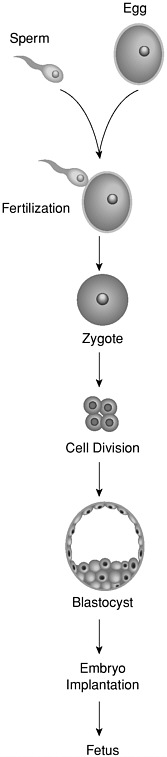
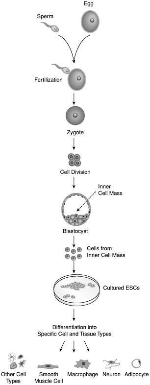
Embryonic stem cells (ESCs) are derived from an early-stage embryo. Fertilization of an ovum by a sperm results in a zygote, the earliest embryonic stage (Figure 1). The zygote begins to divide about 30 hours after fertilization and by the third-to-fourth day, the embryo is a compact ball of 12 or more cells known as the morula. Five-to-six days after fertilization, and after several more cycles of cell division, the morula cells begin to specialize, forming a hollow sphere of cells, called a blastocyst, which is about 150 microns in diameter (one-seventh of a millimeter). The outer layer of the blasotocyst is called the trophoblast, and the cluster of cells inside the sphere is called the inner cell mass. At this stage, there are about 70 trophoblast cells and about 30 cells in the inner cell mass. The cells of the inner cell mass are multipotent stem cells that give rise to all cell types of the major tissue layers (ectoderm, mesoderm, and endoderm) of the embryo. In the past 3 years, it has become possible to remove these stem cells from the blastocyst and maintain them in an undifferentiated state in cell culture lines in the laboratory (NIH, 2001) (Figure 2). To be useful for producing medical therapies, cultured ESCs will need to be differentiated into appropriate tissues for transplantation into patients. Researchers are just beginning to learn how to achieve this differentiation.
Fetal stem cells are primitive cell types in the fetus that eventually develop into the various organs of the body, but research with fetal tissue so far has been limited to only a few cell types: neural stem cells, including neural crest cells; hematopoietic stem cells; and pancreatic islet progenitors. Neural stem cells, which are numerous in the fetal brain, can be isolated and grown in an undifferentiated form in culture, and they have been shown to differentiate into the three main types of brain cells (Brustle et al., 1998; Villa et al., 2000). These cells have been used in rodent models of Parkinson’s disease (Sawamoto et al., 2001; Studer et al., 1998). Neural crest cells arise from the neural tube and migrate from it throughout the developing fetus. They are able to develop into multiple cell types, including the nerves that innervate the heart and the gut, non-neural cells of hormone-secreting glands, pig-
ment cells of the skin, cartilage and bone in the face and skull, and connective tissue in many parts of the body. Neural crest cells from mice have been cultured in the laboratory.
The fetal liver and blood are rich sources of hematopoietic stem cells, which are responsible for generating multiple cell types in blood, but their properties have not been extensively investigated. Although not part of the fetus, the umbilical cord and placenta are also rich sources of hematopoietic stem cells. Tissue extracted from the fetal pancreas has been shown to stimulate insulin production when transplanted into diabetic mice, but it is not clear whether this is due to a true stem cell, a more mature progenitor cell, or to the presence of fully mature insulin-producing pancreatic islet cells themselves (Beattie et al., 1997). Finally, multipotent cells called primordial germ cells have been isolated from the gonadal ridge, a structure that arises at an early stage of the fetus that will eventually develop into eggs or sperm in the adult. Germ cells can be cultured in vivo and have been shown to give rise to multiple cell types of the three embryonic tissue layers (Shamblott et al., 1998).
Adult stem cells are undifferentiated cells that occur in a differentiated tissue, such as bone marrow or the brain, in the adult body. They can renew themselves in the body, making identical copies of themselves for the lifetime of the organism, or become specialized to yield the cell types of the tissue of origin. Sources of adult stem cells include bone marrow, blood, the eye, brain, skeletal muscle, dental pulp, liver, skin, the lining of the gastrointestinal tract, and pancreas. Studies suggest that at least some adult stem cells are multipotent. For example, it has been reported that stem cells from the bone marrow, a mesodermal tissue, can give rise to the three major types of brain cells, which are ectodermal derivatives (Mezey et al., 2000) and that stem cells from the brain can differentiate into blood cells and muscle tissue (Bjornson et al., 1999), but these findings require verification. It is not clear whether investigators are seeing adult stem cells that truly have plasticity or whether some tissues contain several types of stem cells that each give rise to only a few derivative types. Adult stem cells are rare, difficult to identify and purify,
and, when grown in culture, are difficult to maintain in the undifferentiated state. It is because of those limitations that even stem cells from bone marrow, the type most studied, are not available in sufficient numbers to support many potential applications of regenerative medicine. Finding ways to culture adult stems cells outside the body is a high priority of stem cell research.
Additional terms used throughout this report are defined in the Glossary. Although stem cells from all sources are important, the focus of this report is on the characteristics and therapeutic potential of ESCs and adult stem cells that have been at the center of scientific debate.


