


Below is the uncorrected machine-read text of this chapter, intended to provide our own search engines and external engines with highly rich, chapter-representative searchable text of each book. Because it is UNCORRECTED material, please consider the following text as a useful but insufficient proxy for the authoritative book pages.
Di-n-butyl Phthalate John T. James, Ph.D. Johnson Space Center Meclical Sciences Division Houston, Texas PHYSICAL AND CHEMICAL PROPERTIES Di-n-butyl phthalate (DBP) is a colorless to faint yellow viscous liquid with a slight aromatic odor and a strong bitter taste (see Table 3-1) (NIOSH 1994; Grant 1986~. OCCURRENCE AND USE DBP is widely used as a plasticizer to give flexibility to manufactured materials. It is not covalently linked to the plastic polymer, so the potential for leaching from the plastic into contained items such as water or food is high. DBP is also used in insect repellents, lacquers, and rocket propellants. During the NASA/Mir program, DBP was found in the recycled water at an average concentration of 14 micrograms per liter (vigil), with a high of 297 ~g/L (Pierre et al. l 999~. The humidity condensate from one shuttle flight contained DBP at concentrations of 20-42 ~g/L (Straub et al. 1995~. In comparison, in approximately 1,500 samples of ground and surface water in Alberta, Canada, the concentrations of DBP averaged 1 vigil, with a high of 7 ~g/L (Chen and Meek 1994~. In a summary of data on municipal 88
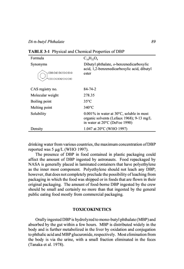
Di-n-buty! Phthalate TABLE 3-1 Physical and Chemical Properties of DBP 89 Formula Synonyms WACO O -CH 2-CH 2-CH 2-CH 3 Em CO 0 -CH 2-CH 2-CH 2-CH 3 CAS registry no. Molecular weight Boiling point Melting point Solubility Density Cl6H22O4 Dibutyl phthalate, o-benzenedicarboxylic acid, 1,2-benzenedicarboxylic acid, dibutyl ester 84-74-2 278.35 35°C 340°C 0.001% in water at 30°C, soluble in most organic solvents (Lefaux 1968~; 9-13 mg/L in water at 20°C (DeFoe 1990) 1.047 at 20°C (WHO 1997) drinking water from various countries, the maximum concentration of DBP reported was 5 ~g/L (WHO 1997~. The presence of DBP in food contained in plastic packaging could affect the amount of DBP ingested by astronauts. Food repackaged by NASA is generally placed in laminated containers that have polyethylene as the inner most component. Polyethylene should not leach any DBP; however, that does not completely preclude the possibility of leaching from packaging in which the food was shipped or in foods that are flown in their original packaging. The amount of food-borne DBP ingested by the crew should be small and certainly no more than that ingested by the general public eating food mostly from commercial packaging. TOXICOKINETICS Orally ingested DBP is hydrolyzed to mono-butyl phthalate (MBP) and absorbed by the gut within a few hours. MBP is distributed widely in the body and is further metabolized in the liver by oxidation and conjugation to phthalic acid and MBP glucuronide, respectively. Most elimination from the body is via the urine, with a small fraction eliminated in the feces (Tanaka et al. 1978~.
9o Spacecraft Water Exposure Guidelines Absorption Absorption of an oral dose of DBP from the gut is related to its hydro- lysis to the monoester derivative (Lake et al. 1977~. This was demonstrated by showing the ability of intestinal tissue from rats, ferrets, baboons, and humans to hydrolyze DBP in vitro. The kinetics and extent of absorption of a single oral dose of DBP by pregnant rats depends on the dose. Rats given 500 milligrams per kilogram (mg/kg) of radiolabeled DBP showed peak radioactivity in the plasma in 1-2 hours (h), whereas rats given 1,500 mg/kg had peak plasma radioactivity in 4-6 h (Saillenfait et al. 1998~. Distribution After an oral dose, DBP and its metabolites are distributed in the blood stream to many organs; however, they are rapidly excreted in the urine and feces. The tissue distribution 24 h after rats were given an oral dose of 14C- labeled DBP at 60 mg/kg showed the following percentages of the dose remaining: intestine, 1.5%; fat, 0.7°/0; muscle 0.3°/0; liver, 0.06%; blood, 0.02%; and kidney, 0.02% (Tanaka et al. 1978~. Radioactivity was unde- tectable in the brain, heart, lung, spleen, testes, prostate, and thymus. After 14 days (~) of administration of DBP at 5 grams (g) per day to pigs in their feed, several tissues were studied for accumulation of DBP (Jarosova et al. 1999~. Phthalates were assayed in the liver, kidneys, lungs, brain, heart, muscle, renal fat, and subcutaneous fat. The highest concentra- tions were found in the muscle and adipose tissue. Similarly, after 14 ~ of administration of 100 mg/d to chickens in their feed, the muscle, skin, liver, and mesenterial fat were assayed for DBP (Jarosova et al. 1999~. The au- thors report that the distribution in those tissues was uniform. Fourteen and 28 ~ after the dosing was stopped, both species seemed to have an accumu- lation of the phthalates in adipose tissue. Saillenfait et al. (1998) reported the tissue distribution of radioactivity following a single oral dose of ~4C-labeled DBP at 500 mg/kg or 1,500 mg/kg to pregnant rats on gestation day 14. The tissues examined were the plasma, kidney, liver, ovary, uterus, placenta, embryo, and amniotic fluid. At the lower dose, radioactivity peaked in each tissue within 1-2 h and declined rapidly, although kidney levels lagged somewhat. Except for plasma, radioactivity on a tissue-weight basis was consistently highest in the kidneys and second-highest in the liver. Radioactivity was almost unde- tectable in any tissue 48 h after the dose was given. At the higher dose, the tissue distribution was similar to the distribution reported for the lower
Di-n-buty! Phthalate . . .. . 91 dose; however, the maximum levels in the tissues appeared 2-6 h after the dosing and then began a slower decline than that reported for the lower dose (Saillenfait et al. 1998~. Metabolism Based on studies with intestinal tissue extracts from a variety of species, the initial step in the metabolism of DBP is hydrolysis to MBP and, presum- ably, n-butano! before the dose is absorbed from the gut (see Figure 3-1) (Lake et al. 1977~. In rats, the hydrolysis seems to continue, because o-phthalic acid has been reported in the urine of rats given an oral dose of DBP (ACGIH 1991~. Alternatively, the remaining busy! group in MBP can be oxidized in the liver, or the MBP can be conjugated to form MBP glucuronide (Tanaka et al. 1978; Foster et al. 1982; Saillenfait et al. 1998~. There are distinct differences in the proportion of metabolites produced by different species. Unconjugated MBP is 3-4 times higher in the urine from rats than in the urine from hamsters, even though each species has comparable esterase in the gut (Foster et al. 1982~. Both species of animals were treated at a dose of 2 g/kg body weight. However, the authors postu- late that the higher level of free MBP may explain the higher susceptibility to testicular toxicity in rats when compared with the susceptibility in ham- sters. The blood disposition of MBP is important because this metabolite is thought to mediate the effects in target tissue. Intravenous doses of DBP in the range of 8-34 mg/kg in rats gave a biphasic blood elimination profile for MBP. This was modeled best with a diffusion-limited' pH-trapping pharmacokinetic model (Keys et al. 2000~. Diffusion limited models postu- late that the MBP must be in the non-ionic form to move between the blood and tissue compartments. The pH-trapping model postulates that un-ionized MBP is "trapped" as MBP- in the tissue, thus preventing its movement back into the blood from the tissue until it becomes un-ionized again. This model was able to give reasonably good predictions of blood MBP for approximately 1 d after oral doses of DBP at 43 mg/kg to 857 mg/kg in rats. The biphasic elimination pattern became more obscure as the concentrations were increased. Elimination The primary route of elimination in rats is the urine, but the feces is also
92 Spacecraft Water Exposure Guidelines WACO ~ -CH 2-CH 2-CH 2-CH AL CO 0 -CH 2-CH 2-CH 2-CH 3 D i-~-bu~l Phthalate [D B P) CH CO H -CH 2-CH 2-CH ~ Cal G n-butanol ~ ~ CO O H ~ ~ CO O H ~ CO O G lucuronide ~ CO O H AL CO O -CH 2-CH 2-CH 2-CH 3 MEL CO O -CH 2-CH 2-CH 2-CH Phthalic Acid 11 Phthalate [~18 P) b1 E P G lucuronide WACO O -CH 2-CH 2-CH O H -CH ~ WACO O -CH 2-CH 2-CH 2-CH ZO H 3-Hydroxy-bu~l Phthalate / /~COOH ~LCo 0 -CH 2-CH 2-CO -CH 4-Hydroxy-blJ~I Phthalate If \rCOOH ~COO-CH2-CH2-CH2-COOH 3-Keto-blJ~l Phthalate 4-Carbox~propyl Phthalate FIGURE 3-1 Metabolism of DBP. Source: ATSDR 1990. an important route of elimination. Radioactivity from ~4C-labeled DBP given to rats orally in 3°/O dimethyl sulfoxide (DMSO) at a dose of 60 mg/kg was, on average, 94°/O eliminated in the urine and feces within 24 h ofthe dose (Tanaka et al. 1978~. Ofthe five animals studied, the fraction in the urine varied from 81% to 98°/O, and the fraction in the feces varied from 1% to SILO. On average, an additional 6% of the radioactivity was eliminated during the second day after dosing (Tanaka et al. 1978~. All of the metabolites shown in Figure 3-1 were found in rat urine except the keto compound; MBP was the predominate metabolite in urine. Hamsters and guinea pigs showed slightly different metabolite profiles in their urine sam- ples (Tanaka et al. 1978~. TOXICITY SUMMARY The toxicity database focuses on the reproductive and developmental
Di-n-buty! Phthalate 93 effects caused by ingestion of DBP. In laboratory animals, the acute and short-term toxicity of the compound is very low by oral ingestion. Well- documented subchronic studies in animals have only recently become avail- able, and no complete reports of a chronic study of DBP could be found. There are no well-documented human oral studies that included a meaning- ful number of subjects. For the purposes of risk assessment, the best studies are those using an appropriate species, animals ofthe appropriate age, doses that produce a NOAEL (no-observed-adverse-effect level) or a clear dose response suitable for benchmark dose (BMD) modeling, and a route of administration that is similar to ingestion in water. Acute Toxicity (1-5 d) Acute oral data available in the NIP chemical registry indicates toxici- ties as shown in Table 3-2. The compound is considered mildly toxic by ingestion. When male rats 3-4 weeks (wk) old were given DBP by oral intubation for 3 ~ at a dose of 2,000 mg/kg, they had testicular injury and a 24% de- crease in testicular weight (Cater et al. 1977~. The weight decrease wors- ened in the 10 ~ after the treatment ceased. The reduced testicular weight was found in rats given 1,000 mg/kg, but not in those given 500 mg/kg for 4 d; however, there was an 1 8°/O loss in testicular weight in rats given DBP at 500 mg/kg for 6 ~ (Cater et al. 1977~. A similar effect was caused on the testes by MBP, but not by n-butano! or o-phthalic acid. The 4-d NOAEL for testicular effects in young rats was 500 mg/kg/d by bolus oral dose. A case report of an acute poisoning after ingestion of approximately 10 g of DBP was described and cited by Le faux (1968~. The man developed nausea and vertigo and was taken to the hospital on the day after ingestion. Albumin and blood cells were noted in the urine; the patient was treated and discharged 14 ~ later with no apparent lasting effects. Whether a person could actually swallow that amount of DBP has been questioned because of its strong and bitter taste (Grant 1986~. Short-Term Toxicity (6-30 d) The effects of~short-term ingestion have been studded by few investiga- tors except for the purpose of understanding the reproductive toxicity, de- velopmental effects, or other specific toxicologic properties of DBP. The
94 TABLE 3-2 Acute Oral Toxicity of DBP Spacecraft Water Exposure Guidelines Type of Dose Species Amount TDLo Human 140 mg/kg LD50 Rat 8,000 mg/kg LD50 Mouse 5,300 mg/kg LD50 Guinea pig 10,000 mg/kg Source: NIP 1991. reproductive and developmental toxicity of DBP is discussed in specific sections below. Groups of five F-344 male rats and groups of five female rats were given DBP in their food at concentrations of 0°/O, 0.6%, 1.2%, or 2.5% for 21 ~ (CMA 1986~. Body weights and food intake were monitored, serum cholesterol and triglyceride were measured (after fasting), and the liver, kidneys, and testes were taken at necropsy for weighing and histopathology. Biochemical parameters indicative of peroxisome proliferation (PP) were assayed in liver tissue. DBP increased the relative liver weights in all DBP-exposed groups; the testes from the high-dose group were 30-40°/O lower in weight and showed severe atrophy upon histopathologic examina- tion. The report indicates that serum cholesterol and triglyceride were decreased in all treated males and only cholesterol was decreased in all treated females, but the decreases were not dose-related (CMA 1986~. Inspection of the data suggests that biologically significant differences related to DBP exposure were not demonstrated by the changes in triglycer- ides and cholesterol. Evidence of PP was found in the livers from all groups oftreated males and in the highest-dose female group. The NOAEL from this study was the lowest dose in the feed, or 0.6%. This was calcu- lated to be equivalent to approximately 620 mg/kg/~. This study was con- ducted according to good laboratory practices (GLPs) promulgated by EPA. It is important to note that there is a direct correlation between the time of ingestion of DBP and the magnitude of the hepatomegaly observed in rats. When 3- to 4-wk-old male rats were given DBP at 2,000 mg/kg in corn oil by gavage for 14 4, the relative liver weights of the exposed rats progressively exceeded the relative liver weights of the controls (Cater et al 1977~. The fraction by which the exposed rat livers exceeded control rat livers was as follows: after 3 4, WHO; after 7 4, 10%; after 10 4, 11%; and after 14 4, 14%.
Di-n-buty! Phthalate 95 Subchronic Toxicity (30-1X0 d) Two thorough rodent subchronic feeding studies have been reported one in F-344 rats and B6C3F~ mice (NTP 1995), and another in Wistar rats (BASE 1992~. Male and female Wistar rats were administered DBP in their food at concentrations of 0, 400,2,000, and 10,000 parts per million (ppm) over a period of 3 months (mo) (BASE 1992~. There were 10 rats per group and gender, and each was evaluated for weight gain, nervous system function, clinical pathology, gross pathology, and histopathology at appropriate times during the study or at the end ofthe exposure period. At 10,000 ppm (750 mg/kg/~) the changes were as follows: decrease in serum triglycerides and triiodothyronine, increase in cyanide-insensitive palmitoyI-CoA-oxidation in the livers of both genders (indicator of PP), transient decrease in red cell indices in males, increase in serum glucose and albumin in males, and in- crease in liver weights in both genders (14% in males and 16% in females). No DBP-related changes were detected at the lower concentrations, hence the NOAEL was 2,000 ppm (150 mg/kg/~. This study was conducted according to Organisation of Economic Cooperation and Development (OECD) guidelines, followed good laboratory practices, and was reported in sufficient detail to be useful for setting a health standard. During a standard 13-wk feeding study, male and female F-344 rats received DBP at 0, 2,500, 5,000, 10,000, 20,000, and 40,000 ppm in their food starting at 5-6 wk of age. Male and female B6C3F~ mice received DBP in their food at half the concentrations received by the rats (NTP 1995~. The findings in rats can be summarized as follows: reduced final body weights in male rats receiving 10,000 ppm or higher and in female rats receiving 20,000 ppm or higher; hepatomegaly in male rats at or above 5,000 ppm and in female rats at or above 10,000 ppm; reduced testicular weight in male rats at 20,000 and 40,000 ppm; slight anemia in male rats at or above 5,000 ppm; reduced cholesterol in male and female rats receiving 10,000 ppm or more; reduced triglycerides in all DBP-exposed male rats and in female rats receiving 10,000 ppm or more; elevated peroxisome enzymes in male and female rats ingesting 5,000 ppm or more; and deple- tion of the germinal epithelium and zinc in testes of male rats given 20,000 ppm or more. If the hypocholesterolemia is not considered an adverse effect, then the NOAEL was 2,500 ppm for F-344 rats. These results and the demonstrated toxic effects of DBP in rats are fully consistent with those reported in the BASF (1992) study.
96 Spacecraft Water Exposure Guidelines Several opportunities for BMD calculations were apparent from the subchronicNTP (1995) study. Selected possibilities are shownin Table 3- 3. The bile acid increases in serum reflect cholestasis (alkaline phospha- tase was also increased), which is most likely related to the hepatomegaly resulting from PP. Because PP is not known to be inducible in humans by phthalates, a risk assessment of this change was not performed. Kidney weights were increased end were clearlyrelatedto DBP exposure; however, despite extensive histopathology, there were no kidney changes noted, so this change was not confirmed as adverse. BMD analyses were conducted on the two indices of testicular injury and on the reduced RBC counts (Ta- ble 3-4, Figures 3-2 and 3-3~. A NOAEL of 176 mg/kg/d was identified from the NIP (1995) study. Mice exposed to DBP for 13 wk exhibited many of the same effects reported for rats (NIP 1995~. Body-weight gains were reduced for males and females in the 5,000 ppm group and above; liver weights were in- creased in males and females ingesting 5,000 ppm or above; a slight anemia was found in females ingesting 20,000 ppm; and microscopic changes in the liver were evident in males receiving 10,000 ppm or above and in females receiving 20,000 ppm. The NOAEL for the effects of DBP on mice ex- posed in this study was 2,500 ppm (353 mg/kg/d for males) (NIP 1995~. Because mice were less sensitive than rats, no attempt was made to do BMD calculations using mice data. Chronic Toxicity (0.5 y to lifetime) The early work of investigators in the U.S., Germany, and France was summarized by Le faux (1968~. The U.S. investigator (C.C. Smith) gave DBP in food to groups of 10 male rats at concentrations of 0.0°/O, 0.01%, 0.05%,0.25%, and 1 .25% for 12 mo starting at 5 wk of age. Those concen- trations are equivalent to 0, 5,25,125, and 600 mg/kg/d, respectively. The three lower doses did not inhibit the growth ofthe rats; however, the highest dose killed half the rats within the first week. No specific lesions could be found at necropsy. Apparently, the survivors in the high-dose group gained weight at a rate comparable to controls. The animals from all groups that survived for 1 y were subjected to gross necropsy and hematologic studies. Both evaluations were negative in all groups except the highest-dose group. The results of this study suggest a NOAEL of 125 mg/kg/d in rats.
97 -i O ~ 0 0 ~ O o o o ° o o o V ^ ~ on ~ o ~ ~ ~ o ~ ~ ~ ~ =_' ~ ~ or or or or Ct V] Em Ad an Cat an 5 an Cat an V] ~ ^ ~ · ~ O ¢ .= as an U. O · - as ¢ Em a~ a~ . u, ~ - ~ ¢ a~ · ~ ~ a~ a~ a~ u, ~ o cd ~ ~ o o o o . . . o o o ~ ~ o . . . o o. o. o o o -H ~ ~ . . . ~ o o Mo ~ ~ o ~ ~ . . . . . . ~ oo ~ ~ o Mo . . . . . . ~ o ~ ~ o ~ ~ ~ ~ ~ ~ ~o ~ Mo ~ ~^ o ~ ~ ~ ~ ~ o o M o ~ ~ ~ ~ ~ E~ ~o .= o c) cd u, c) o ~ E~ ~ ~o cd ~ c) . - . . .~ c) v) v)
98 Spacecraft Water Exposure Guidelines TABLE 3-4 BMD Analysis of Testicular and Hematologic Lesionsa Absolute Weight Atrophy of Germinal of Right Testis Epithelium RBC Count Risk Level 10% 1% 10% 1% 10% 1% BMR 1.3 0.4 0.1 0.01 1.3 0.4 BMD 820 684 652 583 836 611 BMDL 731 591 383 111b 397 102b Model Hill Probit Hill aBased on NTP's ~ 1995) 13 -wk study. bUsed in Rationale section to estimate an acceptable concentration for humans. Abbreviations: BMD,benchmark dose; BMDL, lower confidence limit on the BMD; BMR, benchmark response; NIP, National Toxicology Program; RBC, red blood cell. Source: Data Tom NTP 1995. The study in Germany (G. Bornmann) involved gavage administration of DBP to rats at doses of 0.5 mL/kg and 0.25 mL/kg twice a week for 52 wk. Clinical pathology tests were normal; however, a few of the animals developed sarcomas. The French study (E. Le Breton) involved Wistar rats receiving 0.1, 0.3, and 0.5 g/kg in their diets for 3-5 generations. In a com- plementary study, rats were given the two lowest doses for 21 mo, and the high-dose group was given the DBP-enriched diet for 15 mot The French study concluded that under the test conditions there were no harmful effects of DBP and that the three doses were equivalent to a 70-kg, moderately active human ingesting 60, 180, and 300 mg of DBP per day, respectively. Even though these tests were not conducted according to modern protocols, they seem to suggest that the chronic oral toxicity of DBP is low. Genotoxicity The majority of genotoxicity testing of DBP has given negative results, and that makes sense given that neither of the immediate metabolites of DBP, MBP and n-butanol, are mutagenic (WHO 1997~. The majority ofthe DBP studies have been in vitro, and a wide variety of test systems have been employed. The results ofthose tests have been summarized in review documents, and the reader is referred to those or the original publications for detailed results (WHO 1997; ATSDR 1990~. Many of the negative findings with S. typhimurium seem to be incompletely reported and should
Di-n-buty! Phthalate 1 =, 0.8 a) ~0.6 Cal A 0.4 0.2 o 99 Probit Model with 0.95 Confidence Level Probit BMD Lower Bound I. .o / / ~ ,,' i ~ BMDIL I BMD i . . , ~ 0 200 400 600 800 1000 1200 1400 1600 Dose FIGURE 3-2 BMD analysis ofthe incidence of germinal epithelial atrophy in male rats ingesting DBP for 13 wk (BMDLol = 1 1 1 mg/kg/d). Hill Model with 0.95 Confidence Level 9.4 9.2 a, 9 =8.8 a) ~8.6 ~8.4 8.2 8 7.8 B < MDLI _ 11 \ i \ 1 \ / Hill 0 500 1000 1500 2000 2500 3000 Dose FIGURE 3-3 BMD analysis of RBC data for male rats ingesting DBP for 13 wk ( BMDLol = 102 mg/kg/d).
100 Spacecraft Water Exposure Guidelines be viewed with caution (WHO 1997~. In one of the more thoroughly re- ported studies, Zeiger et al. (1985) reported negative results for strains TA100, TA1535, TA1537, and TA98 using 100-10,000 fig per plate, with and without activation from S9 (derived from the livers of two induced species). The protocol included repeat assays and positive controls. The only positive results in vitro have occurred when metabolic activa- tion using the S9 fraction was not used in the test system. Those results have either been very weakly positive and inconsistent with other results, or they have occurred at cytotoxic concentrations where false positives can be expected (Seed et al. 1 982; Agarwal et al. 1 985; NTP 1 995; WHO 1 997~. For example, Seed et al. (1982) found a 50°/O increase over baseline in the number of mutants from TA100 strain exposed to DPB in liquid suspension at 0.18 mM without S9 activation; however, there was clear evidence of cytotoxicity at this concentration. Also, in the absence of metabolic activa- tion and at concentrations up to 0.03 mg/mL, an equivocal result was ob- tained for chromosomal aberrations in a Chinese hamster fibroblast cell line (Ishidate and Ohashima 1977), but other tests of chromosomal effects have been negative. Neither SCEs nor chromosomal aberrations were induced in Chinese hamster ovary (CHO) cells exposed at concentrations up to 27.8 mg/mL (Abe and Sasaki 1977~. There was an increase in mutant frequency in L1578Y mouse lymphoma cells under conditions of nonactivation and at concentrations above 46 ~g/mL, but that increase occurred at cytotoxic doses (NTP 1995~. False positives are expected from such assays at cyto- toxic concentrations (WHO 1997~. Reproductive Toxicity DBP has received considerable study as a reproductive toxicant to male and female rodents, and it has recently been reported to have weak in vitro estrogen-like activity. End points in males and females can be evaluated separately. It is important to NASA that astronauts not incur adverse effects on their reproductive organs during spaceflight; however, NASA does not anticipate that successful reproductive outcomes during flight are plausible. This means that studies in which animals are fed the test substance during gestation are not considered directly relevant for risk assessment in astronauts; however, adverse reproductive performance or pathology at the end of a subchronic or chronic study is relevant to risk assessment for astro- nauts. It is reassuring that "reproductive-system necropsy data from general toxicity studies can provide a valuable preliminary indication of the likely
Di-n-buty! Phthalate 101 reproductive toxicity of the compound under study twhen evaluated by using more complex tests]" (Chapin et al. 1998~. DBP has been shown to cause reproductive toxicity in male rats, mice, and guinea pigs. Evidence that DBP could cause serious injury to the testes was not reported from early, prolonged feeding studies. However, in the mid-70s, studies demonstrate~profound effects on the testes of rats given only a few doses of DBP at relatively high concentrations (Nikonorow et al. 1973; Cater et al. 1977~. The lesion is characterized by decrease in testicular weight, decrease in diameter of the seminiferous tubes, and atrophy of the germinal epithelium. A loss of zinc through urinary excretion and a reduc- tion of zinc in testicular tissue are associated with DBP-induced injury (Gangoli 1982~. A study of eight di-n-alkyl phthalates showed that those capable of testicular injury were also capable of causing excess urinary excretion of zinc and loss of zinc in testicular tissue (Gangoli 1982~. The mouse, rat, and guinea pig are susceptible to DBP-induced testicular injury at 2,000 mg/kg/d for 10 4, but the hamster is not susceptible at that dose. The lowest dose associated with changes in the adult rat testes is 250 mg/kg/d given by gavage for 15 ~ (Srivastava et al. l 990a). At two higher doses (500 and 1,000 mg/kg/~), testicular weight was decreased, semini- ferous tubes degenerated, and testicular enzymes were changed. At the lowest dose, only a change in one of six testicular enzymes was observed. This was an approximately 20% decrease in acid phosphatase. That study provides an apparent NOAEL of 250 mg/kg/~; however, detailedhistopath- ology data or changes in testicular weights were not given. Changes in sperm counts and acid phosphatase activity were reported and have been used in a BMD analysis (Table 3-5, Figure 3-4~. Acid phosphatase was one of six enzymes measured in testes and found to change significantly with DBP administration. The others were sorbital dehydrogenase, lactate dehydrogenase, y-glutamyl transpeptidase, beta-glucuronidase, and glu- cose-6-dehydrogenase. As discussed below, the changes in acid phospha- tase activity are probably adaptive, whereas the reduced sperm counts are adverse. Based on the NOAELs from the BMD analysis, the reduction in acid phosphatase is a more sensitive end point than the reduction in testicular weights. The question is whether enzymatic changes in the testes are an adverse or adaptive response to DBP exposure. Srivastava et al. (199Oa,b) point out that LDH and SDH are associated with postmeiotic celIs, GGT and beta G are markers for Sertoli celIs, and G6PDH is a marker of spermatogenesis. Acid phosphatase is known to increase as spermatocytes mature to spermatozoa. Relatively small changes (e.g., 20%) in acid phos-
102 Spacecraft Water Exposure Guidelines TABLE 3-5 Testicular Changes in Adult Wistar Rats Given DBP by Gavage for 15 ~ BMD Sperm Counts Dose (mg/kg/d) Testes Acid BMD Acid Phosphatase Sperm Phosphatase Activity in Counts (lo6) 1 0% 1 % 10% 1 onto BMR 1.3 0.4 1.3 0.4 0 200 (24)a 6.2 (0.7) BMD 211 60 413 147 250 160 (19)b 5.8 (1.0) BMDL 134 39 234 70c 500 145 (24)b 4.3 (1~1) Polynomial Polynomial model model 1,000 117 (26)b 1.9 (1~7) Homogeneous Homogeneous variance variance aNumber in parenthesis is the SD, which was calculated from the author's SE and number of subjects. bThese outcomes were considered to be statistically significant by the authors (p < 0.05). Statistical significance was not indicated for any of the sperm counts. CUsed in Rationale section to set an acceptable concentration. Abbreviations: BMD, benchmark close; BMDL,lower confidence limit on the BMD; BMR, benchmark response; SD, standard deviation; SE, standard error. Source: Srivastava et al. 1990a. phatase in the testes do not seem to affect the production of sperm, which is the functional role of the testes. Hence, the reduced sperm counts, not the reduced acid phosphatase activity (which could be considered a marker of testicular effects), will be considered the adverse effect. The underlying mechanism of DBP-induced testicular toxicity is un- known; however, it is known that the initial cellular target, according to ultrastructural studies, is the Sertoli cell and that MBP is likely the chemical mediator (Foster 1997~. The earliest lesion is cytoplasmic vacuolization of the celIs due to enIargement of the cisternae of the endoplasmic reticulum. That might be due to decreased secretion of proteins, disruption of ionic pumps, or changes in cytoskeleton support (Richburg and Boekelheide 1997~. The relationship between Sertoli celIs and germ celIs is complex and intimate. A species-specif~c number of germ celIs, numbering approxi- mately 22 in the rat (Chapin 1997), are associated with and adhere to each Sertoli cell. The mechanism of loss of the germinal epithelium through effects on the Sertoli celIs may include the following: disruption of cell-
Di-n-buty! Phthalate 7 6 a) 5 in o4 Q in tY 3 a) ~ 1 o 103 Polynomial Model with 0.95 Confidence Level JET > \ \ \ \ < i, \ \ - \ \ Polynomial \ - BMDL BMD I I I 0 200 400 600 800 1 000 Dose FIGURE 3-4 BMD analysis of sperm counts in rats given DBP by Savage for 15 d (BMDLo~ = 70 mg/kg/d). to-cell junctions, loss of intracellular fibers in Sertoli cells, reduced semi- niferous fluid secretion due to disruption of Sertoli cell microtubules, dis- ruption of signal transduction (e.g., follicle stimulating hormone receptors on Sertoli cells), failure to provide metabolites (e.g., lactate), or Sertoli-cell directed apoptosis of germinal cells (Richburg and Boekeheide 1997~. DBP has been shown to disrupt androgen-related development of male reproductive indices when administered late in the gestation of rats (My~chreest et al. 1999~. DBP does not elicit the responses expected of an estrogen-like compound in developing females but alters androgen-depend- ent processes in developing males, so the action is presumed to be anti-androgenic. Even though DBP and its active metabolite, MBP, do not interact with the androgen receptor, the effects seen in vivo are typical of androgen receptor antagonists such as flutamide (My~chreest et al. 1999~. When compared to flutamide, DBP produced a lower incidence of prostrate agenesis and hypospadias and induced intra-abdominal testes rather than inguinal testes. According to My~chreest et al. (1999), DBP is the only environmental chemical known to have anti-androgenic effects without interacting with the androgen receptor.
104 Spacecraft Water Exposure Guidelines According to a continuous breeding protocol, CD- 1 mice ingested food containing DBP at 0.0°/O, 0.03°/O, 0.3°/O, and 1.0% for 18 wk (Lamb et al. 1987~. According to this protocol, mice were fed the DBP-spiked food for 1 wk. They cohabitated for an additional 14 wk during which DBP was consumed and the pups discarded. The pups were kept for 3 wk and nursed from a mother receiving DBP in her food. During the latter two periods, reproductive function was measured as the number of litters per breeding pair, number of live pups per litter, pup weight, and pup survival. Even though the body weights ofthe 1.0% males were less than the control body weights, there was not a statistically significant difference in the weights of the testes, epididymis, prostate, or seminal vesicles among the groups (Lamb et al. 1987~. In an NTP continuous breeding study, SD rats were given DBP at 0.1%, 0.5°/O, and 1.0% in their food; this produced daily intakes of 50, 250, and 500 mg/kg (males) and 80, 380, and 800 mg/kg (females) for the three levels, respectively (Wine et al. 1997~. The Fo generation ingested the DBP-spiked food for 14 wk and produced offspring continuously. The last litter (F.) received the same food as their parents until they were mated at about 90 d. The number of live pups per litter from the Fo rats was reduced in all treated groups by 8-1 TWO, and crossover mating demonstrated that this effect was mediated through the females. Sperm parameters and the estrous cycles were not disrupted in any of the groups. The Fat generation males receiving DBP at 1.0% had a higher incidence (~/10) of degeneration of seminiferous tubules than controls (1/10) or middle-dose animals (3/10~. Thus, DBP is a reproductive toxicant in male SD rats when administered at high doses to two generations of animals (Wine et al. 1997~. The results of the NTP study do not yield a firm conclusion as to the NOAEL for reproductive toxicity. The only change observed in the lowest dose Fo animals was an 8°/O reduction in the number of live pups produced per litter. In the mid-dose group, a 15% reduction in live pups per litter was observed, and the pup weights were also reduced by 4°/O (developmental index). The reduced number of live pups was not seen in the crossover breeding experiments or in the second generation; however, the authors attribute this to the greater statistical power of the initial test compared with the follow-up tests. Possible effects on male reproductive organs were indicated by an incidence ofthree out of 10 Fat males showing degeneration of the seminiferous tubules at 0.5°/O. Even though there was a statistically reducednumber of live pups producedby the Fo generation (12.9 in controls and 11.9 in low-dose), this effect was not demonstrated in the Fat matings (14.0 in controls and 15.5 in low-dose); therefore, the lowest dose to fe- males (80 mg/kg/~) was estimated to be a NOAEL in SD rats.
Di-n-buty! Phthalate 105 The ability of DBP and seven other phthalate esters to exhibit estro- genic activity has been evaluated recently using a battery of in vitro and in vivo tests (Zacharewski et al.1998~. Even at doses as high as 2,000 mg/kg, none of the phthalate esters were positive in the two in vivo assays (uterine weights in immature, ovariectomized rats and vaginal epithelial cell cornif~cation in mature, ovariectomized rats). DBP and two other com- pounds exhibited weak estrogen-receptor-mediated estrogenic activity in the in vitro assays. These results suggest that estrogenic activity might not be involved in the reproductive effects of DBP that have been reported in SD rats (Zacharewski et al. 1998; Wine et al. 1997~. Developmental Toxicity DBP given in high concentrations to rodents has been shown to be toxic to developing fetuses, but only at concentrations that approach those that are toxic to dams. Data from ATSDR (1990~ indicate that the NOAEL in mice is approximately 650 mg/kg/d, whereas the NOAEL in rats is near 60 mg/kg/d for decreased pup weight. In a series of studies published since the ATSDR document was written, Ema et al. (1993,1997,2000) showed that pregnant Wistar rats given DBP only during gestation days 7-15 had in- creased fetal malformations at 630 mg/kg/d, but not at 500 mg/kg/~. They also observed that there may be two discrete response periods for skeletal malformations (days 8-9 and day 15) and that for genital effects in male offspring (undescended testes and reduced anogenital distance), the critical time is gestation days 15-17. When dams were given DBP on gestation days 7-15, maternal body weight gains were statistically below controls for the group given 650 mg/kg/d, but not for the group given 500 mg/kg/d (Ema et al. 1993~. When DBP was administered to pregnant rats for only 3 ~ at 500,1,000, or 1,500 mg/kg/d, the dams had reduced weight gains at the two highest doses, whereas a reduced anogenital distance was observed in male offspring from all groups, with greatest effect on pups from dams given the DBP on gestation days 15-17 (Ema et al. 2000~. In another developmental toxicity study, CD rats were given oral doses of DBP at 0,100,250, and 500 mg/kg/d from gestation day 12 to gestation day 21. F~ males showed a number of morphologic effects on their repro- ductive organs, especially at the highest dose (My~chreest et al.1999~. The only effect seen in the young male rats whose mothers received 100 ~See ATSDR (1990) Table 2-2.
106 C) X ~ -i C) E o .O ~ ~ P X O ~ ~ po ~ ~ o . . Ct Ct Ct Ct V V A g U. at . ~ ad U. Ct at so C) at \ o t so V) o at o of to To Ct (~N at V A g U. at at U. Ct at C) at o o o o \ ~ ~ ~ I' t ~ 1 Ct V) o .= a~ ct ~ ~ .s a~ cd s~ ~ ~ o ~ cd s~ s~ ~ o o - ~ ~ d d d O O O O O 00 Ct S~ o o o O~ o O M0 _' .~ X O E~ V) Ct Cq Ct Cd ~ ° ~ ° a~ ¢ .> ~ ·> ~ ~ ~ V) ~ V) ~ V V o \ ~ U~^ U~^ t~ o 9 5 D 3 ° x ~ x 3 9 .5 ~ .= E ~ ~° ~ ~ ~° ~ ~ ^ s ~ .~ ~ I ~^ ¢ ~ O V) O ~ O ~0 a~ Ct ~ ~ O O O O O O ~ ~0
107 Do Cq, Do ~ as ~ ¢ A ¢ . - CO as To .^ ~ ~ ~ ~ 3 ~^ ~ ~.5 ~ 3 3 ~ 1 ~ ~ of , 5 .> ~ _' ~ ~ to ~ 1 ~ so so 1 ~ _I o V) o V) To as ~ ', ~ an ~ ad ~ o ~ o o o o o o ~ o ~ cd (> ad ·= ¢ ¢ Em EM ~ ~ O - D t~ D D O O p Hi B ~ ~.s ~ ~ ad ad Ct of ~^ a a ~ ~ o Do o to a- ~ ~ ·C) ~ ~ ~ . ~ ~ ad O A t~ t~ ~ ~ .O =- == o == o == == ~ =~e ~e ~ ~ o ~ = - = ~ ~ s~ s~ ~ ~ ~ ~ 0 ~ 0 ~ 0 0 ~ 0 ~ ~ ~ ~ ~ ~ V) oo
108 an so ad ~= ~ A, ad r c, r r 3 AL, c) so, ~ O u, ~ r ~ tilt ~ Al c., ~ ~O 5 S #o ED o At U. ad o C) ad C) ad sin . Ct . ~ an an ~ .= O C) ~ . ED Ct an o an ~ an V MY Cd an an To . ~ a, an A > . ~ O c) . d C) Ct C) a C) ~ SON C) . - · - ~ V) an an of on an an d Ad' an an d on on ~0 ~0 3 3$ V ~ V ~ ~ jar jar . O O _' .~ ~ ~ ~ x 0 EN ~ ~ ~ .O ~ ~ ~ d ~ ~ ~ ~ ~ O O V ~ ~ '0 00 ~ ~ ~0 ·- ·- V) V) o an o o U. O SO ~ ~ ~ an O ~ ~ ¢ ~ O O O
Di-n-buty! Phthalate 109 mg/kg/d was delayed preputial separation. A NOAEL was not found in the study (My~chreest et al. 1999~. However, in a follow up study, a NOAEL of 50 mg/kg/d was found in male rats; the most sensitive effect was nipple development (My~chreest et al. 2000~. Spaceflight Effects The physiologic and biochemical changes that occur during spaceflight would not be expected to increase the sensitivity of astronauts to the toxic effects of DBP on the testes or reproduction; however, astronauts may be more sensitive to hematotoxicants because they loose approximately 10% of their red cell mass within the first few days of spaceflight. Synergistic Effects No data were found on DBP synergism with other chemicals. LIMITS SET BY OTHER ORGANIZATIONS AND COMPARISON TO SWEGS Differences Between EPA's 1-d Health Advisory and NASA's 1-d SWEG Both NASA and EPA began with the 500 mg/kg NOAEL for the 4-d exposure reported by Cater at al. (1977~. The 1-d EPA health advisory (HA) applies to a child ingesting approximately 1 L of water and weighing 10 kg, whereas the 1-d SWEG applies to a 70-kg adult ingesting 2.S L of water per day. This accounts for the difference between the HA and the SWEG. The major reason that the HA (50 mg/L) is much lower than the SWEG (1,250 mg/L) is the difference in uncertainty factors. EPA used a factor of 100 (10 for interspecies differences and 10 for intraspecies differ- ences, presumably); only a factor of 10 for interspecies differences was used to estimate the SWEG. There was no intraspecies factor used in the SWEG because the astronaut population consists of healthy adults, so protection of very young persons or ill persons is not required. There is no evidence of genetic differences that would make certain portions of the healthy adult population unusually susceptible to DBP. (See Table 3-7 for SWEGs and Table 3-S for limits set by other organizations.)
110 Spacecraft Water Exposure Guidelines TABLE 3-7 Spacecraft Water Exposure Guidelines for DBP Duration Concentration (mg/L) 1,200 Target Toxicity Testicular injury Testicular injury H e matoto xi city H e matoto xi city 1 d 10 d 100d 80 1,000 d 40 175 Differences Between the 10-d Health Advisory and the 10-d SWEG . EPA (1992) began with findings from the 1 -y rat study by Smith (1953) In which 125 mg/kg/d was the NOAEL. In contrast, NASA began with the data from Srivastava (199Oa) on the reproductive toxicity of DBP in rats. A BRIDLE of 70 mg/kg/d was estimated for reduced sperm counts using doses from 250 to 1,000 mg/kg/d for 15 d and a polynomial fit of the data (Table 3-5). EPA used parameters for a child (10-kg body weight, 1 L/d drinking water), whereas NASA used adult parameters (70-kg body weight, 2.8 L/d drinking water). Furthermore, EPA used an uncertainty factor of 100, and NASA used an interspecies factor of 10. Hence, EPA's 10-d HA of 10 mg/L is much lower than NASA's 10-d SWEG of 175 mg/L. As noted above, NASA is concerned with protecting healthy adults rather than children and ill adults. There is no reason to expect significant genetic differences in humans that would lead to differences in human responses to DBP. Differences Between the Long-Term Exposure Standards The long-term values set by EPA are based on the 1-y rat study report- ing a NOAEL at 125 mg/kg/d (Smith 1953). The 7-y HA was calculated as follows: 7-y HA = (125 mg/kg/d x 70 kg) (100 x 2 L/d) = 44 mg/L (or approximately 40 mg/L). The 70-y drinking water equivalent level (DWEL) of 4 mg/L was calcu- lated in the same way (through the reference dose tRfD]) as the HA, except that an uncertainty factor of 1,000 was applied. NASA's 1,000-d SWEG
Di-n-buty! Phthalate TABLE 3-X Standards and Limits for DBP Set by Other Organ 111 izations Water Equivalent Organization Name of Standard Value (mg/L)a EPA RfD (oral) 0.1 mg/kg/d 3.5 EpAb 1-d HA(child) 50 EpAb 10-d HA (child) 10 EpAb 7-yHA(adult) 40 EPA 70-y DWEL 4 (adult) aAssumes a 70-kg person consuming 2 L of water per day with no other sources of DBP. bHealth advisories published in 1996 by EPA did not include these HAs. Abbreviations: DWEL, drinking water equivalent level; EPA, U.S. Environmental Protection Agency; HA, health advisory; RfD, reference dose. Source: Data from EPA 1992. was also derived from the NOAEL in the Smith (1953) study, except that an exposure time factor of approximately 2.7 was applied, and the uncer- tainty factor~forpossible species sensitivity differences) was only 10, rather than the 100 or 1,000 used by EPA. Based on NOAEL data from the 1-y Smith (1953) study, and applying the spaceflight factor of 3, a 1,000-d AC for hematologic effects was found to be 40 mg/L. A higher volume of water consumption was also used for the SWEG (see below). The 7-y HA of 40 mg/L and the 1,000-d SWEG of 100 mg/L (if the spaceflight factor of 3 is removed) can be viewed as nearly comparable when the exposure times of 7 y for the HA and 1,000 ~ for the SWEG are considered. The SWEG is 2.5 times the HA, but the HA applies to an exposure time 2.5 times that of the SWEG. The 1 00-d SWEG of 80 mg/L was derived from the results of an NIP study (1995) showing a BMDLo~ for hematologic effects at 102 mg/L (Ta- bles 3-3 and 3-4~. Factors of 10 for species differences and 3 for possible susceptibility to reduced red cell mass were included. There is no EPA limit that can be compared directly to this SWEG. RATIONALE The toxicity of DBP is low; however, at high doses, effects on the tes-
112 Spacecraft Water Exposure Guidelines tes, reproductive function, red blood cells, and livers of rats have been repeatedly demonstrated. The testicular lesion is characterized by changes in enzymes associated with testicular function, vacuolization of Sertoli cells, and a loss of germinal epithelium, which is grossly reflected in de- creased testicular weight. Similarly, the changes in the liver involve prolif- eration of the peroxisomes and increases in related enzymes leading to an increase in liver weight. Liver cancer and earlier changes in the rat liver in response to a peroxisome proliferator such as DBP are extremely unlikely to be an appropriate model for human toxicity. The fundamental reason for this difference appears to be quantitative and possibly qualitative differ- ences in peroxisome proliferator-activated receptor-alpha (Holder and Tugwood 1999~. Risk assessment will not be performed on rodent liver lesions, including biochemical lesions associated with cholestasis. A fur- ther note is that rats seem to be much more susceptible than primates to the toxic effects of DBP and other phthalates; however, the species extrapola- tion factor of 10 will be retained for conservatism and because there are few studies involving human subjects. Developmental toxicity was not consid- ered because pregnant astronauts are not expected to fly in the foreseeable future. (See Table 3-9 for summary of ACs.) Ingestion for 1 d The most appropriate study from which to derive a 1-d acceptable con- centration (AC) is Cater et al. (1977~. Young male rats dosed with DBP at 500 mg/kg for 4 ~ did not show the measurable reduction in testicular weight that was observed for slightly longer exposures and at higher doses. Based on this NOAEL, the 1-d AC for testicular injury was calculated as follows: 1-d AC (testicular injury) = (500 mg/kg x 70 kg) (2.8 L/d x 10~; 1-d AC = 1,250 mg/L (rounded to 1,200 mg/L). This assumes a 70-kg person ingesting 2.8 L of water per day (drinking water and food reconstitution). The factor of 10 accounts for possible dif- ferences in species susceptibility between rats and humans. This AC is likely to be conservative because of the use of highly susceptible young male rats, administration of four doses, and the bolus dosing protocol.
113 A ·_I ·_I o o . - A Cal o V A Cal Cal ¢ I o o v C) C) ¢ o C) Ct .; o o o o o ~ . Fat En Ct is, o . C) .O C) Fat ¢ E-° o · C) C) Ct Ct . . x o Ed Cal Ed x .~ To to A to oo i~ ~ ~ ;~ A, y ~~ ~ ~ EN Z ~~ 2 E ~ . . ~ X C) o ~ ~ ~o o . X
114 5 A o V I o o V C) C) ¢ o C) Ct .; o o o o o . Fix Ed Ct at, o I C) ~ .O C) Fin ¢ E-° no ~ sit · C) ~ C) Ct Ct . . x o En Cal En .~ ~ VO ·^ ·^ ¢ ~ 8 o z o C) o .~ .= ~ V, . ~ . ¢ ~ ~ 8 ° ~ ~ ~ Z .~ C) .~ x o ~o oo o - ^ F~ 3 s~ o ~o ~ o Ct o Ct ~ o o .~ o s~ s~ CO . o\ ~ o o Ct C) .= ~ ~ ~ ~ ~° ° C) ; ~ ~o o o~ .~ o o s~ o C) Ct o ~ .~ Ce ~ .e .o o · ~ o ~ Cl g ~ ~ C . ° o ~ . ·g ~:~ ~= m ~,
Di-n-buty! Phthalate 115 Ingestion for 10 d The AC to protect against reproductive toxicity was based on the BMDLo~ value of 70 mg/kg/d (Table 3-5) calculated from reduced sperm counts found in adult rats given 15 daily gavage doses of DBP at 250-1,000 mg/kg/d (Srivastava et al. 1990a). A NOAEL was not indicated by the investigators. The 10-d AC was calculated as follows: 10-d AC (testicularinjury) = (70 mg/kg/d x 70 kg) (2.8 L/d x 10~; 10-dAC=175mg/L. This value is expected to be conservative because the DBP was delivered in bolus gavage doses rather than gradually in feed or water. This conclu- sion is supported by a newly developed PBPK model for DBP and its me- tabolites that suggests diffusion limitations and pH trapping as mechanisms that limit uptake into human tissues (Keys et al. 2000~. Ingestion for 100 d Toxicologic end points for testicular atrophy and reduced red blood cell counts were considered in setting the 100-d SWEG. Reproductive Toxicity Reproductive toxicity in males was evaluated according to the BMDLo~ calculations shown in Tables 3-3 and 3-4. The BMDLo~ for atrophy ofthe germinal epithelium in rats exposed to DBP in their food for 13 wk was found to be 111 mg/kg/d. The 100-d AC was calculated as follows: 100-d AC (testicular injury) = (1 1 1 mg/kg/d x70 kg) (2.8 L/d x 10 x 1.1~; 100-d AC = 250 mg/L. The uncertainty factors were 10 for species extrapolation and 1.1 for extrap- olation from 90 d to 100 d. Hematotoxicity Data from the NTP (1995) subchronic study suggest a slight anemia on
116 Spacecraft Water Exposure Guidelines the basis of clinical hematology parameters. A BMD analysis (Table 3-4) of the data using the polynomial model gave a BMDLo~ of 102 mg/kg/~. This value can be applied to the situation of astronauts as follows: 100-dAC=~102mg/kgx70kg) (2.8L/4X 10X3 X 1.1~; 100-d AC = 77 mg/L (rounded to 80 mg/L). This incorporates a species extrapolation factor of 10, a time-extrapolation factor of 1.1 for extrapolation from 90 ~ to 100 4, and a factor of 3 for presumptive astronaut susceptibility to hematotoxicants. Ingestion for 1,000 d There are no chronic studies that meet modern standards for quality and completeness of reporting. There are basically two choices: (1) extrapolate from 90-d, subchronic rodent data of high quality (i.e., the BMDLo~ of 96 mg/kg/d that gave an AC of 70 mg/L), or (2) use data from the 1-y rodent study of Smith ~ 1953), knowing that the latter has important limitations. To avoid the long and uncertain time extrapolations from a 90-d study to a 1,000-d guideline, the latter approach was initially selected. The NOAEL from the rat exposures was 125 mg/kg/~. The 1,000-d AC to avoidpre- sumed reproductive effects was calculated as follows: 1,000-d AC = (125 mg/kg/d x 70 kg x 2.7) (2.8 L/d x 10~; 1,000-d AC = 114 mg/L. The factor of 10 is for potential species differences and the factor of 2.7 (1,000/365) is to compensate for the exposure time in the study being less than the AC exposure time. This can be rounded to 100 mg/L for reproduc- tive effects; however, an additional factor of 3 was applied for possible interactions between spaceflight effects and hematotoxicity, which was assumed from the hematotoxic findings in the 90-d study (NTP 1995~. 1,000-d AC (hematotoxic effects) = 114 mg/L 3 = 37 mg/L (rounded to 40 mg/L). RECOMMENDATIONS The greatest weakness in the toxicity database is the lack of a chronic
Di-n-buty! Phthalate 117 feed or drinking water study conducted according to modern protocols. Conducting such a study in rodents would be of limited value, but a chronic study inpr~mates would tee very valuable. Because the chronic studies from Smith (1953) and others were reported in such limited detail, using them as the basis for any standard is risky. Additional studies are needed in pr~- mates, including low-exposure studies in human subj ects. The diversity and distribution of the peroxisome proliferator-activated receptor-isotypes in tissues from human populations need to be assessed to more confidently ascertain the relevance of rodent toxicity and cancer data. REFERENCES Abe, S., and M. Sasaki. 1977. Chromosome aberrations and sister chromatic ex- changes in Chinese hamster ovary cells exposed to various chemicals. J. Natl. Cancer Inst. 58: 1635-41. ACGIH (American Conference of Governmental and Industrial Hygienists). 1991. Documentation of TLVs and BEIs, Vol. 1. American Conference of Govern- mental Industrial Hygienists, Cincinnati, OH. Agarwal, D.K., W.H. Lawrence, L.J. Nunez, and J. Autian. 1985. Mutagenicity evaluation of phthalic acid esters and metabolites in S. typhimurium cultures. J. Toxicol. Environ. Health 16:61-9. ATSDR (Agency for Toxic Substances and Disease Registry).1990. Toxicological Profile for Di-n-Butylphthalate. TP-90-10. U.S. Department of Health Human Services, Washington, DC. BASF. 1992. Study on the oral toxicity of dibutyl phthalate in Wistar rats: Admin- istration in the diet over 3 months. Report: ProjectNo.31 S0449/89020. BASF Corporation, Mount Olive, NJ. Cattley, R.C., J. DeLuca, C. Elcombe et al. 1998. Do peroxisome proliferating compounds pose a hepatocarcinogenic hazard to humans? Regul. Toxicol. Pharmacol. 27:47-60. Cater, B.R., M.W. Cook, S.D. Gangolli, and P. Grasso. 1977. Studies on dibutyl phthalate-induced testicular atrophy in the rat: Effect of zinc metabolism. Tox. Appl. Pharmacol. 41:609-618. Chapin, R.E.1997. Germ cells as targets for toxicants. Chapter 10.09 in Compre- hensive Toxicology, Volume 10: Reproductive end endocrine Toxicology. K. Boekheide, R.E. Chapin, P.B. Hoyer, and C. Harris, eds. New York: Elsevier Science Ltd. Chapin, R.E., R.A. Sloane, and J.K. Kent. 1998. Reproductive endpoints in gen- eral toxicity studies: Are they predictive? Reprod. Toxicol. 12:489-494. Chen, P.K.L., and M.E. Meek. 1994. Di-n-butyl phthalate: Evaluation of risks to health from environmental exposure in Canada. Environ. Carcinog. Ecotoxicol. Rev. C12:257-268.
118 Spacecraft Water Exposure Guidelines CMA (Chemical Manufacturers Association). 1986. A 21-day feeding study of di-n-butyl phthalate to rats: Effects on the liver and liver lipids. Report No. 049. Chemical Manufacturers Association, Washington, DC. May 3, 1985. DeFoe, D.L., G.W. Holcombe, D.E. Hammermeister et al. 1990. Solubility and toxicity of eight phthalate esters to four aquatic organisms. Environ. Toxicol. Chem. 9:623-636. Ema, M., H. Amano, T. Itami, and H. Kawaski. 1993. Teratogenic evaluation of di-n-butyl phthalate in rats. Toxicol. Lett. 69: 197-203. Ema, M., A. Harazono, E. Miyawaki, and Y. Ogawa.1997. Developmental effects of di-n-butyl phthalate after a single administration in rats. J. Appl. Toxicol. 17:223-9. Ema, M., E. Miyawaki, and K. Kawashima. 2000. Critical period for adverse ef- fects on the development of reproductive system in male offspring on rats given di-n-butyl phthalate during late pregnancy. Toxicol. Let. 111 :271 -8. EPA (U.S. Environmental Protection Agency). 1992. Drinking Water Criteria Document for Phthalic Acid Esters (Revised Final Report). Report PB92- 173442. U.S. Environmental Protection Agency, Washington, DC. Foster, P.M.D. 1997. Assessing the effects of chemicals on male reproduction; Lessons learned from di-n-butyl phthalate. CIIT Activities 17: 1 -7. Foster, P.M.D., M.W. Cook, L.V. Thomas, D.G. Walters, and S.D. Gangolli.1982. Differences in urinary metabolic profile from di-n-butyl phthalate-treated rats and hamsters. Drug Metab. Dispos. 11 :59-61. Gangoli, S.D.1982. Testicular effects of phthalate esters. Environ. Health Perspect. 45:77-84. Grant, W.M. 1986. Page 317 in Toxicology ofthe Eye, 3rd Ed. Springfield, IL: Charles C Thomas. Holden, P.R., and J.D. Tugwood. 1999. Peroxisome proliferator-activated receptor alpha: Role in rodent liver cancer and species differences. J. Mol. Endocrinol. 22:1-8. Ishidate, M., and S. Odashima. 1977. Chromosome tests with 134 compounds on Chinese hamster ovary cells in vitro-a screening for chemical carcinogens. Mutat. Res. 48:337-354. Jarosova, A., V. Gajduskova, J. Raszyk, and K. Sevela. 1999. Di-ethylhexyl phthalate and di-n-bytyl phthalate in the tissues of pigs and boiler chicks after their oral administration. Vet. Med. (Prague) 44:61-70. Keys, D.A., D.G. Wallace, T.B. Kepler, and R.B. Conolly. 2000. Quantitative evaluation of alternative mechanisms of blood disposition of DBP and MBP in rats. Toxicol. Sci. 53: 173- 184. Lake, B.G., J.C. Phillips, J.C. Linnell, S.D. Gangolli.1977. The in vitro hydrolysis of some phthalate diesters by hepatic and intestinal preparations from various species. Toxicol. Appl. Pharmacol. 39:239-248. Lamb, J.C., R.E. Chapin, J. Teague, A.D. Lawton, and J.R. Reel.1987. Reproduc- tive effects offourphthalic acid esters in the mouse. Toxicol. Appl. Pharmacol. 88:255-269. Lefaux, R. 1968. Practical Toxicology of Plastics. Cleveland, OH: CRC Press.
Di-n-buty! Phthalate 119 Mylchreest, E., M. Sar, R.C. Cattley, and P.M.D. Foster. 1 999. Disruption of the androgen-regulated male reproductive development by di~n-butyl) phthalate during late gestation in rats is different from flutamide. Toxicol. Appl. Pharmacol. 156:81-95. Mylchreest, E., D.G. Wallace, R.C Cattley, and P.M.D. Foster. 2000. Dose-de- pendent alterations in androgen-regulated male reproductive development in rats exposed to di~n-butyl) phthalate during late gestation. Toxicol. Sci. 55: 143-51. Nikonorow M., H. Mazur, and H. Piekacz. 1973. Effect of orally administered plasticizers and polyvinyl chloride stabilizers in the rat. Toxicol. Appl. Pharmacol. 26:253-9. NIOSH (National Institute for Occupational Safety and Health). 1994. Pocket Guide to Chemical Hazards. DHHS Pub. No. 94-116. Department of Health and Human Services, Washington, DC. NTP (National Toxicology Program). l 991. NTP Chemical Repository, Di-n-Butyl Phthalate. Radian Corporation, August 29, 1991. National Toxicology Pro- gram, U.S. Department of Health and Human Services, Washington, DC. NTP (National Toxicology Program). 1995. Toxicity studies of dibutylphthalate (CAS No. 84-74-2) administered in feed to F344/N rats and B6C3F1 mice. Toxicology Series No. 30. U.S. Department of Health and Human Services, Washington, DC. Pierre, L.M., J.R. Schultz, R.L. Saner et al. 1999. Chemical analysis of potable water and humidity condensate: Phase one final results and lessons learned. SAE-ICES Paper 1999-01 -2028. Warrendale, PA: Society of Automotive Engineers. Richburg, J.H., and K. Boekelheide.1997. The sertoli cell as a target for toxicants. Chapter 10.09 in Comprehensive Toxicology, Volume 10: Reproductive and Endocrine Toxicology, K. Boekheide, R.E. Chapin, P.B. Hoyer, and C. Harris, eds. New York: Elsevier Science Ltd. Saillenfait, A.M., J.P. Payton, J.P. Fabry et al. 1998. Assessment of the develop- mental toxicity, metabolism, and placental transfer of di-n-butyl phthalate administered to pregnant rats. Toxicol. Sci. 45:212-224. Seed, J.L. 1982. Mutagenic activity of phthalate esters in bacterial liquid suspen- sion assays. Environ. Health Perspect. 45:111-4. Smith, C.C.1953. Toxicity of butyl stearate, dibutyl sebacate, dibutyl phthalate and methoxyethyl oleate. Ind. Hyg. Occup. Med. 7:310-8. Srivistava, S., G.B. Singh, S.P. Srivistava, and P.K. Seth. 1990a. Testicular toxicity of di-n-butyl phthalate in adult rats; Effect on marker enzymes of spermatogenesis. Ind. J. Exp. Biol. 28:67-70. Srivastava, S.P., S. Srivastava, D.K. Saxena, S.V. Chandra, and P.K. Sith. 1990b. Testicular effects of di-n-butyl phthalate; biochemical and histopathological alterations. Arch. Toxicol. 64:148-152. Straub, J.E., J.R. Schultz, W.F. Michalek, andR.L. Sauer.1995. Further character- ization and multifiltration treatment of shuttle humidity condensate. SAE- ICES Paper 951685. Warrendale, PA: Society of Automotive Engineers.
120 Spacecraft Water Exposure Guidelines Tanaka, A., A. Matsumoto, and T. Yamaha.1978. Biochemical studies onphthalic esters. III. Metabolism of dibutyl phthalate in animals. Toxicology 9: 109- 123. WHO (World Health Organization). 1977. Environmental Health Criteria 189. Di-n-Butyl Phthalate. Geneva: WHO. Wine, R.N., L.-H. Li, L.H. Barnes, D.K. Gulati, and R.E. Chapin.1997. Reproduc- tive Toxicity of Di-n-butylphthalate in a Continuous Breeding Protocol in Sprague-Dawley Rats. Environ. Health Perspect. 105:102-7. Zacharewski, T.R., M.D. Meek, J.H. Clemons, Z.F. Wu, M.R. Fielden, and J.B. Matthews. 1998. Examination of the in vitro and in vivo estrogenic activities o f eight commercial phthalate e sters. Toxicol. S ci . 46: 282 - 93. Zeiger, E., S. Haworth, K. Mortelmans, and W. Speck.1985. Mutagenicity testing of di(2-ethylhexyl~phthalate and related chemicals in Salmonella. Environ. Mutagen 7:213-232.
