


Below is the uncorrected machine-read text of this chapter, intended to provide our own search engines and external engines with highly rich, chapter-representative searchable text of each book. Because it is UNCORRECTED material, please consider the following text as a useful but insufficient proxy for the authoritative book pages.
CHAPTER 7 RADIOTHERAPY SUMMARY Radiotherapy uses ionizing radiation directed at a human or animal body to treat many serious diseases, most notably cancer. High-activity radionuclide sources can be used to create clinical ionizing radiation beams in the form of high-energy gamma rays in teletherapy machines used for external-beam radiotherapy as well as in the Gamma Knife® used for stereotactic radiosurgery. Only four known radionuclides possess characteristics that make them candidates for use in external beam radiotherapy: cesium-137, cobalt-60, europium-152, and radium-226. Europium-152 has not been developed yet for clinical use, and the use of cesium-137 and radium-226 was discontinued for practical reasons and because of safety concerns. Cobalt-60 is currently used in external beam radiotherapy devices found mostly in developing countries. This is because the linear accelerator (linac) is considered a better and more versatile radiotherapy tool, and has largely supplanted cobalt-60 teletherapy devices in the United States and other developed countries. Currently in the United States there are several thousand radiotherapy devices in over 2,400 institutions and clinics (Ballas et al., 2006). Fewer than 250 cobalt-60 teletherapy devices are licensed in the United States and most of those are thought to be in storage for decay, in use for other purposes (such as fixed radiography), or in use for teaching. Radiosurgery is an irradiation technique that uses radiation beams from many directions to treat lesions. It is now used mainly to treat brain lesions with the Gamma Knife®, a device with approximately 200 cobalt-60 sources. Radiosurgery can also be practiced with isocentric linacs as well as with a miniature linac mounted on a robotic arm, but many neurosurgeons and some radiation oncologists believe that Gamma Knife® radiosurgery is superior to linac-based radiosurgery. The cost of radiosurgery with a dedicated linac is similar to that with a Gamma Knife®, while the cost of radiosurgery with a standard linac modified for radiosurgery is much lower. In addition, linac-based radiosurgery is more versatile for development of stereotactic techniques in the treatment of lesions in organs other than the brain, and on a fractionated (rather than a single-treatment) basis. Development of new treatment technologies and shifts in practices in radiotherapy are driven by innovation and decisions about delivery of health care, at both the macrolevel (e.g., insurance reimbursements) and at the personal level (i.e., doctor-patient treatment planning). Medical technology researchers already seek out opportunities to improve nonradionuclide- based radiotherapy and radiosurgery. Because linacs have already largely replaced radionuclide-based teletherapy devices, the committee concludes that teletherapy services, while important, are lesser and declining concerns with respect to radiation source security. It is also possible that the recent rapid growth in Gamma Knife® installations in the United States and the developed world will soon subside, given that a viable alternative is offered with linac- based radiosurgery. 117
118 RADIATION SOURCE USE AND REPLACEMENT INTRODUCTION Radiotherapy is defined as treatment of disease with ionizing radiation. The diseases treated by radiotherapy are mainly malignant; however, on a smaller scale, radiotherapy is also used in treatment of certain benign conditions. Although the mortality rate for all cancer sites combined has been decreasing, age-adjusted incidence rates for all cancers combined have been stable over the most recent periods of analysis (U.S. Cancer Statistics Working Group, 2006), and the incidence of some cancers is increasing. In developed countries, the increasing life expectancy (around 75 years for boys and 80 years for girls born in 2004 in the United States; NCHS, 2006) combined with low birth rate is causing significant aging of the population and making cancer the second most common cause of death in the United States after heart disease. The American Cancer Society recently estimated that about 1,445,000 new cases of invasive cancer will be diagnosed in Americans in 2007 (ACS, 2007), among a population of about 302 million (about 1 in 210 people), according to the U.S. POPClock Projection of the U.S. Bureau of the Census (U.S. Census Bureau, 2007). Thus, in the developed world, cancer is a major health problem, and treatment of cancer is of major importance to modern societies. The cancer rate is lower in developing countries because people in those countries generally die of causes other than cancer at a younger age. Nevertheless, it is estimated that the incidence of cancer worldwide stands at some 10 million new cancer patients per year. Radiotherapy is one of three important cancer treatment modalities; the other two are surgery and chemotherapy. In a modern health care system over 50 percent of cancer patients receive radiotherapy either as primary treatment or in conjunction with surgery and chemotherapy. There are two main types of radiotherapy: external beam radiotherapy and brachytherapy (also referred to as curietherapy or endocurie therapy). In a typical radiotherapy department about 80 percent of treatments are done with external beam radiotherapy, the rest with brachytherapy. External beam radiotherapy is delivered with the radiation source at a certain distance from the patient, whereas brachytherapy is carried out with radiation sources placed directly into the patient, either into a body cavity (intracavitary brachytherapy) or surgically into a body organ (interstitial brachytherapy). During the past 100 years of radiotherapy many types of equipment have been designed and used for external beam radiotherapy. Modern external beam radiotherapy is delivered with: ⢠x-ray machines producing kilovoltage x-ray beams, typically used for treatment of superficial (skin) lesions; ⢠teletherapy machines producing megavoltage gamma-ray beams, typically used for treatment of deep-seated lesions; ⢠linacs producing megavoltage x-ray beams, typically used for treatment of deep- seated lesions and megavoltage electron beams typically used for treatment of superficial (skin) lesions. Brachytherapy sources are most commonly gamma emitters manufactured in the form of a sealed source encapsulated into a special container to prevent leakage of the radioactive material. Because the activity of brachytherapy sources is relatively low (on the order of 0.37 TBq [10 Ci] or less), they are Category 3 sources and as such are outside of this studyâs scope. Teletherapy external beam sources, on the other hand, are of very high activity (on the order of 370 TBq [10,000 Ci]), warranting Category 1 classification. Linacs are of interest as a viable, safe, and practical alternative to teletherapy machines. The International Atomic Energy Agency (IAEA) in Vienna, Austria, has recently published a textbook, Radiation Oncology Physics: A Handbook for Teachers and Students, which covers in detail the technical and safety issues related to modern external beam
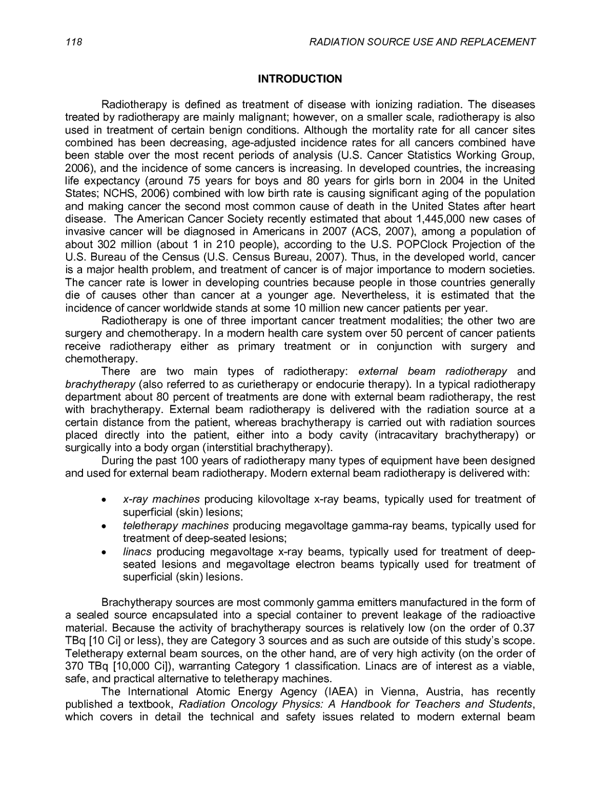
RADIOTHERAPY 119 radiotherapy and brachytherapy (IAEA, 2005c). Many other textbooks have been published on the technical and clinical aspects of radiotherapy (e.g., Hendee et al., 2004; Khan, 2003; Williams and Thwaites, 2000; Johns and Cunningham, 1984). STANDARD EXTERNAL BEAM RADIOTHERAPY Since the inception of radiotherapy soon after the discovery of x-rays by Roentgen in 1895, the technology of radiation production has been aimed toward ever higher photon energies and intensities and more recently toward computerization and intensity-modulated radiation therapy (IMRT). During the first 50 years of radiotherapy, the technological progress was relatively slow and mainly based on x-ray tubes, Van de Graaff generators, and betatrons. All of these units were heavy and bulky and not suitable for isocentric mounting. The first truly practical megavoltage therapy machine was the cobalt-60 teletherapy machine developed by Canadian physicist Harold E. Johns in the early 1950s (Johns et al., 1951). The invention of cobalt-60 teletherapy provided a tremendous boost in the quest for higher photon energies in a compact unit and placed the cobalt-60 unit into the forefront of radiotherapy for a number of years, mainly because it incorporated a radioactive source that is characterized by features extremely useful for radiotherapy. Standard e-beam cobalt-60 radiotherapy is carried out with teletherapy machines that are loaded with high-activity (Category 1) gamma-ray sources. The source is most often mounted isocentrically, allowing the beam to rotate about the patient at a fixed source-axis distance (SAD). Modern teletherapy machines have a SAD of 80 cm or 100 cm. Figure 7-1 shows a photograph of a typical modern teletherapy machine using the cobalt-60 radionuclide as the source of radiation. The main components of a teletherapy machine are: ⢠radioactive sealed source; ⢠source housing, including shielding, beam collimator, and source movement mechanism; ⢠gantry and stand in isocentric machines or a housing support assembly in stand- alone machines; ⢠patient support assembly (treatment couch); ⢠machine operating control console. Gamma radiation is obtained from specially designed and built sources that contain a suitable, artificially produced, radioactive material. The parent source material undergoes a beta-minus decay resulting in excited daughter nuclei that attain their ground state through emission of gamma rays (gamma decay). The important characteristics of a radionuclide used for external beam radiotherapy are: ⢠high gamma-ray energy (of the order of 1 MeV) for better penetration into tissue; ⢠high specific activity (of the order of 100 Ci/g) to achieve adequate dose rate with a relatively small diameter source (source size affects the uniformity of the therapy beam); ⢠relatively long half-life (of the order of several years) to avoid the need for frequent and costly source replacements.
120 RADIATION SOURCE USE AND REPLACEMENT FIGURE 7-1 A modern, isocentrically mounted cobalt-60 teletherapy machine, manufactured by MDS Nordion, Ottawa, Ontario. SOURCE: Image courtesy of MDS Nordion. The basic physical properties of the two gamma-emitting radionuclides (cobalt-60 and cesium-137) that have proven useful in external beam teletherapy and a potential source for teletherapy (europium-152) are listed in Table 7-1. Of the three radionuclides, cobalt-60 is the most widely used, because it offers the most practical approach to external beam radiotherapy, considering the energy of emitted photons, half-life, specific activity, means of production, and safety. Cobalt-60 and europium-152 come in metallic form encapsulated in a special source container, while cesium-137 is used in the form of cesium-137 chloride encapsulated in a special source container. The use of cesium-137 for external beam radiotherapy was discontinued during the 1980s despite its attractive half-life. There are two reasons for this: (1) security of cesium sources is a major concern and, in addition, (2) cesium-137 has a relatively low specific activity. The low specific activity implies a relatively low source output and large source diameter; this effectively precludes a teletherapy machine SAD larger than 50 cm, yet in modern radiotherapy a minimum SAD of at least 80 cm is the accepted norm. Note that cesium-137 is still used in blood irradiators and research irradiators because in these machines the distance between the source and the irradiated object is relatively short, of the order of only 10 to 25 cm. A cobalt-60 teletherapy source is typically a cylindrical stainless-steel capsule containing many hundreds of tiny, high-activity cobalt metal pellets and sealed by welding. A double- welded seal is used to prevent any leakage of the radioactive material from the source container. The typical diameter of the cylindrical teletherapy source is between 1 and 2 cm, the height of the cylinder is about 2.5 cm. For a given activity, a smaller source diameter yields a smaller physical penumbra,1 making for a sharper beam edge; but higher specific activity sources are more expensive. Often a diameter of 1.5 cm is chosen as a compromise between the cost and penumbra size. The cost of a cobalt-60 teletherapy source is on the order of $200/TBq ($7.5/Ci). 1 The penumbra is the spread of the beam beyond the idealized beam shape.
RADIOTHERAPY 121 TABLE 7-1 Physical Properties of Radionuclides Used In External Beam Radiotherapy Radioactive Source Property Cobalt-60 Cesium-137 Europium-152a Half-life (yr) 5.3 30 13.4 Specific activity (Ci/g) 1,100b (300c) 88b (30c) 180b (~150c) Photon energies (MeV) 1.17 and 1.33 0.662 0.6 and 1.4 Specific gamma rate constantd 1.31 0.33 1.06 Î [R â m 2 / (Ci â hr)] Specific air-kerma rate constante 309 78 250 Î AKR [ μGy â m 2 / (GBq â hr)] Half value layer HVL (cm Pb)f 1.1 0.5 1.1 59 151 Means of production Co + n Fission Eu + n in reactor by-product in reactor a Europium-152 has not yet been developed for clinical use. b Theoretical specific activity: a = (NA ln 2) / (t1/2 A) . c Practical specific activity is smaller than the theoretical specific activity because the source is not carrier- free; i.e., the source contains a stable nuclide in addition to a radioactive nuclide (example.g., cobalt- 59 mixed with cobalt-60). d Exposure rate achievable for a given activity of the source. e Dose rate in air for a given activity of the source. f Thickness of shielding (centimeters of lead) required to reduce the radiation by a factor of 2. SOURCE: Provided by the committee. To facilitate interchange of sources from one teletherapy machine to another and from one radionuclide production facility to another, standard source capsules have been developed for use around the world. Teletherapy sources are usually replaced within one half-life of installation; however, financial considerations may result in longer source use. Teletherapy machines are designed to enable on-site replacement of sources by trained technicians (the machines are not returned to the manufacturer for resourcing). The housing for the source in a teletherapy machine is called the source head. It consists of a steel shell with lead for shielding and a mechanism for bringing the source in front of the collimator opening to produce the clinical gamma-ray beam. Two different methods are in use for moving the teletherapy source from the BEAM OFF into the BEAM ON position and back: (1) a source in a sliding drawer and (2) a source in a rotating cylinder. Both methods incorporate a safety feature that terminates the beam (i.e., moves the source into the OFF position) automatically in case of power failure or emergency. When the source is in the BEAM OFF position, a light appears in the BEAM ON position above the collimator opening, allowing an optical visualization of the radiation field, as defined by the machine collimators and any special shielding blocks.
122 RADIATION SOURCE USE AND REPLACEMENT Typical teletherapy source activities are on the order of 185 to 370 TBq (5,000 to 10,000 Ci), and provide a typical dose rate on the order of 1 to 2 Gy/min at a distance of 80 cm from the teletherapy source. Often the output of a teletherapy machine is stated in Rmm (roentgens per minute at 1 m) as a rough guide to the machineâs source strength. The housing for the source in a teletherapy machine is called the source head. It consists of a steel shell with lead for shielding and a mechanism for bringing the source in front of the collimator opening to produce the clinical gamma-ray beam. Two different methods are in use for moving the teletherapy source from the BEAM OFF into the BEAM ON position and back: (1) a source in a sliding drawer and (2) a source in a rotating cylinder. Both methods incorporate a safety feature that terminates the beam (i.e., moves the source into the OFF position) automatically in case of power failure or emergency. When the source is in the BEAM OFF position, a light appears in the BEAM ON position above the collimator opening, allowing an optical visualization of the radiation field, as defined by the machine collimators and any special shielding blocks. Some radiation will escape the unit even when the source is in the BEAM OFF position. This head leakage typically amounts to less than 0.01 mSv/hr (1 mR/hr) at 1 m from the source. International regulations require that the average leakage of a teletherapy machine head be less than 0.02 mSv/hr (2 mR/hr) at 1 m from the source. Linacs as Alternatives to Cobalt-60 Teletherapy Machines Linacs were developed after 1945 concurrently by two groups: W.W. Hansenâs group at Stanford University in the United States (Ginzton et al., 1948) and D. D. Fryâs group at Telecommunications Research Establishment in the United Kingdom (Fry et al., 1947). Both groups were interested in linacs for research purposes, used 3,000 MHz as the design frequency, and profited heavily from the microwave radar technology developed during World War II. The potential for use of linacs in radiation therapy became apparent in the 1950s, and the first clinical linac was installed at the Hammersmith Hospital in London (Miller, 1953). During subsequent years, the linac eclipsed the cobalt-60 machine and became the most widely used radiation source in modern radiotherapy, with several thousand machines in clinical practice around the world today. In contrast to a cobalt-60 unit that provides only two gamma rays with energy of 1.17 MeV and 1.33 MeV (average, 1.25 MeV), a medical linac accelerates electrons to megavoltage kinetic energies in the range from 4 to 25 MeV and can produce two types of radiation beams for use in external beam radiotherapy: electron beams and x-ray beams. Electron-beam radiotherapy linacs accelerate electrons to megavoltage energies, extract them from the accelerating waveguide and transporting them through a beam-forming network to produce a clinical electron beam. X-rayâbeam radiotherapy uses an accelerated beam to strike a target in which some of the kinetic energy is transformed into photons. As is explained in Chapter 4, these photons, referred to as bremsstrahlung x-rays, are subsequently formed into a clinical x-ray beam with the help of a flattening filter and special collimators. In a linac, the electrons are accelerated following straight trajectories in special evacuated structures called accelerating waveguides. Electrons follow a linear path through the same relatively low potential difference a large number of times; hence, linacs fall into the class of cyclic accelerators just like other cyclic machines that provide curved paths for the accelerated particles (e.g., betatrons, microtrons, synchrotrons). The high-power radiofrequency fields, used for electron acceleration in the accelerating waveguide, are produced by devices called magnetrons and klystrons. Various types of linac are available for clinical use. Some provide x-rays only in the low megavoltage energy range (4 or 6 MV), and others provide both x-rays and electrons at various
RADIOTHERAPY 123 megavoltage energies (see, e.g., Figure 7-2): typically two photon energies (6 and 18 MV) and several electron energies (e.g., 6â22 MeV). In comparison with cobalt-60 teletherapy machines, linacs are much more versatile for radiotherapy, and their only disadvantages are that they are significantly more expensive to purchase, operate, calibrate, and maintain (costs are discussed later in this chapter). Linacs are usually mounted isocentrically and the operational systems are distributed over five major and distinct sections of the machine: 1. gantry, 2. gantry stand or support, 3. modulator cabinet, 4. patient support assembly (treatment couch), 5. control console. Cobalt-60 Teletherapy Machine Versus Clinical Linac In comparison to cobalt-60 machines, linacs have become very complex in design because of: 1. multimodality capabilities (see IGRT, below) that have evolved and are available on most modern machines; 2. increased use of computers in the control systems of these machines; 3. added features, such as high-dose-rate modes, multileaf collimation, electron arc therapy, and dynamic motion (i.e., while the beam is ON) of the collimators (dynamic wedge), the leaves of a multileaf collimator (IMRT), and the gantry and couch (dynamic stereotactic radiosurgery). FIGURE 7-2 Modern dual x-ray energy linac manufactured by Varian; the gantry, gantry stand, and the patient support assembly are shown. SOURCE: Image courtesy of Varian Oncology Systems, Palo Alto, CA.
124 RADIATION SOURCE USE AND REPLACEMENT Despite the clear technological and practical advantages of linacs over cobalt-60 machines, the latter still occupy an important place in the radiotherapy toolbox, mainly because of considerably lower capital, installation, and maintenance costs of cobalt-60 machines compared to linacs. In the developing world, the cobalt-60 machines, because of their relatively lower costs, simplicity of design, and ease of operation, are likely to play an important role in cancer therapy for the foreseeable future. Many modern features of linacs, such as multileaf collimators, dynamic wedges, and dynamic operation, can also be installed on modern cobalt-60 machines to allow, at a lower cost, a sophistication in treatment similar to that of linacs. Manufacturers of cobalt-60 machines have been slow in reacting to new technological developments in radiotherapy, conceding preeminence to linac manufacturers even in jurisdictions that would find it much easier and more practical to run cobalt-60 machines as compared to linacs. Economic Considerations: Megavoltage Linac Versus Cobalt-60 Teletherapy Machine for Standard Radiotherapy Cobalt-60 machine (isocentric, no beam stopper) Capital cost: $750,000 2 Operating expense : $50,000/year including. Equipment-related costs only, consisting of (1) maintenance and servicing of equipment (estimated at 4% of capital cost per year = $30,000/year) PLUS (2) cost of source replacement every 5 years (new source cost: $ 100,000 or $20,000/year) Output calibration: Every six months. Linac (low-energy, isocentric, single photon: 6 MV) Capital cost: $2,250,000 Operating expense: $150,000/year (equipment only) Equipment-related cost only, consisting of maintenance and servicing of equipment through either a service contract (estimated at 6.7% of capital cost) or in-house maintenance engineering crew (also estimated at 6.7% of capital cost). Output calibration: twice per week, that is, 104 times per year. Linac (high-energy, isocentric: 6 and 18 MV, and 5 electron energies) Capital cost: $4,000,000 Operating expense: $300,000/year (equipment only) Equipment-related cost only, consisting of maintenance and servicing of equipment through either a service contract (estimated at 7.5% of capital cost) or in-house maintenance engineering crew (also estimated at 7.5% of capital cost). Output calibration: Twice per week. One additional expense difference is the disposition of the spent cobalt-60 source upon decommissioning of the device. As noted in Chapter 2, some source manufacturers and 2 Note that the operating expenses provide an estimate for only the annual cost of equipment maintenance and servicing and do not include the personnel cost, such as cost of physics calibration, nor do they include the cost of radiotherapists (radiotherapy technologists) who operate the equipment during the dose delivery to patients.
RADIOTHERAPY 125 distributors will take back spent cobalt-60 sources for a fee, and use the pellets to balance the activity of newly irradiated pellets in the manufacture of new sources. Others simply dispose of them. The cost cited above for source replacement includes the cost of removal of the spent source when a new source is purchased. The cost to the owner of the radiotherapy device for disposal of the last source at decommissioning is in the range of $20,000 to $80,000, depending on the condition and origin of the source. STEREOTACTIC RADIOSURGERY From an obscure irradiation technique practiced in the 1960s and 1970s in only a few specialized centers around the world, stereotactic irradiation has during the past 20 years developed into a mainstream technique practiced in most major radiotherapy centers around the world. Stereotactic irradiation is the term used to describe focal irradiation techniques that use multiple, non-coplanar radiation beams and deliver a prescribed dose of ionizing radiation to preselected and stereotactically localized lesions primarily in the brain, although progress has been made recently in extending the technique to other parts of the body. As in standard radiotherapy, the use of multiple beams aimed at the targeted tumor from different directions concentrates the radiation dose in the target and leaves the surrounding tissues with a relatively lower dose. In standard radiotherapy these multiple beams are usually coplanar; in radiosurgery they are non-coplanar. The main characteristics of stereotactic irradiation are: 1. Total prescribed doses are of the order of 10â50 Gy and the planning targets are small with typical volumes ranging from 1 to 35 cm3. 2. The requirements for positional and numerical accuracy in dose delivery are ±1 mm and ±5 percent, respectively. 3. Treatment usually is delivered in a single dose or a reduced number of fractions. The dose in stereotactic irradiation may be delivered through a stereotactic implantation of radioactive sources (stereotactic brachytherapy) or, more commonly, with one or several external radiation sources (stereotactic external beam irradiation). With regard to dose fractionation, stereotactic external beam irradiation is divided into two categories: 1. Stereotactic radiosurgery: Total dose is delivered in a single session. 2. Stereotactic radiotherapy: Similarly to standard radiotherapy, the total dose is delivered in multiple fractions, often fewer in number than in standard external beam radiotherapy. From a technical point of view, there is essentially no difference between stereotactic radiosurgery and stereotactic radiotherapy, and often the term radiosurgery is used to describe both techniques. Essentially any radiation beam that has been found useful for external beam radiotherapy has also found use in radiosurgery (cobalt-60 gamma rays, megavoltage x-rays, proton and heavy charged particle beams, and even neutron beams). Equipment Used for Stereotactic Radiosurgery and Stereotactic Radiotherapy In addition to a suitable radiation source, in contrast to standard radiotherapy, stereotactic irradiation requires sophisticated specialized equipment and techniques as well as
126 RADIATION SOURCE USE AND REPLACEMENT more stringent quality assurance measures. The general list of the specialized equipment is as follows: stereotactic frame, imaging equipment, target localization software, and three- dimensional treatment planning system. ⢠Stereotactic frame defines a fixed coordinate system for an accurate localization and irradiation of the planning target volume. In addition, the stereotactic frame is also used for patient setup on the treatment machine and for patient immobilization during the actual treatment procedure. Figure 7-3 shows two commercial stereotactic frames. ⢠Imaging equipment (computed tomography (CT), magnetic resonance (MR), digital subtraction angiography) is used for visualization, definition, and localization of the structures, lesions and planning target volumes. ⢠Target localization software is used in conjunction with the stereotactic frame system and imaging equipment to determine the coordinates of the target in the stereotactic frame reference system. ⢠Treatment planning system is used for calculation of three-dimensional dose distributions for the radiosurgical treatment. The three-dimensional dose distribution is superperimposed on the patient's anatomical information. The combined use of stereotaxy and irradiation in treatment of certain brain diseases was introduced in the early 1950s by Swedish neurosurgeon Lars Leksell who also coined the term radiosurgery to describe the technique (Leksell, 1951). Leksell initially used 200 kVp x-rays to deliver, in a single session, a high radiation dose (of the order of 100 Gy) to an intracranial target. He approached the target from several directions to focus the dose on the target within the brain and spare the surrounding vital structures. Radiosurgery based on kilovoltage x-rays was discontinued in the late 1950s but the idea of focal brain irradiation was carried over to other, more suitable radiation beams, first to protons from cyclotrons (Larsson et al., 1958; Lawrence et al., 1962; Kjellberg et al., 1968) then to focused cobalt-60 gamma rays (Leksell, 1968), and more recently to megavoltage x-rays from linacs (Lutz et al., 1988; Podgorsak et al., 1987; Colombo et al., 1985; Hartmann et al., 1985; Betti and Derechinsky, 1984). FIGURE 7-3 Two commercial stereotactic frames: (a) Leksell stereotactic frame with attachments for biopsy and (b) OBT frame attached to patient in preparation for linac-based radiosurgery. SOURCE: Images provided by the committee.
RADIOTHERAPY 127 In 1974, Larsson proposed linacs as viable radiation sources for radiosurgery (Larsson et al., 1974). In 1984, Betti and Derechinsky from Buenos Aires reported on the development and clinical application of the linac-based multiple non-coplanar converging arcs technique. Soon thereafter in 1985, Colombo and colleagues introduced the technique clinically in Vicenza (Italy) while Hartmann and colleagues introduced it in Heidelberg, Germany. In 1986, Harvard University in Boston and McGill University in Montreal were the first two institutions to use linac-based radiosurgery in North America. Harvard adopted the multiple non-coplanar converging arcs technique (Lutz et al., 1988), while McGill developed its own radiosurgical technique, referred to as dynamic stereotactic radiosurgery (Podgorsak et al., 1987). Gamma Knife® The Gamma Knife® (Elekta, Stockholm, Sweden) is a radiosurgical device that has been associated with, and dedicated to, radiosurgery for the past 40 years. Despite great technological advances during this time, the fundamental design and principles of the Gamma Knife® have not changed much since the Swedish neurosurgeon Lars Leksell introduced the prototype unit in 1968 (Leksell, 1968). The unit incorporates 201 cobalt-60 sources housed in the central body of the unit. These sources produce 201 collimated beams directed to a single focal point (machine isocenter) at a source-focus distance of about 40 cm. The final definition of the circular-beam field size is provided by one of four helmets delivering circular fields with nominal diameters between 4 and 18 mm at the machine focal point (isocenter). The main components of the Gamma Knife® (see Figure 7-4) are: ⢠radiation unit with upper hemispherical shield and central body; ⢠operating table and sliding cradle; ⢠set of four collimator helmets providing circular beams with diameters of 4, 8, 14, and 18 mm at the isocenter; ⢠control unit. Each of the Gamma Knife® cobalt-60 sources is in the form of a steel capsule with a diameter of 1 mm and a height of 20 mm, containing 20 cobalt-60 pellets. The capsule is inserted into another steel capsule, which is enclosed by a bushing and loaded into the central body of the machine. Each source bushing assembly is aligned with its precollimator (6.5 cm of tungsten alloy), stationary collimator (9.25 cm of lead), and the final collimator (6 cm of tungsten alloy) on one of the four helmets. A newly loaded Gamma Knife® has a total activity of the order of 222 TBq (6,000 Ci) and all individual source activities are within 5 percent of an average source activity, which is of the order of 1.11 TBq (30 Ci). The dose rate at the center of a spherical water-equivalent phantom with a radius of 8 cm placed with the sphere center into the isocenter of the Gamma Knife® is on the order of 3 Gy/min. This rate will decrease to 50 percent of its original value during one half-life of the cobalt-60 radionuclide (5.26 years). A Chinese company, GammaStar® Medical Group, Ltd., is introducing its own cobalt-60 radiosurgery device, called Gyro Knife. This device is the functional equivalent of the isocentric linac dedicated to stereotactic radiosurgery, described below. The Gyro Knife therapy head pivots the 220- to 260-TBq (6,000- to 7000-Ci) source on an axis, using a multileaf collimator to direct the radiation beam at the tumor, while the gantry rotates the therapy head around the patientâs body. Unlike Gamma Knife®, this device can be used for tumors in any part of the body.
128 RADIATION SOURCE USE AND REPLACEMENT FIGURE 7-4 A Gamma Knife® installation showing the main body of the unit containing 201 cobalt sources (at 30 Ci = 1.11 TBq each source), the treatment couch, and a collimator helmet attached to the treatment couch. The inset shows an up-close image of the automatic positioning system used to position the patient for treatment. SOURCE: Image courtesy of Elekta. Nonradionuclide Replacements for Cobalt-60 Radiosurgery There are currently three options for nonradionuclide alternatives to cobalt-60 radiosurgery: Stereotactic radiosurgery based on a standard isocentric linac, stereotactic radiosurgery with dedicated isocentric linac, and a miniature linac on a robotic arm (CyberKnife®).3 Each of these is discussed below. Stereotactic radiosurgery based on a standard isocentric linac In contrast to Gamma Knife® which is dedicated solely to stereotactic radiosurgery, linac- based radiosurgery uses a standard isocentric linac with tight mechanical and electrical tolerances, modified for radiosurgery. This means that radiosurgery can be performed on such a linac on top of the daily routine radiotherapy patient load. The required modifications to a standard linac for use in radiosurgery consist of: ⢠Supplementary collimation, either in the form of a set of collimators to define the small diameter circular radiosurgical beams or a micro-multileaf collimator (micro- MLC), to define the small area irregular fields. ⢠Remotely controlled motorized couch or treatment chair rotation. ⢠Couch brackets or a floor stand for immobilizing the stereotactic frame, i.e., the patient, during treatment. ⢠Inter-locked readouts for angular and height position of the couch. ⢠Special brakes to immobilize the vertical, longitudinal, and lateral couch motions during treatment. 3 One other technique, proton beam radiation therapy (also called proton therapy), can also be used to treat the same tumors that are treated with a GammaKnife® but at two or three times the expense. Proton therapy is in some ways superior to standard radiation therapy techniques, but is not discussed here because of the clear cost difference from linac and gamma radiosurgery.
RADIOTHERAPY 129 Isocentric linac-based radiosurgical techniques currently fall into two categories: (i) multiple non-coplanar converging arcs and (ii) dynamic stereotactic radiosurgery. Each linac- based technique is characterized by a particular set of individual rotational motions of the linac gantry and the patient support assembly (couch or chair) from given start to stop angles. Of the two approaches, the multiple converging arcs technique is the more common. In the multiple non-coplanar converging arcs technique the patient is stationary on either the treatment couch or chair, while the gantry moves through a given arc. In the dynamic stereotactic radiosurgery technique, both the gantry and the patient rotate simultaneously during the dose delivery (the gantry moves 300 degrees, from 30 to 330 degrees, and the couch moves 150 degrees, from â75 to +75 degrees; see Figure 7-5). Stereotactic Radiosurgery with Dedicated Isocentric Linac Lately, linacs dedicated solely to stereotactic radiosurgery have become commercially available. Some still use stereotactic frames and operate essentially in the same manner as the standard linacs modified for radiosurgery; others (e.g., Novalis®, BrainLab) actually dispense with the stereotactic frame and achieve high precision without a frame using a mask for immobilization and on-line x-ray imaging of internal structures. Orthogonal stereoscopic x-ray sources are placed below the floor level in the treatment room, and ceiling-mounted amorphous silicon flat-panel detectors provide diagnostic-quality imaging of the patient anatomy in the treatment position. These sophisticated frameless techniques use a micromultileaf collimator for shaping of irregular fields and allow intracranial as well as extracranial stereotactic irradiation with equipment and operating costs similar to those for a Gamma Knife®. Figure 7-6 shows a linac (Novalis®, BrainLAB) installation dedicated to stereotactic radiosurgery. FIGURE 7-5 Patient receiving linac-based dynamic stereotactic radiosurgery on a Clinac 18, a linac manufactured in the 1970s by Varian Medical Systems. More recent medical linacs have been developed specifically for stereotactic radiosurgery incorporate image-guidance and beam-shaping technologies.
130 RADIATION SOURCE USE AND REPLACEMENT FIGURE 7-6 Linac installation dedicated to stereotactic radiosurgery (Novalis®, BrainLAB). The isocentric linac operates at 6 MV; imaging is carried out with two x-ray sources installed below the treatment room floor and two ceiling-mounted amorphous silicon flat-panel detectors. SOURCE: Image courtesy of BrainLAB; Heimstetten, Germany. Miniature Linac on a Robotic Arm (CyberKnife®) The CyberKnife® radiosurgery system (see Figure 7-7) provides a different approach to image-guided dose delivery utilizing an on-line orthogonal pair of digital x-ray imagers, a patient CT data set fused with MR and/or positron emission tomography images and a miniature linac mounted on an industrial robotic arm. This broadens the range of traditional stereotactic radiosurgery: Because it monitors and tracks the patient position continuously and uses on-line images for finding the position of the target in the treatment room coordinate system, the CyberKnife® allows frameless radiosurgery; that is, it can operate without a rigid and invasive stereotactic frame. The device directs the radiation beam into the target with a reported dose- delivery accuracy on the order of 1 mm. Because the linac is mounted on a robotic arm, it also can perform frameless radiosurgical dose delivery to extracranial targets, such as the spine, lung, and prostate, by using the body skeleton or surgically implanted fiducial markers as frame of reference for targeting purpose. Gamma Knife® Versus Linac-Based Radiosurgery The introduction of linac-based radiosurgery in radiation oncology departments during the late 1980s has very rapidly transformed radiosurgery from an obscure technique practiced in only a few specialized neurosurgery departments around the world into a mainstream radiotherapeutic technique. This stimulated great advances in technical and clinical utility of radiosurgery. However, the introduction of radiosurgery into radiation oncology departments has also caused some problems and differences of opinion between neurosurgeons, who were the inventors and until then the principal users of radiosurgery, and radiation oncologists, who are the professionals trained and licensed in treatment of disease with ionizing radiation and are quite comfortable with the clinical use of isocentric linacs.
RADIOTHERAPY 131 FIGURE 7-7 CyberKnife® installation consisting of miniature linac mounted on a robotic arm, treatment couch, and imaging device: ceiling-mounted x-ray tubes and floor-mounted image intensifiers. SOURCE: Image courtesy of Accuray, Sunnyvale, CA. Radiation oncologists, on the one hand, embraced the new linac-based radiosurgical techniques with great enthusiasm, but had some reservations about the use of single high-dose irradiation in radiosurgery in contrast to the multifractionated schemes used in conventional radiotherapy. The neurosurgeons, on the other hand, have had previous favorable experience with Gamma Knife® radiosurgery and have expressed concern about the mechanical stability of isocentric linacs when used in radiosurgery. An unstable linac isocenter could adversely affect the accuracy of dose delivery and result in substandard treatments in comparison to treatments provided by the 201 stationary beams from the Gamma Knife®. These concerns are valid, and clearly not all isocentric linacs are suitable for conversion to radiosurgery. However, there is no question that a well-designed, well-aligned, and properly maintained isocentric linac will have a stable and small enough isocenter sphere (of the order of 1-mm diameter) making it suitable for use in radiosurgery. The debate on the relative merits of Gamma Knife® versus linac-based radiosurgery continues, but one thing is clear: the Gamma Knife® incorporates some 200 cobalt-60 sources, each with an activity of 1.11 TBq (30 Ci), whereas linac-based radiosurgery does not use any radioactive material. The general consensus among radiation oncologists and medical physicists is that linac- based radiosurgical treatments with regard to treatment outcomes are equivalent to those provided by the Gamma Knife®. However, linac-based techniques, in comparison, with Gamma Knife® techniques, are more complicated and slower, but have greater potential for new technical and clinical developments, such as intensity modulation, extracranial application, on-line imaging, fractionated as well as image-guided dose delivery, and automatic patient repositioning. Informal examination of the usage and publications in this area suggests that the majority of neurosurgeons and some radiation oncologists believe that the Gamma Knife® is superior to any linac-based radiosurgical technique. During the past decade this apparent consensus has resulted in over 100 Gamma Knife® installations in the United States, many of them installed in neurosurgery departments.
132 RADIATION SOURCE USE AND REPLACEMENT Economic Factors: Radiosurgery with Gamma Knife® Versus Radiosurgery with Isocentric Linac The introduction of linac-based stereotactic radiosurgery techniques in the 1980s has not only stimulated rapid growth in clinical radiosurgery, it has also started a heated debate on the clinical and economic merits of the linac-based versus Gamma Knife® approach. A comparison among the three commonly used approaches to stereotactic radiosurgery (Gamma Knife®, dedicated linac, and modified standard linac) is as follows: Gamma Knife® Capital cost (machine): $4,000,000 Capital cost (bunker): $2,000,000 Annual operating cost (equipment and infrastructure only): Amortization (10 percent of capital cost): $600,000 Service contract (8 percent of equipment capital cost): $320,000 Source exchange (20 percent of cost at 5 years): $200,000 Total annual operating cost (infrastructure and equipment): $1,120,000 Radiosurgery with a dedicated linac Capital cost (machine): $4,000,000 Capital cost (bunker): $2,000,000 Annual operating cost (equipment and infrastructure only): Amortization (10 percent of capital cost): $600,000 Service contract (8 percent of equipment capital cost): $320,000 Total annual operating cost (infrastructure and equipment): $920,000 Radiosurgery based on modified standard isocentric linac Capital cost (machine): machine already available for standard radiotherapy Capital cost (bunker): already available for standard radiotherapy use Cost of linac modification for radiosurgery: $50,000 Cost of micro-MLC and radiosurgical treatment planning system: $600,000 Annual operating cost (radiosurgical equipment only): Amortization (10 percent of modification cost): $65,000 Service contract (8 percent of $600,000): $48,000 Total annual operating cost (equipment): $113,000 Just as in the comparison of radiotherapy costs, an additional expense difference is the decommissioning cost of the Gamma Knife®, including disposition of the last set of spent cobalt- 60 sources. This cost is estimated to be $50,000 to $70,000. Undoubtedly, the clinical utility of the Gamma Knife® is well proven; however, radiosurgery based on the Gamma Knife® is also significantly more expensive than that based on a modified linac, assuming, of course, that the linac is used for standard radiotherapy and carries radiosurgery as additional load to the standard treatment load. Departments with large neurosurgical patient loads require dedicated stereotactic radiosurgery equipment and then a choice between a Gamma Knife® and dedicated linac must be made. In this situation, the costs for infrastructure, equipment, servicing, and operation of a Gamma Knife® and dedicated linac are very similar, except that the Gamma Knife® costs must also include expensive cobalt-60 source replacement after every 5 years of operation and eventual decommissioning costs for disposal of the last set of sources.
RADIOTHERAPY 133 VETERINARY RADIOTHERAPY Radiotherapy is also used in veterinary medicine, essentially with same equipment as that used in human radiation oncology. The U.S. Veterinary Cancer Society lists about 70 therapy machines in clinical service. There are no cesium-137 teletherapy machines on the list of veterinary service machines, although it is possible that a veterinary school still has one, but the schools are not included in this tally. FINDINGS In the United States and other developed countries, cobalt-60 teletherapy machines have during the past two decades largely been supplanted by linacs which are considered a better and more versatile tool for provision of standard and advanced radiotherapy. In developing countries, cobalt-60 teletherapy machines will likely continue to play a major role in radiotherapy because of their significantly lower capital and operating costs in comparison with linacs. Radiosurgery is a special irradiation technique practiced mainly with the Gamma Knife® which uses around 200 cobalt-60 sources in combination. The use of Gamma Knife® for radiosurgery is not essential, because the technique can also be practiced with isocentric linacs as well as with a miniature linac mounted on a robotic arm. However, many neurosurgeons and some radiation oncologists believe that Gamma Knife® radiosurgery is superior to linac-based radiosurgery. The cost of radiosurgery with a dedicated linac is similar to that with a Gamma Knife®, whereas the cost of radiosurgery with a standard linac modified for radiosurgery is only a small fraction of that incurred with a Gamma Knife®. In addition, linac-based radiosurgery has greater potential than the Gamma Knife® for development of stereotactic techniques in the treatment of small localized lesions not only in the brain but also in other organs of the human body and not only on a single treatment basis but also on a fractionated basis, both under daily image guidance. Development of new treatment technologies and shifts in practices in radiotherapy are driven by innovation and decisions about delivery of health care, both at the macrolevel (e.g., insurance reimbursements) and at the personal level (i.e., doctor-patient treatment planning). Medical technology researchers seek out opportunities to improve nonradionuclide-based radiotherapy and radiosurgery. Because cesium-137âbased teletherapy is no longer practiced in the United States and there are only a few cobalt-60 teletherapy machines left in clinical operation in the United States, the committee concludes that teletherapy services, while important, are lesser and declining concerns with respect to radiation source security. It is also possible that the recent rapid growth in Gamma Knife® installations in the United States and the developed world will soon subside given that a viable alternative is offered with linac-based radiosurgery. An important issue is the end-of-life source disposal, which currently is somewhat costly and cumbersome. This encourages storage of spent machines including Co-60 sources in places that may be poorly secured.
