


Below is the uncorrected machine-read text of this chapter, intended to provide our own search engines and external engines with highly rich, chapter-representative searchable text of each book. Because it is UNCORRECTED material, please consider the following text as a useful but insufficient proxy for the authoritative book pages.
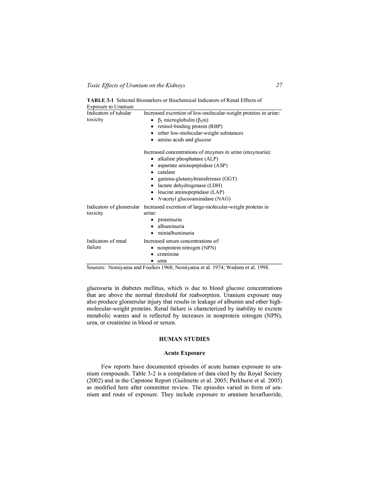
3 Toxic Effects of Uranium on the Kidneys The U.S. Armyâs Capstone Report (Guilmette et al. 2005; Parkhurst et al. 2005) identifies the kidneys as the organs most sensitive to the chemical toxicity of uranium. This chapter reviews the kidney literature that is pertinent to a health risk assessment of exposure of soldiers to depleted uranium (DU) in com- bat situations (other health end points are considered in Chapters 4-7). It is not a comprehensive review of the uranium literature; many reviews are available (Voegtlin and Hodge 1949, 1953; Berlin and Rudell 1986; ATSDR 1999; IOM 2000; WHO 2001; The Royal Society 2001, 2002; Marshall 2005). Those re- views and the Capstone Report generally base exposure guidelines on relation- ships between estimates of the renal burden of uranium and renal injury and measurement of toxic end points reflected by changes in biomarkers in animal models and humans. This chapter therefore focuses on animal and epidemi- ologic studies in which kidney burdens of uranium are reported or sufficient information is provided to calculate the kidney burdens. The findings are used in Chapter 8 to consider whether the renal uranium-concentration thresholds cho- sen for the Capstone health risk assessments are consistent with the scientific literature. BACKGROUND Acute uranium exposure produces degeneration of renal tubular epithe- lium followed by regeneration. Biomarkers or biochemical indicators of effects of uranium exposure on various aspects of renal function are available, and those cited in animal and human studies are listed in Table 3-1. Tubular cell injury is associated with increased excretion of low-molecular-weight proteins and other substances that are normally reabsorbed by tubular cells. Enzymuria reflects release of enzymes from injured cells. Glucosuria due to renal tubular dysfunction occurs with normal blood glucose concentrations, in contrast with 26
Toxic Effects of Uranium on the Kidneys 27 TABLE 3-1 Selected Biomarkers or Biochemical Indicators of Renal Effects of Exposure to Uranium Indicators of tubular Increased excretion of low-molecular-weight proteins in urine: toxicity ⢠β2 microglobulin (β2m) ⢠retinol-binding protein (RBP) ⢠other low-molecular-weight substances ⢠amino acids and glucose Increased concentrations of enzymes in urine (enzymuria): ⢠alkaline phosphatase (ALP) ⢠aspartate aminopeptidase (ASP) ⢠catalase ⢠gamma-glutamyltransferease (GGT) ⢠lactate dehydrogenase (LDH) ⢠leucine aminopeptidase (LAP) ⢠N-acetyl glucosaminidase (NAG) Indicators of glomerular Increased excretion of large-molecular-weight proteins in toxicity urine: ⢠proteinuria ⢠albuminuria ⢠minialbuminuria Indicators of renal Increased serum concentrations of: failure ⢠nonprotein nitrogen (NPN) ⢠creatinine ⢠urea Sources: Nomiyama and Foulkes 1968; Nomiyama et al. 1974; Wedeen et al. 1998. glucosuria in diabetes mellitus, which is due to blood glucose concentrations that are above the normal threshold for reabsorption. Uranium exposure may also produce glomerular injury that results in leakage of albumin and other high- molecular-weight proteins. Renal failure is characterized by inability to excrete metabolic wastes and is reflected by increases in nonprotein nitrogen (NPN), urea, or creatinine in blood or serum. HUMAN STUDIES Acute Exposure Few reports have documented episodes of acute human exposure to ura- nium compounds. Table 3-2 is a compilation of data cited by the Royal Society (2002) and in the Capstone Report (Guilmette et al. 2005; Parkhurst et al. 2005) as modified here after committee review. The episodes varied in form of ura- nium and route of exposure. They include exposure to uranium hexafluoride,
TABLE 3-2 Renal Effects of Acute Exposure to Uranium in Humans 28 Peak Renal Route of Chemical Uranium Renal Exposure Form Subjects Intake (mg) (µg/g)a Effectsb Outcome Reference Ingestion Acetate 1 male 8,500 100 +++ Acute renal failure; Pavlakis et al. 1996 glucosuria persisted beyond 32 wk of observation Skin (burn) Nitrate 1 male 130 35 +++ Peak renal tubular Zhao and Zhao 1990 dysfunction after 7 d; normal after 1 mo Inhalation Tetra-fluoride 1 male 920 10c ++ Renal dysfunction, Zhao and Zhao 1990 including increased NPN and proteinuria and aminoaciduria, 15 mo after exposure but gradual return to normal Injection Nitrate 2 males 16 6 + Increased NPN, urinary Luessenhop et al. 1958 1 female 11 4 + catalase, albumin 6 2 + Skin (burn) Nitrate 1 male 10 3 ++ Albuminuria persisting Butterworth 1955 for 3 wk after exposure Injection Nitrate 2 males 5 1.8 â No abnormalities Luessenhop et al. 1958 1.4 â Inhalation Hexa-fluoride 3 males 40-50d 4 + Albumin and casts in Kathren and Moore 4 + urine after accident; no 1986 1.2 + effects 38 y after exposure
Inhalation Hexa-fluoride 1 adult male 24 2.5 + Transient proteinuria and Fisher et al. 1990 glucosuria 11 adult males 6-18 0.05-1.9 + Transient proteinuria 19 adult males 6-18 0.05-1.9 â No abnormalities Ingestion Nitrate 1 adult male 470 1 + Transient albuminuria; Butterworth 1955 complete recovery within 24 h Inhalation Hexa-fluoride 1 adult male 20 1 â Apparently well 9 d after Boback 1975 exposure a Modeled estimates. b Severe clinical symptoms indicated by +++; biochemical indicators of renal dysfunction indicated by ++ (for protracted effects), + (for transient ef- fects), andâ(no detectable effects). c Kidney concentration of uranium peaked 60 d after exposure at 10 µg/g, whereas urinary concentration of uranium and indicators of renal dysfunction peaked about 2 mo after exposure. Capstone Report indicted that these events occurred much later than predicted from ICRP models for uranium tetra- fluoride and suggested that exposure of kidney was more chronic than acute. d Potential exposure range was wide. Sources: Adapted from Royal Society 2002 and Guilmette et al. 2005. 29
30 Risks to Military Personnel from Exposure to Depleted Uranium uranium tetrafluoride, uranyl nitrate, and uranyl acetate. Exposure was by inges- tion, skin contact, inhalation, and injection. The cases presented in Table 3-2 are arranged in descending order of peak renal concentration. Peak renal concentra- tions are calculated estimates provided in the Royal Society report and the Cap- stone Report. The highest renal concentration was the result of the deliberate ingestion of uranyl acetate, which resulted in acute renal failure that required dialysis (Pavlakis et al. 1996) and was followed by slow recovery. Glucosuria persisted beyond the 32-wk observation period. Zhao and Zhao (1990) described a man originally reported in the Chinese literature (Wu and Fan 1982) who was burned over 70% of his body with hot (108°C) uranium oxide. Renal function worsened over the next 6 d, and the pa- tientâs condition became critical 7 d after the accident. Uranium exposure was due primarily to absorption from the skin. Renal tubular function recovered within a month. The peak renal concentration of uranium was estimated to be 35 µg/g 5 d after the accident. Zhao and Zhao also described a patient who experienced an accidental in- halation exposure to uranium tetrafluoride powder for about 5 min. The lung deposition calculated in the Royal Society report was 915 mg. A week after ex- posure, the renal effects included proteinuria, increased NPN, and amino- aciduria. Concentrations of uranium increased over the first 2 mo after exposure and gradually declined to background concentrations about 3 y after exposure. The peak renal concentration of uranium was estimated to be 10 µg/g 60 d after exposure, and the urinary uranium and indicators of renal dysfunction peaked at about the same time. The Capstone Report indicated that those events occurred much later than predicted by the International Commission on Radiological Pro- tection (ICRP) models for uranium tetrafluoride, so renal exposure might have been more chronic than acute. Nevertheless, renal-function test results gradually returned to normal. In a study of radiation treatment for brain tumors, the tolerable intravenous dose of uranyl nitrate was investigated in five comatose or semicomatose pa- tients (Luessenhop et al. 1958). The three patients with the highest estimated peak renal concentrations of uranium (2-6 µg/g) had renal dysfunction, includ- ing increased NPN, increased urea, proteinuria, and increased urinary catalase excretion (Royal Society 2002). Butterworth (1955) reported a case of dermal exposure to hot uranyl ni- trate. The Royal Societyâs calculated peak renal concentration of uranium was about 3 µg/g 10 d after the accident. Albuminuria persisted until the third week after exposure; this suggested glomerular injury, but there was no indication of tubular dysfunction. Kathren and Moore (1986) evaluated the renal status of two of three peo- ple who were seriously injured by an accidental release of uranium hexafluoride. Uranium intake of the three was estimated to be about 40-50 mg, but peak renal concentrations were not estimated. Published and unpublished data indicate that all had albuminuria, which is evidence of renal injury. However, normal renal
Toxic Effects of Uranium on the Kidneys 31 function was found in two of the three examined 38 y after the accident. The authors noted an anomalous pattern of urinary excretion of uranium, which they attributed to pulmonary edema caused by hydrogen fluoride, a hydrolysis prod- uct of uranium hexafluoride. The pulmonary edema may have caused longer retention of uranium in the lungs. Thus, acute exposure to uranium hexafluoride might not be typical of or necessarily applicable to study of exposure other ura- nium compounds, including DU. Fisher et al. (1990) reported data on a group of 31 workers exposed to uranium hexafluoride and hydrolysis products after the rupture of a large ship- ping container. The peak renal uranium concentrations calculated from the au- thorsâ own biokinetic model (not an ICRP model) ranged from 0.05 to 2.5 µg/g. The worker with the highest concentration also had high concentrations of pro- tein (over 200 mg/dL) and glucose (1+ to 3+) in the urine in the first two sam- ples submitted 4 d after the accident. Eleven other workers were reported to have positive tests for protein in at least one urine specimen during the first 20 d after the accident. None of the remaining 19 workers had abnormal tests of renal function. As in the Kathren and Moore (1986) study, the relevance of this study for predicting effects of DU is uncertain. Butterworth (1955) also described an experiment in which a healthy vol- unteer ingested 1 g of uranyl nitrate to help to establish excretion. He experi- enced an illness that lasted about 24 h. Albuminuria was found in two urine samples taken 2-4 h after dosing but not in any other samples taken for 7 d thereafter. Peak renal uranium concentration was estimated by the Royal Society (2002) to be 1 µg/g 1 d after ingestion. Boback (1975) described four subjects exposed to uranium-ore dust and six others exposed to uranium hexafluoride. Table 3-2 includes data on one per- son who had a uranium intake of 20 mg and peak renal uranium concentration estimated to be 1 µg/g, but biochemical indicators of renal dysfunction were negative in all subjects. Two subjects were evaluated 38 years later (see Kathren and Moore [1986] described above). The cases listed in Table 3-2 show a relationship between renal effects and peak renal concentrations after acute exposure. There is uncertainty in the con- clusions that can be drawn from those data, because the renal concentrations are modeled estimates and it is unclear whether the same models were used to de- rive them. The only episode of acute renal failure noted came after ingestion of a large quantity (8,500 mg) of uranyl acetate, but even in this extreme case, as in all others, there was eventual recovery. The question of whether uranium expo- sure can cause chronic or irreversible renal disease is still open and recognized in other reviews, including the Capstone Report. In small numbers of subjects, transitory biomarkers of renal effects have been observed at renal uranium concentrations as low as 1 µg/g (Butterworth 1955; Kathren and Moore 1986; Fisher et al. 1990). Fisher et al. (1990) reported that 11 workers had transitory proteinuria but observed no other biomarkers as- sociated with kidney damage. They noted that proteinuria is only one indicator of tubular dysfunction and that other positive indicators must be present to show
32 Risks to Military Personnel from Exposure to Depleted Uranium kidney damage. Proteinuria can be induced by many other causes, such as infec- tion, exercise, dehydration, and stress (Carroll and Tempte 2000; Kashif et al. 2003), which were also experienced by the people in the acute-exposure studies. The lack of quantitative measurements of renal uranium in individual cases that demonstrate proteinuria creates uncertainty with regard to the association of uranium with this effect in these individuals. Chronic Exposure Studies of people chronically or continuously exposed to uranium for months or years should provide the most relevant information for predicting potential chronic renal effects of exposure to retained fragments of DU. How- ever, the evidence to date is sparse. There are many epidemiologic studies of morbidity and mortality from chronic renal disease in workers who have inhaled uranium dust from mining, processing, and conversion of processed uranium into milled and fabricated metal products. Reviews by other organizations do not suggest any increase in morbidity or mortality from chronic exposure. For ex- ample, the Royal Society (2002) analyzed seven studies of deaths from chronic renal disease among uranium workers, but there was no specific evidence as to whether the disease was related to uranium exposure or to some other disease. The total of 85 deaths in the seven studies was 18% less than would have been expected from renal-disease mortality in the general population. Evaluation of epidemiologic studies of occupational exposure by the Agency for Toxic Sub- stances and Disease Registry (ATSDR 1999) did not identify an increase in deaths from renal disease. The following are brief reviews of reports of exposure and renal effects af- ter three forms of chronic exposure: inhalation of yellowcake or ore by uranium process workers, ingestion of drinking water by the general population, and chronic exposure of Gulf War veterans due to systemic release of uranium from embedded fragments. Exposure information and renal effects are summarized in Table 3-3. Occupational Studies Thun et al. (1985) evaluated renal function in 39 uranium-mill workers and 36 local cement-plant workers of equivalent age, sex, and race. Mean uri- nary concentration of uranium measured in 1975 was 65.2 µg/L (median, 20 µg/L) and in 1981 was 7.2 µg/L (median, 6 µg/L). Biologic characteristics were measured in 1981 when the urinary uranium was below the action level for all workers. A relationship was found between the clearance of β2m relative to that of creatinine and the length of time that a worker had potential exposure to solu- ble uranium. The increase in urinary β2m was found to be due to both an in- crease in serum concentration and reduced renal tubular reabsorption. Renal
TABLE 3-3 Renal Effects of Chronic Exposure to Uranium in Humans Urinary Uranium Renal Uranium Reference Exposure Concentration Concentration Renal Effects Thun et al. 1985 Occupational: Mean/median: Up to ~ 1 µg/ga Mild increase in aminoaciduria; 36 workers, <10 y; 1975, 65.2/20 µg/L; β2m serum creatinine normal 2 workers, >10 y; 1 worker, 1981, 7.2/6 µg/L >20 y Zamora et al. 1998 Drinking water: Range: ~ 0.1 µg/ga Positive correlation of uranium low-dose group, <1 µg/L; 0.01-2.58 µg/L intake with urine concentrations of high-dose group, â¥1 µg/L; (from Zamora et glucose, ALP, β2m, and increased males, 2-410 µg/d; al. 2002) LDH (7 males) females, 2-570 µg/d Kurttio et al. 2002 Drinking water: Mean/Median: Not determined Positive correlation of urinary 325 persons; mean daily 0.424/0.078 µg/L uranium exposure with fractional intake, 39 µg (7-224 µg) excretion of calcium and phosphate; positive correlation of uranium in drinking water and daily intake with fractional calcium excretion; no association with glucose or β2m; possible bone effect (see Kurttio et al. 2005) Kurttio et al. 2006 Drinking water: Not reported Not determined Battery of urinary enzyme and 95 men and 98 women; biochemical tests of renal median concentration, 25 µg/L; cytotoxicity and function interpreted maximum, 1,500 µg/L as evidence of no effect; however, glucosuria was noted (Continued) 33
34 TABLE 3-3 Continued Urinary Uranium Renal Uranium Reference Exposure Concentration Concentration Renal Effects Squibb et al. 2005 Embedded metal fragments Mean of samples Highest to date: Increase in RBP in 2001 and in most and inhalation of DU oxides; collected 1993-2001: 0.95 µg/g recent assessment in 2003 uranium released to blood, 0.025- 0.01-38.5 µg/g of 0.7 mg for 6 y; 9.29- creatinine 190 mg for 10 y McDiarmid et al. 2007 Embedded metal fragments, 0.002-44.1 µg/g Not determined No increase in RBP in 2005; inhalation exposure to DU oxides of creatinine battery of tests of glomerular and tubular function showed no evidence of effect a Kidney concentrations from Capstone Report as calculated by Royal Society (2002) with ICRP model.
Toxic Effects of Uranium on the Kidneys 35 uranium concentration was calculated in the Capstone Report to be up to 1 µg/g. The study suggests that urinary β2m and amino acids may be sensitive bio- markers of uranium renal toxicity. Russell et al. (1996) reviewed the histopathology of kidneys from seven long-term uranium workers whose uranium intake ranged from a few to a few hundred milligrams. No differences were found between that group and a com- parison group of six autopsied adults who had neither known kidney disease nor exposure to uranium. There were no distinguishing features in either group, only a continuum of changes from normal to slight abnormalities that might be ex- pected in the general population. Chronic kidney disease has been examined in other uranium-worker co- horts (see Table 3-4 and Appendix B for details of the individual studies). Over- all results were compatible with no renal effect of uranium, although three stud- ies (all with small numbers of renal-disease cases) showed suggestive excesses of chronic renal effects. That the number of chronic renal-disease cases in all nine studies was not excessive (68 observed vs 85 expected) suggests that the renal effects, if any, are small. However, the small number of cases and the sparseness of the information on exposure concentrations and solubility of workplace uranium compounds mean that there are substantial uncertainties in the risk assessment. Renal-cancer mortality or incidence has been evaluated in cohorts of uranium-processing workers, and the results have been uniformly negative (see Chapter 6). Drinking-Water Studies Zamora et al. (1998) compared the effects of uranium on renal function in two Candian communities, one of which had private wells supplied by ground- water whose uranium content was higher than the Canadian drinking-water guidelines. Subjects were divided into a low-exposure group (drinking water contained uranium at <1 µg/L) and a high-exposure group (2-781 µg/L). Time of residence of the low-exposure group was 1-33 years, and of the high- exposure group 3-59 years. The urinary glucose concentrations differed between the high- and low-exposure groups, and the glucose concentrations increased with uranium intake. Increases in glucose, alkaline phosphatase (ALP), and β2m also correlated positively with increased uranium intake and are evidence of cell toxicity. There was also an increase in lactate dehydrogenase (LDH) excretion in seven subjects in the high-exposure group, but it was not directly related or co- ordinated with uranium intake. The authors suggest that the glucose, creatinine, and total-protein data indicate that the segment of the nephron most at risk of injury is the proximal tubule rather than the glomerulus. The Royal Society (2002) calculated the renal uranium concentration to be about 0.1 µg/g from ingestion of uranium at 80 µg/L of drinking water. That estimate is unexpectedly low. Urinary uranium was not reported in the study, but in a separate study on
36 Risks to Military Personnel from Exposure to Depleted Uranium TABLE 3-4 Standardized Mortality Ratios (95% Confidence Intervals) and [Observed Numbers of Deaths] from Renal Diseases in Uranium Workers Chronic Nephritis or Chronic Renal Study Failure Reference Colorado plateau uranium-mill workers 1.35 (0.58-2.67) [8]a Waxweiler et al. 1983; (with no history of uranium mining) Pinkerton et al. 2004 TEC/Y12 (1943-1947): Oak Ridge 0.77 (0.53-1.08) [30] Polednak and Frome 1981 uranium conversion and enrichment, all workers TEC/Y12 (1943-1947): Oak Ridge 0.60 (0.29-1.09) [9] Polednak and Frome 1981 uranium conversion and enrichment, alpha and beta chemistry departments Y12 (1947-1974): Oak Ridge uranium 0.97 (0.31-2.27) [5] Checkoway et al. 1988; metal production and recycling Loomis and Wolf 1996 Mallinckrodt uranium-processing 1.88 (0.75-3.81) [6] Dupree-Ellis et al. 2000 workers Portsmouth gaseous diffusionb 0.54 (0.11-1.56) [3] Brown and Bloom 1987 Savannah River nuclear-fuel production 0.27 (0.04-0.89) [2] Cragle et al. 1988 c Springfields, UK: mortality 0.61 (0.31-1.08) [10] McGeoghegan and Binks 2000a Capenhurst, UK: 235U enrichment plant 1.82 (0.58-4.39) [4] McGeoghegan and Binks mortalityc 2000b Total Observed/Expected Casesd 68/85 a Waxweiler et al. (1983) indicate that three cases were associated with prostatic obstruc- tion or prostatic cancer and probably represent secondary renal disease. b Data not given for âSubcohort I,â so include entire cohort. c Data given only for those classified as radiation workers. d Sums do not include row labeled âTEC/Y12 (1943-1947): Oak Ridge uranium conver- sion and enrichment, alpha and beta chemistry departments,â because those workers were already included in the TEC/Y12 row for all workers. gastrointestinal absorption of uranium in the same communities, Zamora et al. (2002) reported urinary concentrations of 0.01-2.58 µg/L (converted from mi- crograms per day by 1.4 L/24 h). Kurttio et al. (2002) measured uranium concentrations in drinking water and urine in 325 persons who had used drilled wells for drinking water. Urinary and serum concentrations of calcium, phosphate, glucose, albumin, creatinine, and β2m were measured to evaluate possible renal effects. Uranium concentra- tions over 300 µg/L in drinking water were associated with increased calcium fractional excretion. Urinary calcium and phosphate excretion were greater in persons in the high-uranium-excretion group (over 300 µg/L) than in those with low uranium excretion (under 2 µg/L). Uranium exposure was not associated
Toxic Effects of Uranium on the Kidneys 37 with changes in indicators of renal glomerular function (creatinine clearance or urinary albumin) or with urinary β2m, the biomarker of tubular dysfunction. Later, the authors provided evidence that uranium may affect calcium and phos- phate metabolism in bone but not in the kidneys (Kurttio et al. 2005). In a more recent study of 95 men and 98 women with continuous uranium intake from drinking water, no evidence of renal cytoxicity or functional effects was re- ported even at relatively high exposures (median uranium concentration, 25 µg/L; maximum concentration, 1,500 µg/L) (Kurttio et al. 2006). However, cu- mulative uranium intake was associated with increased urinary glucose excre- tion, which may reflect an effect on the glucose transport site. Gulf War Cohort Studies Because early animal studies demonstrated the sensitivity of the kidneys to uranium exposure, special attention was paid to renal-function assessments in a cohort of 74 Gulf War veterans exposed to DU from embedded metal fragments. Results of the first examination, in 1993-1994, showed no evidence of a rela- tionship between urinary uranium excretion and clinical measures of adverse renal function (serum creatinine, calcium, phosphate, and uric acid; and urinary creatinine and β2m) (Hooper et al. 1999). Urinary concentrations of uranium have been measured every 2 y since 1993. In a recent assessment, mean urinary uranium concentrations ranged from 0.01 to 38.5 µg/g of creatinine (Squibb et al. 2005). Renal concentrations were estimated from urinary excretion with the ICRP model (ICRP 1995a). In eight of the 16 soldiers, predicted renal urinary concentration peaked before the last measurements; that indicated that net accumulation of uranium in the kidney was no longer occurring. Uranium concentrations that were increasing and that were highest at the last measurement time suggest that net tissue accumulation was still occurring. Estimated renal uranium concentrations in those veterans 10 y after the war are as high as 0.95 µg/g (Squibb et al. 2005). Other biomarkers of the early effects of uranium on proximal tubular cells were added in the 1997 health-surveillance examinations. In addition to an ab- sence of changes in the standard clinical markers of renal function, there have been no observed statistically significant increases in urinary excretion of the proteins that serve as biomarkers of proximal tubular cell damageâN-acetyl glucoaminidase (NAG) and ALPâin the high-exposure group in any of the sur- veillance examinations. Increases have been observed in one of the low- molecular-weight proteins, retinol-binding protein (RBP), a marker of decreased proximal tubular protein reabsorption. Those increases were observed in both the 2001 and 2003 examinations although they were not significant in 2003, and they were not observed in 2005 (McDiarmid et al. 2004a, 2006, 2007). The lack of overt renal damage is consistent with recent modeling studies reported by Squibb et al. (2005), but the authors suggest that the small increases in RBP excretion observed in 2002 and 2003 may reflect the slow accumulation of ura-
38 Risks to Military Personnel from Exposure to Depleted Uranium nium in the veteransâ kidneys in excess of the higher urinary excretion of ura- nium. ANIMAL STUDIES Acute Toxicity Exposure concentrations that produce renal injury depend on the solubility of the uranium compound and the route of exposure. Study of the renal changes that follow injection of uranium compounds provides a model relating kidney burden to acute renal-cell injury and repair. Diamond et al. (1989) studied kid- ney effects after multiple intraperitoneal injections of uranyl fluoride in rats at two doses (cumulative dose, 0.66 or 1.32 mg/kg of body weight). Earlier studies by Morrow et al. (1982) showed that inhaled and parenterally administered uranyl fluoride results in nearly identical patterns of distribution and excretion of uranium. Injury and death of cells of the proximal renal tubule began about 3 d after injection and were followed by evidence of renal tubular dysfunction. Light microscopy revealed swelling and vacuolation of epithelial cells progress- ing to necrosis. Histologic changes were accompanied by glucosuria, increased excretion of amino acids, and transient enzymuria with increased excretion of aspartate aminotransferase (AST), LDH, NAG, ALP, catalase, and leucine aminopeptidase (LAP). The magnitude and duration of the increased excretion of LDH were greater than those of the increases in excretion of AST and NAG; this suggests that LDH may be the most sensitive enzymatic biomarker. The lowest observed-adverse-effect-level (LOAEL) was 0.7-1.4 µg/g and peaked when renal uranium concentrations were between 3.4 and 5.6 µg/g, followed by progressive reversal of both morphologic and functional effects, returning to normal about 3 wk after the last injection. Subchronic Toxicity Oral Exposure Gilman et al. (1998a) administered uranyl nitrate in drinking water at 0.96, 4.8, 24, 120, or 600 mg/L to weanling male and female Sprague-Dawley rats for 28 and 91 d. There were no significant changes after 28 d (kidney dose, 0.92 µg/g), but both male and female rats exposed for 91 d had degenerative changes in tubular cells at the lowest concentration (kidney dose, 0.42 µg/g). Changes in tubular epithelial cells included apical nuclear replacement and vesiculation, cytoplasmic vacuolation, and tubular dilatation. At 600 mg/L, males showed only tubular changes (residual kidney concentration, 2.12 µg/g), whereas fe- males showed irreversible glomerular sclerosis and interstitial fibrosis indicative of a chronic nonreparable nephropathy. Residual uranium concentration in the
Toxic Effects of Uranium on the Kidneys 39 kidneys of female rats was 1.67 µg/g. There was no apparent explanation of the difference in sensitivity between the kidneys of male and female rats. In studies of New Zealand white rabbits, males were administered uranyl nitrate in drinking water at 0.96, 4.8, 24, 120, or 600 mg/L for 91 d (Gilman et al. 1998b). Females were similarly exposed for 91 d at 4.8, 24, or 600 mg/L. The 91-d LOAEL in males was 0.96 mg/L and in females it was 4.8 mg/L. However, the residual renal uranium concentration at the LOAEL was 0.04 µg/g in males and 0.019 µg/g in females; thus, male and female rabbits might have different pharmacokinetic characteristics. Rabbits appear to be more sensitive than rats (Gilman et al. 1998a) to ingestion of uranyl nitrate. Reversibility of uranyl nitrate-induced renal injury was studied in male New Zealand white rabbits exposed at 24 or 600 mg/L in drinking water for 91 d followed by various recovery periods (Gilman et al. 1998c). Histologic changes in the renal tubular cells of rabbits that received 600 mg/L were not reversed after a 91-d recovery period. Urinary glucose, protein, and LAP were maximally increased during the first week of the recovery period and then gradually de- clined. The renal uranium concentration was 3.48 µg/g after 91 d of treatment, and uranium was completely excreted during the 91-d recovery period. Gluco- suria persisted beyond the recovery period. Dermal Exposure Dermal application of soluble uranium compounds (uranyl nitrate hexahy- drate, uranyl acetate hexahydrate, and ammonium uranyl tricarbonate) produced renal toxicity in rabbits, guinea pigs, rats, and mice (de Rey et al. 1983). Pro- teinuria continued for up to 10 d and was followed by recovery to control val- ues. Rabbits had increased blood NPN at doses over 270 mg/kg. There was mi- croscopic evidence of renal damage in the animals that died, but the kidneys were histologically normal in the ones that survived, and this suggested that re- nal tubular cells had regenerated. Chronic Toxicity Implanted Depleted-Uranium Pellets The results of long-term effects of implanted DU pellets in animal models have particular relevance to understanding of the effects of embedded fragments in Gulf War veterans. Pellmar et al. (1999a) implanted DU in rat muscle. The greatest concentrations of uranium were found in the kidneys and bone from 1 d after pellet implantation until 18 mo after implantation. No nephrotoxcity oc- curred in rats after 12 mo despite renal uranium accumulation exceeding 5 µg/g and excretion in the urine at 1µg/mL. Urinary excretion of LDH, NAG, protein, and glucose; creatinine clearance; and fractional excretion were not altered after 6 or 12 mo of exposure.
40 Risks to Military Personnel from Exposure to Depleted Uranium Inhalation Exposure Comparisons of renal burdens due to acute ingestion and acute inhalation show that inhaled uranium compounds generally result in higher burdens in the kidneys than ingestion of the same amount of uranium compounds (Chen et al. 2004). The renal effects of chronic or long-term inhalation of natural-uranium dust, as might occur in occupational exposure, have been studied in rats, dogs, and monkeys (Leach et al. 1970, 1973). No histologic changes were found in the kidneys immediately after inhalation of natural-uranium dust for up to 5 y and after followup periods of up to 1 y in rats and 75 mo in dogs and monkeys (see Table 3-5). Monkeys had the highest renal burdens. Renal burdens were greater in dogs than in rats. No evidence of uranium toxicity was found in the records of body weights, mortality, various hematologic measures, or histologic examina- tions of the kidneys. Beginning sometime during the first postexposure year and lasting until the fifth, some uranium-exposed monkeys consistently had higher blood NPN concentrations than controls; NPN concentrations were normal at the end of the postexposure followup. In the absence of abnormal renal histologic characteristics, the significance of that finding is unclear. Biomarkers of renal tubular function were not measured; renal effects were evaluated only through histologic examination. Mechanisms of Uranium Renal Toxicity The mechanisms whereby uranium produces injury to the cells of renal tu- bules and glomeruli are not fully understood. Various hypotheses are discussed by Leggett (1989) and Diamond and Zalups (2005). Autoradiography of rats administered [232U] uranyl nitrate showed that uranium concentrates in the outer stripe of the outer medulla and along the medullary rays penetrating the inner cortex; this is consistent with the location of the pars recta of the proximal tu- bule. It has been suggested that uranium combines with bicarbonate, citrate, and plasma proteins in the blood. At low pH, the complexes split, and the resulting uranyl ion may combine with proteins and be deposited on the surfaces of tubu- lar epithelium and cause renal-cell damage (Basset et al. 1948). Brady et al. (1989) have shown that exposure of rabbit renal cells to uranyl nitrate in vitro inhibited both sodium-dependent and sodium-independent adenosine triphos- phate use. Uranyl nitrate absorbed in the cytoplasm of tubular epithelium may impair mitochondrial oxidative phosphorylation. The resulting impairment of energy metabolism may contribute to the tubular dysfunction and ultimately to cellular degeneration. Leach et al. (1984) noted in animal studies that glucosuria was the most sensitive indicator of renal injury associated with exposure to uranium. Gluco- suria is also the most persistent abnormality during recovery from DU toxicity in rabbits (Gilman et al. 1998c) and in humans (Pavlakis et al. 1996). Glucosuria
Toxic Effects of Uranium on the Kidneys 41 TABLE 3-5 Renal Effects of Chronic Inhalation of Uranium Dioxidea in Experimental Animals Number Animal Species and Sex Duration Renal Burden Renal Effects Wistar rats 80 females Exposure for 1 y 0.8 µg/g None (Study 1) Wistar rats 120 females Exposure for 1 y 1.1 µg/g None (Study 2) Beagles 72 females, Exposure for 5 y 5.8 µ/g None 5 males Rhesus monkeys 20 females Exposure for 5 y 13 µg/g Increased NPN, 5 males significance or cause unknown; normal renal histology Wistar rats 6 Followup for 1 y 1.2 µg/g None (Study 1) Wistar rats 6 Followup for 1 y 0.9 µg/g None (Study 2) Beagles 5 Follow up for 75 mo 2.1 µg/g None Rhesus monkeys 5 Followup for 75 mo 1.7 µg/g None a 5 mg/m3 for 6 h/d 5 d/wk. Sources: Leach et al. 1970, 1973. Modified table reprinted with permission; copyright 1970, 1973, Lippincott Williams & Wilkins. was noted in subjects exposed to uranium in drinking water in the absence of other indicators of renal toxicity (Kurttio et al. 2006). Recent studies indicate that uranium has a direct dose-dependent and pH-dependent inhibitory effect on the rat kidneyâs brush-border membrane vesicles (Goldman et al. 2006). The pathogenesis of glomerular injury is not well understood, but Dia- mond and Zalups (2005) have suggested that uranium decreases outer renal cor- tical blood flow and glomerular perfusion and may induce acute renal failure. Leggett (1989) reviewed several possible mechanisms of uranium-induced glomerular injury, including evidence that uranium produces structural altera- tions that decrease the glomerular surface area available for filtration. In vitro studies of isolated glomeruli exposed to uranium bicarbonate suggest that a re- duction in glomerular filtration rate may result from glomerular contraction and disorganization of the cytoskeleton (Mirto et al. 1999; LâAzou et al. 2002). McDonald-Taylor et al. (1992) found thickening of the glomerular base- ment membrane in rabbits exposed to uranyl nitrate in drinking water (at 24 and 600 mg/L), as measured with electron microscopy. In addition, thickness in- creased during 45-d and 91-d recovery periods, and this suggested that the glomerular effect may become chronic or even progressive in contrast with the regeneration of injured tubular cells. Progression of ultrastructural glomerular
42 Risks to Military Personnel from Exposure to Depleted Uranium changes not discernible with light microscopy may reflect continued retention of even small concentrations of uranium during postexposure followup. Renal Uranium Concentration and Renal Tubular Dysfunction The renal uranium concentrations sometimes found after acute exposure suggest that minimal transient effects (such as proteinuria and albuminuria) may occur after exposure at concentrations as low as 1 µg/g (Kathren and Moore 1986; Fisher et al. 1990). Renal effects have also been reported at renal concen- trations around 1 µg/g in workers with chronic occupational exposure to ura- nium (Thun et al. 1985) and in Gulf War veterans with embedded DU fragments (Squibb et al. 2005). The Royal Society (2002) report also noted that transient renal effects occurred in humans at renal concentrations of 1 µg/g and that the trend for chronic exposures is toward greater renal effects with lower renal con- centrationsâpossibly as low as 0.1 µg/g. The duration of exposure may be an important factor. Groups with longer exposure appear to have the greatest ef- fects. How those findings compare with the renal uranium concentration thresh- olds chosen for the Capstone health risk assessments is presented in Chapter 8. SUMMARY ⢠The primary target of uranium in the kidney is the proximal tubule, but glomerular effects also may occur. ⢠Biomarkers of tubular effects include enzymuria and increased excre- tion of low-molecular-weight proteins, amino acids, and glucose. ⢠Biomarkers of glomerular effects include urinary excretion of high- molecular-weight proteins (albuminuria) and increased blood creatinine or NPN. ⢠Glucosuria is the most persistent tubular biomarker during recovery from acute uranium exposure in animals and humans. ⢠Transient biomarkers of renal effects (such as proteinuria and albumin- uria) have been observed at peak renal uranium concentrations as low as 1 µg/g.
