


Below is the uncorrected machine-read text of this chapter, intended to provide our own search engines and external engines with highly rich, chapter-representative searchable text of each book. Because it is UNCORRECTED material, please consider the following text as a useful but insufficient proxy for the authoritative book pages.
Appendix B Review of the Prevalence Literature Myopia prevalence refers to the proportion of individuals in a population who are myopic at a given time. Two major issues in measuring prevalence concern the ability to correctly classify individuals as myopic and nonmyopic and the manner in which the observed group was selected from the population at large. Classification criteria are described in Chapter I, in the section on interpreting the myopia literature. Chapter 4 contains the working group's recommendations for classification standards for future studies. AGE In most populations, age is the most important determinant of the distribution of refractive error, at least during the first three decades of life. Prevalence studies necessarily take a cross-sectional view of the patterns of refraction in relation to age. The characteristics of this pattern depend on the longitudinal development of refraction within the population, secular trends in refraction, and selective (survival) factors operating from one age group to the next. If two of these are known or can be reasonably assumed, and if sampling is valid, then inferences about the third can usually be maple from cross-sectional data. Age patterns of myopia prevalence seem to be used in two major areas of inquiry. The first concerns the progression of myopia and changes of refraction with age. An excellent example of this kind of interpretation of cross-sectional data is provided by Hirsch (1952) in a study of 9,552 schoolchildren, ages 5 to 14, in the vicinity of Los Angeles, California. In the context of universal education, one would expect little selection- or refraction-associated attrition in such a study. There is also little reason to expect a major secular trend over the 10 years separating the 5-year-olds from the 14-year-olds. This study uses a sophisticated statistical analysis that addresses the problems of making longitudinal inferences from cross-sectional data, presenting both means and centiles. The reporting of the percentage of students having various refractive states by age and by gender also makes possible the estimation of the prevalence of myopia based on various degrees of refractive error. Kalogjera (1979) studied 483 children ages 3 to Tin Central Zagreb, Yugoslavia. Ex- aminations, consisting of skiascopy under cycloplegia, were done in connection with vacci- nations. Refractive error was reported in 1.00 diopter (D.) intervals. About 3.4 percent of eyes had a negative refractive error of -1.00 D. or more. There was no apparent trend with age, but the numbers were small. Hirsch (1952) found that the percentage of children with a negative refractive error 45
46 greater than -1.00 D. increased from 0.6 percent in 5- and ~year-olds to 5.4 percent in 13- and 14-year-olds. The prevalence of any negative refractive error increased from 6.8 percent in ~ and ~year-olds to 23.9 percent in 1~ and 14-year-olds. Kempf et al. (1928) examined 1,860 Washington, D.C., schoolchildren in 1924. Refractive error was determined by retinoscopy under cycloplegia. The percentage of right eyes with a negative refractive error equal to or greater than -0.25 D. rose from 1.4 percent among ~ and 7-year-olds to 9.1 percent among 12- and 13-year-olds and also in those 14 years and older. In Finland, Laatikainen and Erkkila (1980) studied 411 schoolchildren; 1.9 percent of 7- and ~year-olds had a negative refractive error equal to or greater than -0.50 D., increasing to 21.8 percent among 14- and 15-year-olds. Most studies of older children and young adults show a continued increase in the prevalence of myopia into the third decade. Johansen (1950) studied Danish students ages 12 to 15 and found an increased prevalence of myopia with increasing age. Goldschmidt (1968) studied Danish schoolchildren ages 13 to 14 and found that 9.7 percent had either a previously ascertained myopia, reduced visual acuity, or a school record of use of glasses accompanied by optimum vision achieved by spherical concave lenses in one or both eyes. Among military recruits ages 18 to 25, 14.5 percent wore glasses with a negative correction. The author concluded that about one-third of myopes become myopic after their fourteenth year. Fledelius (1983) studied patients referred for ophthalmological evaluation of general disease, excluding referrals for glasses prescriptions. Among nondiabetics, the highest prevalence occurred in the age group 26 to 35 . Two surveys conducted by;the U.S. government contain data on older children and young adults. Angle and Wissmann (1980a) reported on an analysis of data from the United States Public Health Service Health Exarn~nation Survey, cycle Ill, summarized Originally by Roberts and Slaby (1974~. They found a slight increase in the prevalence of myopia with age, from 29.9 percent at 12 to 33.2 percent at 17. Sperduto et al. (1983) analyzed a subset of the data in the National Health and Nutrition Examination Survey (NHANES) collected between 1971 and 1972 and reported by Roberts and Roland (19783. Although Sperduto et al. report little variation with age, the data show an increase in prevalence of myopia from'24 percent among 12- to 17-year-olds to 27.7 percent among 18- to 24-year-olds, followed by a drop to 2~25 percent in the older age groups. Whereas the differences may not be statistically significant, they are consistent with other observed age patterns. Adults over age 40 were studied in Israel by Hyams et al. (1977~. The study was done in conjunction with a glaucoma screening program, and refractive error was determined from the subjects' own glasses. This methodology was felt to be valid by the authors because of the availability and use of ocular care. The authors report no change in the prevalence of myopia from the age of 40 to age 70 and a decrease thereafter. Many studies that report mean refraction other than prevalence suggest that the prevalence of myopia reaches a peak at about age 20 to 25, declines slightly to age 45, and then begins to rise very slightly again (Brown and Kronfeld, 1929; Jackson, 1932; Pendse and Bhave, 1954; Sorsby et al., 1960~. The pattern of marked increases in the prevalence of myopia during childhood largely reflects longitudinal growth patterns. Interpretations of the various patterns of plateau or decline after age 30 are discussed below in the section on secular trends. Whatever the source of the age- related patterns of myopia prevalence, it is clear that age must always be considered a potential confounder in comparisons of myopia prevalence between populations or subpopulation groups. This confounding could probably not be controlled by a single linear term in a regression mode! if the age range of subjects is very wide. By contrast,
47 populations of schoolchildren, a common source of data on myopia prevalence, may have sirn~lar numbers of children at all age levels, in which case there would be no confounding if the age range of the compared groups ~ the same. Controlling for age may also not control for age-associated phenomena, such as developmental maturation. Thus, differences between populations in childhood may not necessarily imply differences in the ultimate prevalence of myopia. GENDER The relationship of myopia prevalence with gender appears to be less consistent, less well documented, and less predictive than that with age. Peckham et al. (1977) studied 11,179 children in England, Scotland, and Wales. There was no significant difference between the sexes in the occurrence of reacquired myopia," defined as poor distant vision, satisfactory near vision, ant] a deterioration of two lines in distant visual acuity in children between ages 7 and 11. In California, Hirsch (1952) found a more negative (myopic) refraction among boys than among girls ages 5 to 6, shifting to more myopia among girls at age 13 to 14. The age pattern depends on the parameter of refraction being considered. For example, for myopia in excess of 1.00 D., the percentage of girls became slightly higher than the percentage of boys starting at age 7 to 8, markedly higher at 11 to 12, and only slightly higher again at 13 to 14. By contrast, both the mean and median refraction are higher (less myopic) for girls than for boys until ages 13 to 14. Hirsch attributes this pattern to the earlier onset of puberty in girls. Kempf et al. (1928) reported a slightly higher proportion of myopes among boys than among girls for all ages, including 14 and older. Angle and Wissmann (1980a), in their analysis of data from the 1966-1970 National Health Examination Survey, found a higher prevalence of myopia among females than males ages 12 to 17. Whether this varied with age is not stated. Sperduto et al. (1983) reanalyzed data on people ages 12 to 54 from the 1971-1972 National Health and Nutrition Examination Survey. They reported higher prevalences in women than men through ages 25 to 34. Goldschmidt (1968) found a higher prevalence of myopia in girls than in boys among Danish schoolchildren ages 13 to 14 but cautions that this does not imply that females are in general more myopic, since these results may reflect differences in maturational development between boys and girls at that age. Krause et al. (1982) found a higher prevalence of both myopia and hyperopia in female than in male Finnish schoolchildren, on the basis of a questionnaire reporting ophthalmologist visits and records of refraction; they also pointed out the connection with puberty. Studies of Eskimo and American Indian populations are also inconsistent in their gender patterns. Alsbirk (1979) reported a higher prevalence of any negative refractive error as well as a negative refractive error greater than -2.00 D. among men than among women over age 40; it was statistically significant for any negative refractive error. Myopia prevalence was somewhat higher for women than for men ages 15 to 39, but this was not statistically significant. `~7- - I --my - ~ ~ 1 ~ {< ~_~\ ~ , ~ , . .. more myopia among females, largely during adolescence. Among Belcher Island Eskimos, however, Woodruff and Samek (1976) report a lower mean refraction for males than for females. Since the standard deviation of refractive error is similar for males and females in this study and there are no high refractive errors, the prevalence of myopia is also probably higher in males, but this comparison may be confounded by age. Since participation rates for women tend to be higher than for men in these studies, observed gender differences may also be due to differences in selection. lace ruII anti cameos My data on Ontario lnd~an populations suggest · ~. --cat cat -
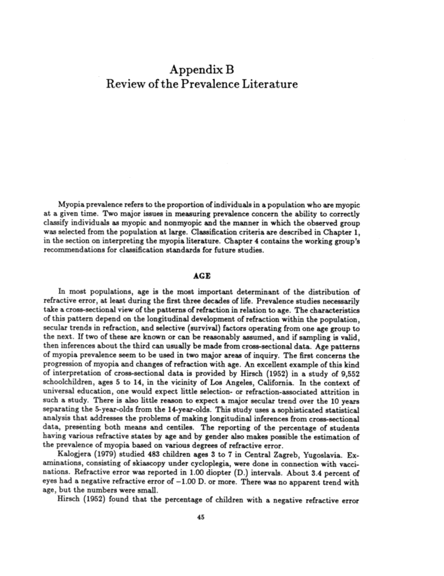
48 TERRITORY OF HAWAII DIAGNOSIS PERCENT 30 25 20 15 10 5 0 30 25 20 15 10 5 0 PERCENT ALL RACES 100,377 HAWAIIAN 2,758 PART HAWAIIAN 2O,375 PORTUGUESE 6,562 PUERTO RICAN 1,764 SPANISH 193 OTHER CAUCASIAN 5,353 CHINESE 5,621 JAPANESE 41,684 KOREAN 1,036 FRENCH 9,732 OTHERS 5,299 FIGURE B-1 Myopic errors by race. This figure represents eyes; in many individuals both eyes have the same defect, so the percentage on a case basis would be somewhat more than half of the figures given here. Source: Adapted from Crawford and Hammond, 1949. RACIAL AND ETHNIC PATTERNS Asians The original data from prevalence studies in Asian countries are not among the articles reviewed. Sato (1957) refers to prevalence rates as high as 67 percent among high school students in Japan. In Hawaii, Crawford and Hammond (1949) screened nearly 100,000 schoolchildren for a defect in vision or ocular balance. The classification of a student as myopic was based on the subsequently reported refractive error from an ophthalmologist or optometrist. As indicated by Figure B-1, myopia was found to be most common among the Chinese students, followed by Japanese and Korean students. Hawaiians, part-Hawa~ians, and a group designated as "others" had the lowest prevalences of myopia. Jews Two studies comparing Jewish and non-Jewish subpopulations report more myopes among Jews. Kantor, summarized by Sorsby (1932), compared the prevalence of myopia among Jews and White Russians (Byelorussians). Among Jews the prevalence of myopia was 14.75 percent; among White Russians it was 4.93 percent; among the subgroups who worked as type compositors, the prevalence of myopia was 22.8 percent for Jews and 6.1
49 percent for White Russians. The methods for selecting the sample were not specified, nor were those for estimating myopia. In I`ondon, Sourasky (1928) studied Jewish and non- Jewish schoolchildren. Among boys ages 8 to 13, visual defects were more common in Jews than in non-Jews and unrelated to age. He also examined case records and found a higher proportion classified as myopes among Jews than among non-Jews. In Israel, Hyams et al. (1977) studied men and women over 40, mostly of eastern European origin, in communal settlement. The prevalence of any negative error was found to be 18.4 percent, the prevalence of a negative error greater than -~.00 D. was 11.6 percent. These rates may be underestimates because of the use of subjects' glasses to determine refractive error. Shapiro et al. (1982a) reported on students ages 18 and 25 at the Hebrew University in Jerusalem. A negative refractive error of -0.25 D. or more in at least one eye was found in nearly 13 percent of those who had visited the university health service. There is, of course, concern that university students who register at the university health service may not be representative of the general student population and, furthermore, that prevalence of myopia in university students is likely to be higher than in the general population. Taken together, these studies do not seem to provide strong evidence supporting the belief that myopia prevalence is unusually high among Jews. The values reported are not noticeably higher than reported values for other Caucasian adult populations, and the validity of the internal comparisons is uncertain. Blacks Two sets of data document a lower prevalence of myopia in blacks than in whites in the United States: the National Health Examination Survey of youths ages 12 to 17, reanalyzed by Angle and Wissmann (1980a), and the National Health and Nutrition Exarn~nation Survey, of which a subset ages 12 to 54 was reanalyzed by Sperduto et al. (1983~. The two reports from Africa that were reviewed had very different findings. Abiose et al. (1980) surveyed 5,220 postprimary schoolchildren (ages 12-20) in Kaduna, Nigeria, to determine the need for ocular care. Of the 5,220 children screened for visual acuity, visual defects, and any ocular pathology, 886 were referred for further examination, and 744 of these were actually examined. Of the 140 children who were refracted, the records of 127 were available for analysis, and among those 79 were found to be myopic: these 79 represent less than 2 percent of the original screeners. Even allowing for the possibility of missing many cases, and despite the absence of a clear criterion for myopia, this finding represents an unusually low prevalence for students ages 12 to 20. The authors' conclusion that myopia was common in this population is based on the proportion of myopia among students refracted. McLaren (1960) reported on two surveys of related tribes conducted in Tanganyika, one near Mvumi and the other near Mwanza. One major difference between the two areas was that, whereas Mwanza had not recently experienced any serious food shortages, the area around Mvumi had experienced frequent famines, the last of which occurred about five years before the survey. Thus, the children of Mvumi were survivors of a time of nutritional deprivation. Refraction was determined by retinoscopy under cycloplegia and a mean of the four values (two horizontal and two vertical meridians) of the two eyes was calculated, from which 2.00 D. (1.00 D. for distance and 1.00 D. for cycloplegia) were subtracted. Estimating from a histogram of the refraction distribution, the proportion of children with a negative refractive error of -1.50 D. or more appears to be about 7 to 10 percent, somewhat higher among the Mvumi than the Mwanza. This probably corresponds
50 to myopia greater than -0.25 D. after readjusting for one of the corrections. These data, though difficult to compare with other studies in the absence of age-specific rates, do not suggest an extremely low prevalence of myopia in what was then Tanganyika. ~nerican Indians Three studies of the Americans Indians were reviewed. Jones (1908) reported on 289 Indians ages 14 to 22, from about two dozen different tribes who attended the Hampton Normal and Agricultural Institute. The criteria for myopia included visual acuity of less than 20/20 or symptoms accompanied by myopia diagnosed by refraction under cyclople- gia. Myopia was found in 10 percent and astigmatism in 26 percent of eyes. Wick and Crane (1976) examined 398 Sioux schoolchildren in Grades 1 through S. Retinoscopy was performed without cycloplegia, using a cartoon for fixation: 13 percent of the eyes were found to have a negative refractive error of -0.25 D. or more. In addition, high degrees of corneal astigmatism are reported. Heard et al. (1976) studied 420 Zuni schoolchildren in grades K through 12. Mean and median spherical refractive errors for the right eye based on retinoscopy were reported. The mean spherical refractive error was +0.87 D. in grade K through 6, and -0.62 D. in grades 7 to 10. There was no mention as to what, if any, means were used to relax accommodation. Wick and Crane concluded that mean refractive error is hyperopic compared with a study of Caucasian schoolchildren. However, there was no discussion of the comparability of these two studies. They also reported an unusually high degree of astigmatism. Overall, while these studies do not indicate a pattern of either very high or very low prevalence of myopia, they do suggest the possibility that astigmatism is unusually common among American Indians. Australian Aborigines Taylor (1980) studied visual acuity and the Attribution of refractive errors in 161 Australian aborigines in eight communities and 152 Australians of European origin, 20 to 30 years of age. Approximately 4.8 percent of the aborigines had a negative refractive error greater than -0.75 D., compared with 13.5 percent of the Europeans; no aborigine had myopia of greater than -4.00 D. No mention was made of the level of literacy in either of the two groups; however, visual acuity was determined using an illiterate E chart. It was specified that the areas sampled had only recently come under European influence and that traditional values and lifestyles of the aborigines were maintained. T=E TEl:NDS Evidence about secular time trends of myopia prevalence is of two types: comparisons of myopia prevalence at two different times and inferences from the age patterns of myopia prevalence. Actual comparisons over time are limited by differences between studies in methods of measurement and the selection of study subjects. For example, Ask (1925) described a drop in the prevalence of myopia in Sweden from 1885 to 1920, which he attributed to changes in the school curriculum and "hygienic measures." However, there may also have been a change in the selective nature of the school population during that time. Denmark Table ~1 presents data regarding time trends in Danish schoolchildren ages 12 to 14.
51 TABLE B-1 Studies of Myopia Prevalence Among Danish Schoolchildren, 1884-1968 Study Year Number Age Criterion Class Prevalcncc (years) (D.) (%) B jcrrum 1884 198 13- 14 > - 1.50 Philipsen 1884 210 13-14 >-1.50 Normal school 10.6 Normal school 12.4 (advanced) Johansen 1950? 12-15 >-0.50 Rural school 8.2 (boys) Goldschmidt 19629,243 13-14 > -1.50 All streams 6.0 Goldschmidt 19623,226 13-14 > -1.50 Academic stream 8.4 Goldschmidt 1962486 13-14 >-1.50 Grammar school 10.7 In 1962, Goldschmidt (1968) surveyed 9,243 children born in 1948 who were residing in Copenhagen. The criteria for myopia included a previously ascertained myopia or reduced visual acuity-or a school record of the use of glasses and, in addition, an examination in which the optimal visual acuity was achieved by use of spherical concave lenses in one or both eyes. By these criteria, 9.7 percent of students attending normal schools were found to be myopic. Goldschmidt compares his results with those of Bjerrum and Philipsen (1884), who report approximately 11.5 percent myopia among schoolchildren ages 13 and 14 in 1884. This is believed to correspond approx~rnately to a myopia greater than 1.50 D. The prevalence of myopia of this degree in Goldschmidt's study was 6.2 percent. However, Goldschmidt felt that Bjerrum and Philipsen may have overestimated the prevalence of myopia, since 60 percent of their population came Tom `'higher schools," probably resulting in an overrepresentation of the more academically oriented students. When these data from the earlier study are compared with the prevalence Goldschmidt found in academic classes (8.4 percent) and grammar schools (10.7 percent), there is much less discrepancy. Johansen (1950) studied boys ages 12 to 15 in seven schools outside Copenhagen. He found that 8.2 percent of boys exhibited a negative refractive error greater than or equal to -0.50 D. based on reduced visual acuity in either eye plus an "ordinary eye examination" for "simple myopia." These results are very close to the 8.5 percent myopia reported in boys by Goldschmidt.
52 TABLE B-2 Studies of Myopia Prevalence Among Danish Men, 1882-1983 55 Myopic Military Recruits Tscherning (1882)a Goldschmidt (1968)b excluding refractive error of -1.5 D. or less ReferralsC Fledelius (1983) d: refractive error -0.25 D. or greater excluding mild myopia 8.3 14.5 9.2 32.6 10.3 aProbably refractive error greater than 1.5 D. bAges 18-25; negative refractive error based on subjects' own glasses. CAges 16-25. dInciudes a high proportion of diabetics. Table B-2 represents data on young men in Denmark. In 1964, Goldschmidt (1968) surveyed 3,651 Danish young men ages 18 to 25 who attended the medical board for conscription. Approximately 14.5 percent had a negative correction based on the refractive value from subject's own glasses. He compared these data with those reported by Tscherning (1882), who found a prevalence of myopia of 8.3 percent among rn~litary recruits in 1882, probably corresponding to a negative refractive error of at least -1.5 D. Upon excluding negative refractive errors of -1.5 D. or less, the prevalence of myopia among the 1964 recruits was 9.2 percent, and after stratification by occupational category, even this small difference disappeared. Fledelius (1983) compared the data from his 16- to 25-year-old age group with those reported by Tscherning and Goldschmidt. His study population was comprised of referrals for ophthalmological evaluation of general disease (see methods above) and included females and a high proportion (20 percent) of diabetics. His reported prevalence of negative refractive error of greater than or equal to -0.25 D. was 32.6 percent (diabetics and nondiabetics combined). However, upon removal of lower degrees of myopia (degree not specified but probably a negative error smaller than -2.00 D.), the reported prevalence was 10.3 percent, a figure quite close to those of Tscherning and Goldschmidt. United States Table B-3 summarizes the four sets of data providing information regarding time trends in prevalence of myopia in the United States: (1) A survey of Washington, D.C., schoolchildren ages 6 to 14 and over (Kempf et al., 1928~. (2) A survey of California schoolchildren ages 5 to 14 (Hirsch, 1952~.
53 TABLE B-3 Myopia Prevalence Among Children and Adults in the United States, 1924-1972 (Percentage) i Myopic Error (D.)_ _ Throughout Throyghout U.S.C U.S.a Washington, D.C.a Californiab 1966-70 1971-72 Age 1924 - 1952 % eyes (right eye) - > -.25> 1.0 > 1.1 > -.12 D. > -1.O D. > -.12D. > -.] 2D. 6 7 2.11.2 0.9 8 10.4 0.9 9 16.4 1.9 10 5.42.4 1.6 11 21.2 4.4 12 29.9 13 9.13.9 3.2 31.5 14 23.9 5.4 31.2 24.0 , 15 31.9 ( 16 17 32.2 ~ 18-24 - - 27.7 25-34 24. aKempf et al. (1928). bHirsch (1952). CRoberts and Slaby (1974~; Angle and Wissmann (1980a). dRoberts and Roland (1978~; Sperduto et aL (1983). (3) The 196~1970 Health Examination Survey (HES) Cycle Ill of noninstitutionalized youths ages 12 to 17, reanalyzed by Angle and Wissmann (1980a). (4) A subset ages 12 through 54 of the 1971-1972 National Health and Nutrition Examination Survey (NHANES), which was reanalyzed by Sperduto et al (1983~. Kempf et al. (1928) collected data on 1,860 Washington, D.C. schoolchildren in 1924. All the children were Caucasian and ranged in age from 6 to over 14 years. The measure of refractive error was based on retinoscopy under homatropine cycloplegia. In order to examine whether the requirement of parental permission for the administration of cyclo- plegia might have resulted in a selection bias, the authors compared the visual acuity of the children in the study with that of nearly 1,000 children In the same schools whose parents refused to permit the use of cycloplegia and with the visual acuity of groups of children tested in South Carolina, Maryland, Delaware, and New York. The distributions of visual acuity based on the Snellen eye chart were similar for all three groups. The paper presents extensive, detailed distributions, usually of the right eye alone or the right and left eye separately, of various measures, including visual acuity with and without cycloplegia, refractive errors in the vertical and horizontal axes, and astigmatism, using various criteria for classification. From these, the prevalence of myopia based on various criteria of refrac- tive error can be estimated. Categories of refraction are designated by a single number at .
54 quarter diopter intervals. The prevalence of negative refraction error of -0.25 D. or more and greater than -1.00 are presented in Table B-3. Hirsch (1952) examined 9,552 schoolchildren In a number of towns in the vicinity of Los Angeles, California. The measure of refractive error was by skiametry, with accommodation "relaxed physiologically" and reported as spherical equivalents. Means, medians, 7th, 25th, 75th, and 93rd percentiles, ranges in diopters for various percentages, and the percentages of cases having various refractive states are presented for the right eye for males and females at various ages. The percentage of children with myopia in excess of 1.00 D. is also presented by age and gender. For the purposes of Table ~3, the rates for males and females were averaged to obtain overall estimates of myopia prevalence in excess of 1.00 D. and the prevalence of any negative refractive error. From 1966 through 1970, 6,768 youths ages 12 to 17 were examined as part of the Health Examination Survey, Cycle Ill (Roberts and Slaby, 1974~. There was a 90 percent participation rate among subjects selected as a probability sample of noninstitutionalized youths. While there were no explicit criteria for myopia, 42 percent of those surveyed had a visual acuity less than 20/20 in one or both eyes, and 82 percent of these (i.e. 34 percent of those surveyed) required negative correction or showed "some evidence of myopia." This suggests an overall prevalence of myopia of 34 percent based on reduced visual acuity plus any negative correction on either a trial lens or the subject's own lens for either eye. Since the emphasis of this report is on visual acuity and the adequacy of the current correction, it is difficult to find basic age-specific prevalence information on myopia. However, these data were reanalyzed by Angle and Wissmann (19SOa), who reported the age-specific prevalence of myopia (Table B-3) as a percentage of all eyes, based on a combination of: (1) the subject's own corrective lens having a negative spherical equivalent, (2) uncorrected visual acuity less than 20/20 and improved by a negative sphere trial lens, and; (3) no vision problem not correctable with a lens. It would appear from the description that an eye with any negative spherical equivalent would be counted as myopic if the individual either wore corrective lenses or failed the visual screen for one or both eyes, but that many eyes with a small negative refractive error (up to -0.50 D.) might escape detection. In 1971-1972, 9,263 people ages 4 to 74 were screened to detect refractive errors and motility defects (Roberts and Rowland, 1978~. These individuals were from a Location subsample of the National Health and Nutrition Examination survey; there was a 71.6 percent response rate. There was no classification of Myopia as such. In fact, different data were collected on different individuab depending on whether they brought corrective lenses with them, what their corrected or uncorrected visual acuity was, and whether they were in a subsample designated for retinoscopy. Figure ~2 is a flow chart illustrating the data collection procedure inferred from the report of this study. It provides a useful guide to the categorization of subjects by refractive error. Sperduto et al. (1983) published a reanalysis of 5,282 of these individuals ages 12 to 54, basing their classification of myopia on the refractive error from the subjects' lenses adjusted according to their visual acuity if their usual visual acuity was at least 20/40 and improved with pinhole testing, or usual visual acuity less than 20/40 and negative trial lens or retinoscopic refraction. Fifteen percent of individuals were not classified; the authors believe that that underestimated the prevalence of myopia by about 1 percent, but they do not refer to any dioptric criterion. A comparison between the Washington, D.C., schooIchildrenin 1924 at ages 12 to over 14 (Kempf et al., 1928) with the 1952 California schoolchildren at ages 11 to 14 (Hirsch, 1952) shows a higher prevalence of myopia (3.2 versus about 5 percent) greater than 1 D. in the latter. This difference is statistically significant at the 5 percent level based on a chi-square test under the assumption that Hirsch's age categories contain approximately
l 55 Wears glasses or contact lenses full or part time by history - yes (52%) - - brought glasses or contacts- no (59.3%) yes (40.7%) lensometer (90% in ages 6-74) (45% ages 4 & 5) + visual acuity assessment, <20/50 corrected >20/50 no further information (correction from glasses accepted) FIGURE B-2 NHANES data collection procedure. Source: Adapted from Roberts and Rowland, 1978. no (48%) visual acuity assessment, uncorrected <20/50 - - - >20/50 no further information nutritional detailed examines examines (1 9%) (81%) refraction with trial lens, spherical with 0.5 dineriments retinoscopy . to determine best VA and characteristics of the lens required J equal numbers (these are not given in his paper). A comparison based on a criterion of myopia of greater than or equal to 1.00 D. would lead to an underestimation of the difference between them. Other problems of comparability between these two studies include: (1) Relaxation of accommodation: Kempf used cycloplegia, while Hirsch used convex lenses to relax accommodation. (2) Summary of meridians: Hirsch reports spherical equivalents; Kempf et al. classified compound astigmatism according to the spherical correction (i.e., the axis with the smaller correction) and simple astigmatism according to the abnormal axis and excluded cases of mixed astigmatism from the analysis. These differences would yield a lower percentage of myopes in the Kempf et al. study compared to that of Hirsch. (3) Reported categories of refractive error: Kempf et al. reported refractive error as 0.00, -0.25, -0.50, etc.; the actual cutoff point for each category is unclear. Hirsch reported categories as a range 1 D. wide (e.g., ~1, 1-2, 2-3~. Although one of the tables and the text indicate that the Tower value was not actually included in each
56 category, prevalence estimates from Kempf et al. including -1.00 D. are included in Table B-3 to provide for a more ~conservative" comparison. (4) Population: the D.C. schoolchildren were all Caucasian, while the racial de- mographics of the California schoolchildren were not stated. In extracting and summarizing Hirsch's data, overall age-specific prevalences were taken to be the mean of the age- and gender-specific prevalences, whereas the estimates from Kempf et al.'s studies were based on the actual numbers of males and females in each age category. However, in no age category did either gender comprise less than 46 percent or more than 54 percent of the population. No other demographic characteristics of the population of either study are specified. - an--r --- Comparison of Kempf et al.'s myopia greater than or equal to -0.25 D. with Hirsch's prevalence for all myopia (9 versus 24 percent) suggests the possibility of an even larger increase in low-order myopia at all ages, but the comparability of classification criteria is even less secure. Myopia greater than -1.00 D. in younger children appears to be about the same in both studies. Due to the vagueness in the estimation of myopia prevalence from the national surveys, these data are difficult to interpret. An underestimation of myopia prevalence by 1 percent by Sperduto et al. (1983) does not explain the difference between his estimate of 24 percent of right eyes in 1971-1972 and Angle and Wissmann's estimate of 29.9 to 33.2 percent of eyes in 1966 1970. Furthermore, in their article on myopia and corrective lenses, Angle and Wissmann (1980a) state, "Cycle Ill of the Health Examination Survey showed that 23.6% of eyes free of a problem that cannot be corrected by lenses have a corrective lens with a negative spherical equivalent correction for myopia." Whether these discrepancies are due to criteria of classification, methods of classification, the use of a subset of the data, or some other factor is not clear. It is unlikely that there was a drop in myopia prevalence of 5 percent between the late 1960s and the early 1970s. If the 24 percent estimate is to be believed, the data suggest that the U.S. myopia prevalence among 12- to 17-year-olds in 1970 wan similar to-that observed by Hirsch in California in 1952. Japan Sato (1957) references a number of studies that suggested a major increase in the prevalence of myopia in Japan between about 1911 and 1937, a reported drop in the prevalence of myopia during World War II, and then a subsequent increase. Canada Of particular interest are the data on changes in myopia prevalence with the introduction of schools to previously isolated communities. Although not fully documented by surveys, it has been generally accepted that, until recently, myopia was rare among Eskimos in Canada. Surveys in East Greenland (referenced by Alsbirk, 1979) and the Belcher Islands (Woodruff and Samek, 1976) indicate that myopia prevalence remains low among Eskimos in areas that are isolated and with no universal public education. The results of surveys among Canadian Eskimos and American Indians are described as showing an epidemic of myopia in the younger population. Boniuk (1973) examined 951 northwestern Ontario Indians at Sioux Lookout in 1970 1971. The project was service-related and selection was by self-referral, although most school-age children were examined routinely. The highest proportion of the population
s7 examined was 50 percent in Big Trout Lake and Fort Hope. Of those examined 53 percent were between the ages of 4 and 19. Moderate myopia, defined as -~.00 to -5.00 D. spheres based on retinoscopy under cycloplegia, was the most prevalent refractive error, peaking to over 50 percent in the second and third decade and then falling off markedly. The unusually high myopia prevalence reported in this study may be in part due to the use of positive cylinder. In this form the spherical component will, by convention, be -more negative when expressed as a sphere combined with a positive cylinder (Woodruff and Samek, 1977~. Morgan and Munro (1973) examined 2,833 Eskimos and 844 Indians in the Yukon and Northwest Territories. These data were collected as part of a survey of selected settlements to determine eye care needs. Moderate myopia was defined as a refractive error of -1.00 to -5.00 D. based on retinoscopy under cycloplegia. The Eskimos and Indians were found to have similar patterns of myopia, peaking to a prevalence of about 30 percent at ages 15 to 20 and then dropping precipitously to under 10 percent at about age 30. Woodruff and Samek (1977) examined 4,018 Cree Indians of northern Ontario (approx- imately 60 percent of the total population) in 1970 and 1971 as part of a program providing vision screening and care. Nearly all children in any school or community were included, whereas adults were more likely to have been self-selected. Refractive error was based on subjective and retinoscopic refraction expressed as the equivalent spherical refractive state (sphere plus 1/2 cylinder) for each eye; 47 eyes with a negative error greater than -7.00 D. were excluded. Internal inconsistencies in the reported total numbers and proportions make it difficult to summarize the results of the study; however, the general pattern is one of highest rates of myopia occurring among adolescents and young adults, with a considerable reduction after age 30. The reduction is almost certainly a result of reviewing cross-sectional data and does not reflect longitudinal changes but rather a change in the prevalence over different generations. The authors present figures comparing the percentage of myopic per- sons by age with Morgan's Sioux Lookout data. The peak prevalence for myopia from their data appears to be under 15 percent occurring between ages 10 and 14, a considerably lower and earlier peak than that shown by Morgan. The authors interpret this difference to reflect the fact that their sample is less selected than that of Morgan. The criteria for myopia for this figure are not stated, however, and a percentile table of spherical equivalent refractive states indicates a much higher prevalence of myopia: the prevalence of a negative refractive error greater than -1.00 D. was over 50 percent in the 17- to 2~year-old age group, falling below 25 percent after age 30 and below 15 percent after age 35. This pattern, of a peak in myopia prevalence followed by a rapid decline, is what one might expect to see with a secular trend of increasing myopia. However, it could also be an artifact of sampling, and it is of concern to note that the pattern is less striking in studies with higher participation rates. . ~ RichIer and Bear (1980a) refracted 971 persons representing about 80 percent of the population of three isolated communities in western Newfoundland in 1974. The population was all Caucasian and there was no formal compulsory education in the area prior to 1949. Retinoscopy was performed with fogging to relax accommodation and then redefined subjectively to determine the maximum convex or minimum concave correction required for distant acuity of 20/20. Data for the right eye were presented. Myopia was defined as any negative refraction (i.e. myopia of any amount). The proportion of myopes peaked at ages 15 to 19 at 63.9 percent. By age 40 it had fallen below 20 percent and by 50 to about 10 percent. The authors compared the age curves for the prevalence of myopia in populations with the recent introduction of public education to those in which there had been public education for a long time, the latter showing a much more gradual decrease in myopia prevalence with age (Figure ~3~.
- o ~ 60 LL ~ 40 100 _ 80 _ 20 _ n 0 10 20 58 Newfoundland Alaska At ~Ontario ,~/ ~United States - an' / \\ ~Greenland 30 40 50 60 70 80 AGE IN YEARS FIGURE B-3 Changes with age in proportion of subjects who are myopic in a comparison of five studies. Source: Adapted from Richler and Bear, 1980. Apart from secular trends and sampling bias, possible explanations for these findings are differential attrition among myopes and spontaneous regression of myopia. There is little evidence to support either of these possibilities. ACTIVITIES An association of myopia prevalence with certain activities including reading, is sug- gested by its association with some occupations, with education, and with reading measures. Reading Angle and Wissmann (1980a) used grade in school, reading test scores, and reported time spent reading magazines, books, and newspapers in a typical day as measures of near work and reading. All three were positively associated with myopia prevalence. Further- more, the authors estimated refractive status for each individual and regressed myopia on the social variables of age, sex, race, income, and region. Upon adding the measures of near work to each regression equation, the regression coefficient for each social variable was reduced. The authors concluded that at least some of the social patterns of myopia variabil- ity can be explained by the measures of near work. This study has been criticized because refractive error was not measured directly but approximated from other measures, and be- cause there was no control for other confounding, especially by age and ethnic background, when assessing the impact of the near-work variables (Taylor, 1982~. Near Work When Richier and Bear (1980a) measured the refractive status of 971 people in three communities of Newfoundland, they also obtained data on near work, measured as hours per day spent at tasks requiring focusing of the eyes at a distance of 20 inches or less, as reported by the subject, and education measured in years at last completed grade. Results
s9 were presented separately for five age strata. Refraction and near work were significantly negatively correlated (indicating a positive correlation between near work and myopia) for all age strata except age 60 and over. These correlations remained significant after controlling for age, sex, and education. Occupation The prevalence of myopia is not uniform over occupational groups. Kantor, for example "summarized by Sorsby, 1932), reported a higher prevalence of myopia in type compositors than noncompositors, both among Jews and White Russians. Goldschmidt (1968) alludes to many studies in Europe illustrating associations with categories of occupation, which are based on education and quantity of close work. His data on military conscripts and those of Tscherning (1882), when classified according to occupation, revealed a prevalence of myopia that varied from a low of under 3 percent in category six intended to include men who use their eyes least for close work (e.g., farm laborers, seamen, unskilled and semiskilled workers) to a high of over 30 percent in category one, which was comprised mainly of students. Goldschmidt (1968) also describes reports of specific occupations in which workers have a high probability of having myopia. For example, women employed in finding and repairing weaving faults in a clothing mill, textile workers, and compositors have all been found to have a high frequency of myopia, which tends to increase with the numbers of years spent in the trade. This increase in myopia prevalence could be due to the effect of the work, but it could also be explained by increasing loss of nonmyopes from the trade with increased age. Kinney et al. (1979) summarized three studies concerning the association of the occupa- tion of submariners with the development of myopia. This possible association is interesting but unconfirmed. Provines et al. (1983) found that navigators were more likely to become myopic than pilots. Since these studies concern the development of myopia in visually selected groups, they do not address myopia prevalence. ACADEMIC ABILITY In addition to Angle and Wissmann (1980a), several other authors, including Krause et al. (1982) in Finland and Peckham et al. (1977) in England, Scotland, and Wales, have reported associations between myopia and academic ability. Academic ability, however mea- sured, is probably related to both intelligence and time spent reading. Dalton (1943) studied nearly 6,000 California schoolchildren and found no difference in academic achievements between children with sufficiently defective vision to notify their parents and a random sample of children with normal vision. Since reduced visual acuity alone is an inadequate indicator of myopia, the results of the study are essentially uninformative. Rosner and Belkin (1987) recently conducted a nationwide survey in Israel noting the refractive error and intelligence scores among 157,748 males age 17-19 years. This repre- sents a largely unselected study population since all Jewish males age 17-19 undergo medical examination to check fitness for military service. Refractive error was measured only for those who had less than 20/25 vision in either eye (assuming this criterion screened for my- opia). They found that both "years of schooling" and "intelligence" weighed anDroximatelY equally in their significant positive correlation with myopia. .. ..
60 OTHER CHARACTERISTICS Socioeconomic Status Angle and Wissmann (1980a) and Sperduto et al. (1983) both report an association between myopia prevalence and income in the United States. Krause et al. (1982) report an association with myopia with social classes in Finland. Peckham et al. (1977) report that the prevalence of mvonia in 11-Years-olds was higher for children in "nc~n-man,~al familiar" than ~v _ __ be_ ~ in "manual families." In addition, they report a higher prevalence in small families than in large families, higher in the first child compared with subsequent children, and higher in children whose parents showed an interest in school progress compared with children whose parents were not interested. Urb~n/Rural Residence Jain et al. (1983) and Paritsis et al. (1983) report a higher prevalence of myopia among urban residents. Angle and Wissmann (1980a) report no relation between the prevalence of myopia and the degree of urbanization of the place of residence. Height and Weight Several investigators (Johansen, 1950; Pendse and Bhave, 1951; Krause et al., 1982) report an association of greater myopia prevalence or negative refractive error with height and weight in children. It has been suggested that, as with gender differences, this is due to an association of both variables with clevelopmental maturity. Goldschmidt (1968) studied the relationship of' myopia and height among 3,511 conscripts. He found that the myopes were significantly taller than nonmyopes in the group as a whole, but that there was no significant difference within occupational category. Low B~rthwei~ht FIedelius (1980) examined a subset of members of two cohorts, one of premature infants with low birthweight and one of full-term infants, who had reached age 18. An effort was made to identify all individuals who had become myopic by using school records. The 18- year incidence of myopia was 17.6 percent for the low-birthweight group and 13.1 percent for the full-term group. The author regards these as minimum incidences, because some individuals with myopia may not have been recorded in school medical records. The higher incidence in the Tow-birthweight group appeared to be due to 16 cases designated as having "myopia of prematurity." When these 16 cases were excluded, the proportion of myopia in the two groups was quite similar. Genetic Factors In an analysis of family variations in refractive error and optical components of the eye, Alsbirk (1979) found an apparent Tower inheritability for refractive error than for axial length, corneal curvature, or chamber depth. Siblings showed a greater similarity with respect to refractive error than did parents and children. ~:~ ~u`^v.~ ~.~ -~" "~ The authors proposed that the influence of a common familial environment best explained these findings, although genetic factors are theoretically also possible.
61 Goldschmidt (1968) provides an extensive review of the literature on genetics in myopia as well as his own investigation. He concludes that genetic factors are important in the etiology of myopia, but that there are several types of myopia with different genetic patterns. Basu and dindal (1983) studied myopia among the Dawoodi Bohras of India, a highly inbred group. They examined members of 92 families, in each of which at least one member had myopia. Myopia was found to be associated with consanguinity and low birth order. In addition to the absence of specification of selection criteria or definition of myopia, ages were not specified, and it is therefore uncertain whether the compared groups had similar age distribution. Geographic Correlates Daubs (1984) used data collected from 1967 to 1970 in 16 U.S. states of the mode! reporting area to look for correlates of the incidence of malignant myopia. This study is analogous to early dental caries research and, like dental caries, malignant myopia was found to be inversely related to annual hours of sunshine, distance to the seacoast, and fluoride and calcium levels. The author also mentions his clinical impression that myopic patients have more dental defects.
