
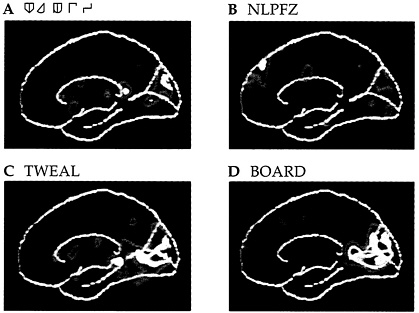
PLATE 1. As a research subject is shown a series of images on a screen that run from letter-like graphics (A) to a meaningless string of letters (B) to something that is pronounceable but is not a word in English (C) to a real English word (D), several sites come into action or drop out of participation. A positron emission tomographic (PET) image shows activity in the occipital lobe that increases sharply when the graphics become real words. Source: Marcus Raichle, Washington University School of Medicine, St. Louis, Mo.
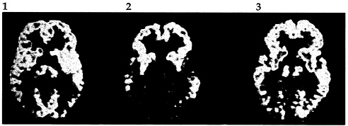
PLATE 2. Treatment with AZT can help in the management of AIDS-related dementia, as is demonstrated by these three PET images. In the healthy brain (1), levels of activity are relatively even through the frontal, temporal, and occipital lobes. Brain activity of a patient with AIDS-related dementia (2) is uneven, with the occipital region and part of the temporal lobes much less active. After 13 weeks of treatment with AZT (3), brain activity has returned significantly toward the normal pattern. Source: National Cancer Institute, Bethesda, Md.
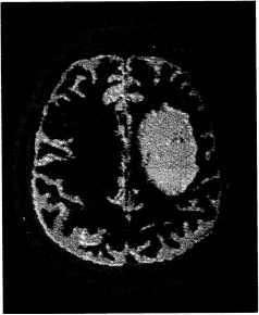
PLATE 3. Magnetic resonance imaging (MRI) is an invaluable tool in clinical practice today, as well as in neuroscientific research. Here, the crosssectional image of a patient's head clearly discloses the shape and location of a tumor (the egg-shaped yellow mass in the right hemisphere). Healthy brain tissue appears in gray, and the skull and scalp in pink. Such images contribute to precise diagnosis and, when necessary, the planning of neurosurgery.
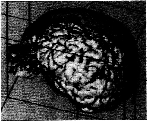
PLATE 4. In further MRI of the patient shown in Plate 3 , the surface of the brain is revealed by selectively “cutting away” the scalp.
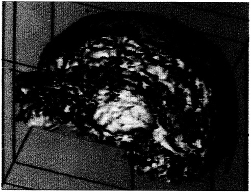
PLATE 5. The brain itself is then cut away, in imaging terms, slice by slice, until the image plane arrives at the tumor. Source for Plates 3-5 : H. E. Cline et al., 1990. Journal of Computer Assisted Tomography 14(6):1037–1045.
PLATE 6. The positron emission tomographic (PET) scan of a research subject undergoing a panic attack shows very high activity (white area) in a portion of the limbic system, the “emotional brain.” What this image of just one hemisphere cannot show, however, is another striking feature of the brains of people subject to panic attacks: an asymmetry between the two hemispheres, with the left side less active than the right. PET has been useful as a research tool in revealing general patterns of brain activity that characterize panic disorder. Source: Marcus Raichle, Washington University School of Medicine, St. Louis, Mo.
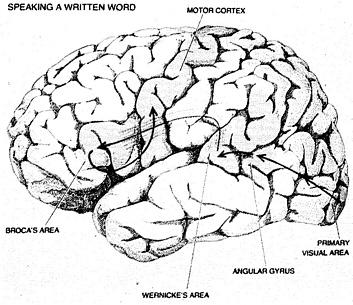
PLATE 7. The simple act of pronouncing a written word is the outcome of an intricate path of signals through several parts of the brain. Recognizing the word calls for the participation of the primary visual area and the visual association areas (in and around the angular gyrus). Then, for the production of speech, Broca's area becomes active; finally, the signals arrive at the motor cortex, which sends instructions for movement to the lips and tongue and larynx. Source: Marcus Raichle, Washington University School of Medicine, St. Louis, Mo.
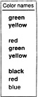
PLATE 8. At several years of age, humans perform the unusual feat of developing an entirely new visual pathway in their brain: the ability to process written information. The pathway, reinforced by training, can become very strong, even overriding other forms of visual information. To test the dominance of this pathway, try saying aloud the colors in the list. Which word presents itself: the color in which the word appears, or the color name that is written? This test demonstrates the Stroop effect, a well-known psychological phenomenon (see page 120 for additional discussion). Source: Marcus Raichle, Washington University School of Medicine, St. Louis, Mo.


