Questions? Call 888-624-8373

HARDBACK
list:$34.00
Web:$30.60
add to cart

PDF BOOK
your price: $26.50
add to cart

Rights & Permissions

topleft topright

Brucellosis in the Greater Yellowstone Area (1998)
Board on Agriculture (BOA)

Page
187
bottomleft bottomright

The following HTML text is provided to enhance online readability. Many aspects of typography translate only awkwardly to HTML. Please use the page image as the authoritative form to ensure accuracy.


PLATE 1. (upper) Drawing of the microscopic appearance of Brucella abortus in placental exudates and placental membrane surfaces (lower) of a cow that had aborted. Bacteria are in and around leukocytes in pus. Source: Poppe 1929.

Page
187

Below are the first 10 and last 10 pages of uncorrected machine-read text (when available) of this chapter, followed by the top 30 algorithmically extracted key phrases from the chapter as a whole.
Intended to provide our own search engines and external engines with highly rich, chapter-representative searchable text on the opening pages of each chapter. Because it is UNCORRECTED material, please consider the following text as a useful but insufficient proxy for the authoritative book pages.

Do not use for reproduction, copying, pasting, or reading; exclusively for search engines.

OCR for page 187
PLATE 1. (upper) Drawing of the microscopic appearance of Brucella abortus in placental exudates and placental membrane surfaces (lower) of a cow that had aborted. Bacteria are in and around leukocytes in pus. Source: Poppe 1929.

OCR for page 188
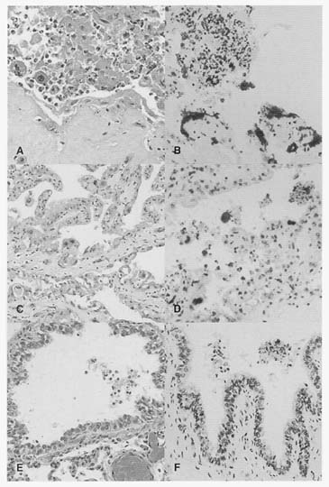
PLATE 2. Histology (A,C,D) and immunohistochemistry (B,D,F) of tissues from the uterus and retained placenta from an aborting bison cow found in March 1995 adjacent to the YNP. A. Necrosis and exudate, placenta. B. Strong labeling of bacteria in trophoblastic epithelial cells. C. Non-necrotic areas of the placenta. D. Lesser amounts of bacteria in non-necrotic areas of the placenta. E. Lung: hyperemia with necrotic debris in a large bronchiole (center). F. Bacterial debris in bronchiolar lumen are stained. Source: Courtesy J. Ryhan.

Representative terms from entire chapter:

trophoblastic epithelial