
HOWARD M. TEMIN
December 10, 1934–February 9, 1994
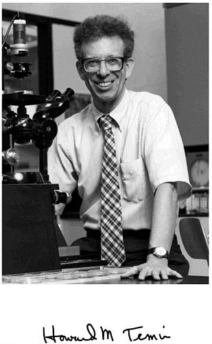
BY BILL SUGDEN
HOWARD TEMIN LOVED knowledge, its acquisition, and its sharing. He pursued research where the logic of his experiments led him, independently of the scientific community’s initial skepticism toward his findings. He applied his expertise to improve public health policy to minimize smoking and to maximize benefits from research on the human immunodeficiency virus (HIV).
This recounting of Howard Temin’s scientific career reflects my appreciation of his work as a tumor virologist. We shared lunch on Tuesdays for 20 years and gradually grew to be friends. Howard classified himself as a virologist and taught his students to be virologists. He taught the prominent course on animal virology on the University of Wisconsin’s Madison campus for 30 years. (I filled in for him when I first came to the McArdle Laboratory for Cancer Research while he traveled to Stockholm to accept the Nobel Prize. Typically, he insisted that I give several lectures before he left so that he could gauge whether I could lecture adequately and to coach me. I passed his test and I teach that course today.)
During the first week I was in McArdle, Howard invited me to have lunch with him, Paul Kaesberg, and Roland Rueckert. These lunches were squeezed in for one-half hour before the weekly seminar on tumor virology—a training
ground for formal presentations by graduate students and postdoctoral fellows that Howard had organized. I learned much from and about Howard at those lunches. He usually determined the subjects to be discussed but wanted our contributions. He valued science enormously while savoring the peccadilloes of its practitioners. These lunches, our frequent discussions about our faculty colleagues in McArdle, and our shared participation on many graduate student committees led us from formal collegiality to informal friendship. The uncited interpretations and motivations I ascribe to him come from these times together.
Many of us who grew to know Howard professionally valued him personally. Rayla Greenberg Temin warmly described his rich personal life.1 Here I shall outline the depth and breadth of his scientific contributions. It is these contributions, coupled with his commitment to reason in all facets of his professional life, that made Howard Temin a major force in biology during the latter half of the twentieth century.
THE PROVIRUS HYPOTHESIS
Quite early in life Howard Temin was a devotee of science in general and biology in particular. He published his first paper at the age of 18 in 1953. He began the research he would follow professionally as a graduate student with Renato Dulbecco in Cal Tech in 1957. Not long before, Dulbecco and Margarite Vogt2 had developed a method to plaque poliomyelitis virus in cell culture; that is, they learned how to enumerate infectious virus particles in a stock of virus by detecting one focus of dead cells per infectious particle. The infected cells were lysed by expanding rounds of infection, viral replication, and cell death. This assay was the foundation for quantitative studies of lytic viruses in cell culture. As Howard began work in Dulbecco’s group, Mannaker and Groupé3 described an assay in which Rous
sarcoma virus (RSV) altered the morphology of cells infected in culture. Here each focus of infection did not result from ensuing rounds of cell death but rather in survival of cells with altered phenotypes of shape. This in vitro assay was exciting because it could reflect in vitro the known ability of RSV to cause cancers in vivo soon after its inoculation into newborn chicks. Howard, working with Harry Rubin, a postdoctoral fellow in Dulbecco’s group, refined this assay and used it to investigate “morphological transformation” of cells by RSV in culture.
Temin and Rubin (1958) refined the assay for RSV by overlaying chick embryo fibroblast (CEF) cells with agar soon after their exposure to dilutions of a virus stock. This overlay minimized the spread of progeny virus from an initially infected cell to distant cells, thereby confining the progeny from initial infections each to a single focus. The agar also helped to restrain the infected cells, which became less adherent to their initial site. Overlaid, infected cells yielded foci that enlarged exponentially with time, and this enlargement resulted primarily from division of the infected cells (1958). These foci arose linearly as a function of the dilution of the virus stock assayed over a large range of dilutions; these results indicated that a single particle of RSV is competent to initiate a focus (1958).
Rubin and Temin (1959) used this quantitative focus assay for RSV to analyze its initiation of infection radiologically. They compared the sensitivities to exposure to X rays and ultraviolet (UV) light of RSV and New Castle disease virus (NDV) in initiating or maintaining infection. NDV infects CEF cells only lytically and has an RNA genome, as RSV was then thought to have. (Bather4 found RNA in semi-purified RSV. Crawford and Crawford5 developed an isopycnic method to purify RSV further and confirmed its genome as being RNA). They found that infection by NDV was resistant to
previous exposure of the host cells to X rays or UV light, while infection by RSV was exquisitely sensitive to similar treatments. Once cells were infected with RSV, their ability to produce progeny virus had an intermediate sensitivity to treatment with X rays. The exquisite sensitivity of RSV to initiate infection of radiologically treated cells equaled that of treated CEF cells to form colonies. This finding indicated that initiation of infection by RSV shared a common radiosensitive target with cell division, in marked contrast to the radiologically resistant, cytocidal NDV. The reduced sensitivity to radiological treatment of RSV production after establishment of its infection, coupled with its exquisite sensitivity for initiation of infection, appeared similar to those of the temperate phage (1959). These studies led them to hypothesize that “the genome of the Rous sarcoma virus must be integrated with that of the cell before virus production can begin” (1959). This hypothesis drove much of Howard Temin’s research for the next 11 years.
Temin and Rubin6 established the previously suspected model that cells in which RSV infection is established pass on to their progeny the capacity to release RSV. They analyzed single infected cells in microdrops and found both that the cells divided and that the daughter cells could release virus. This observation underscored the difference between RSV and lytic viruses such as EMC and NDV. RSV infection did not dramatically alter its host cell’s survival, but it did affect it genetically; lytic viruses merely killed their host cells. This realization provided another similarity to the temperate phage, and a difference. Temperate phage pass on phage genomes from infected parent to infected daughter cell; however, they do not continuously release progeny phage.
In 1959 Harry Rubin moved to Berkeley, while Howard Temin continued his work at Cal Tech. Howard characterized
isolates of RSV that conferred distinctive morphologies on their infected cells. He infected CEF cells that he had cloned to demonstrate that the distinctive cellular morphologies resulting upon infection with different isolates of RSV reflected genetic contributions of the viruses (1960). The capacity to endow an infected host cell with one morphology could mutate, the mutant virus then conferring on the cells it infects a new, distinctive morphology. Howard concluded that “the virus becomes equivalent to a cellular gene controlling cellular morphology.” He also contemplated the possibility that the ability of RSV to control the morphology of cells infected in vitro was related to the virus’s tumorigenic capacity in vivo.
In 1960 Howard moved to the McArdle Laboratory for Cancer Research, where he carried on his research for the rest of his life. By this time an appreciation of the functions of cellular RNAs was crystallizing. Two groups published their findings, indicating that ribosomal RNAs were stable, structural elements of ribosomes, whereas short-lived RNAs conveyed information from DNA to the ribosomes to encode protein synthesis.7,8 This short-lived RNA is messenger RNA (mRNA). The other component of cellular RNA was transfer RNA (tRNA), small stable RNAs required to move amino acids to the ribosomes. The recognized functions of cellular RNAs did not include long-lived transfer of information. The means by which RSV, an RNA tumor virus, could stably affect the heritable morphology of infected cells was therefore enigmatic both for Howard and the scientific community at large.
At the McArdle Laboratory Howard continued to study RSV infection genetically. He isolated and characterized cells infected with RSV arising after a low multiplicity of infection, which were morphologically altered but did not in general produce virus. Virus production could be rescued
by infection of these converted non-virus-producing (CNVP) cells with a different strain of RSV. These CNVP cells induced tumors in susceptible chicks, as did virus-producing cells (1963,1). These experiments separated virus production from virus-mediated morphological transformation of the infected host cell and tumorigenicity. They demonstrated that RSV-infected cells could maintain the information for producing virus in the absence of such production. The information in the infected cell necessary to produce virus was designated the provirus.
Howard analyzed the mechanism of infection by RSV biochemically. He assessed actinomycin D as an inhibitor of RSV infection and production. Actinomycin D had been recently shown to inhibit DNA-dependent RNA synthesis,9 but not the replication of some RNA viruses.10 At low concentrations (0.1 to 0.2 µg/ml), actinomycin reversibly inhibited infection by and production of RSV (1963,2). Concentrations up to 10 µg/ml had no effect on lytic infections by NDV (1963,2). Thus the effect of this inhibitor varied dramatically for the lytic RNA virus NDV and the transforming RNA tumor virus, RSV. Howard “suggested that the template responsible for synthesis of viral (RSV) nucleic acid either is DNA or is located on DNA” (1963,2). This unorthodox suggestion was a logical outgrowth of Temin’s genetic and biochemical studies of RSV replication in cell culture. Similar experiments demonstrating the sensitivity of RSV infection to treatment with actinomycin D were also reported by other groups.11 Temin tested his suggestion directly with two recently developed methods to detect specific RNA/DNA hybrids. He labeled RSV RNA with tritiated uridine by propagating infected cells in labeled medium, isolating released virus, and purifying its genomic RNA. This labeled RNA was then hybridized to cellular DNAs isolated from uninfected and infected cells. The results of
these experiments indicated that more DNA in infected cells was detected by its hybridization to RSV RNA than DNA in uninfected cells (1964). The specific c.p.m. of labeled RNA hybridized to cellular DNAs were extremely low—too low to be compelling today. However, the small signals were consistent with the provirus of RSV being DNA, as Howard had hypothesized.
Howard analyzed the effects of serum on the proliferation of CEF cells in culture and recognized that the removal of serum inhibited the cells’ proliferation.12 He used this insight to manipulate cultures of cells such that they were partially synchronized within their proliferative cycle. He infected these partially synchronized cells with RSV and found that the capacity of infected cells to support virus production required the infected cells to pass through mitosis (1967). He found this requirement for mitosis when cultures were partially synchronized by removal of serum, by treatment with excess thymidine, or by treatment with colchicine (1967). In all cases, RSV was produced from infected cells after they passed through mitosis. These experiments did not allow him to distinguish between a requirement for mitosis to form the provirus or to activate the provirus to allow production of progeny virus. Additional experiments with synchronized cells did support the hypothesis that the provirus was composed of DNA. Howard treated cells arrested largely in the G2 phase of the cell cycle with cytosine arabinoside, an inhibitor of DNA synthesis. This treatment inhibited formation of the provirus as reflected by a failure to produce progeny RSV; the inhibition was abrogated by simultaneous treatment with deoxycytidine (1967). This and related observations indicated that formation of the provirus required DNA synthesis, but that DNA synthesis need not occur during the S phase of the cell cycle. The accumulated findings from Howard’s experiments analyzing infection by RSV in
cell culture were convincing to him, but still did not persuade virologists in general.
The years 1967 and 1968 were watershed occasions for animal virology; at least three findings with DNA and RNA viruses would eventually contribute to Howard’s appreciation of the life cycle of RSV and to its general acceptance by virologists. Joe Kates and Brian McAuslan,13 working with rabbit poxvirus, a member of the same family of DNA viruses as is smallpox, demonstrated that purified viral core particles contain a DNA-dependent RNA polymerase. The activity was detected only after intact viral particles were disrupted, was inhibited by actinomycin D, and synthesized RNA homologous to rabbit poxviral DNA. (It has since been demonstrated that poxviral DNA-dependent RNA polymerase is encoded by the virus and related to similar cellular enzymes.) In 1968 Aaron Shatkin and J.D.Sipe identified an RNA-dependent RNA polymerase in cores of reoviruses.14 Reoviruses contain multiple distinct segments of double-stranded RNA within their inner core. Shatkin and Sipe found that they needed to remove the virus’s outer protein shell to detect the polymerase activity, which was dependent on the addition of all four ribonucleoside triphosphates and synthesized RNAs homologous to reoviral genomic RNA. Thus, by the end of 1968 it was evident that both a DNA-and an RNA-containing animal virus house a template-dependent RNA polymerase activity.
Studies with the DNA tumor viruses, simian virus 40 (SV40) and polyoma (Py) in 1968 demonstrated that these tumor viruses affected their transformation of cells in culture by maintaining their DNA genomes integrated into their host cell’s chromosomes. Joe Sambrook—a postdoctoral fellow with Howard’s former mentor Renato Dulbecco—led a team of researchers who isolated chromosomal DNAs from SV40-and Py-transformed cells and showed by nucleic acid hybridi-
zation that these cellular DNAs had integrated copies of the viral DNAs.15 Two approaches were critical to the success of their experiments. In one approach the isolated cellular DNAs were separated on alkaline sucrose gradients by velocity sedimentation, which minimized any fortuitous, non-covalent association of viral DNAs with large chromosomal DNAs. In all experiments the integrated viral DNAs were detected by hybridization with viral RNAs synthesized in vitro with DNA-dependent RNA polymerase isolated from E. coli. This latter approach permitted the radiolabeled RNA synthesized in vitro with purified viral DNA templates to be of high specific activity. The results from the hybridization experiments were thus compelling and demonstrated that certain DNA tumor viruses maintained their genomes as DNA integrated into the virally transformed host’s chromosomes.
As these results with different animal viruses were being appreciated, David Boettiger began working as a graduate student at the McArdle Laboratory with Howard Temin. Colleagues at McArdle had been working previously with various halogenated nucleotides. Charlie Heidelberger had synthesized fluorodeoxyuracil (5-FU) and pioneered its use in chemotherapy for certain human cancers. Waclaw Szybalski had studied bromodeoxyuridine (5'-BUdR) and had shown that, on its incorporation into the DNA of bacteria, DNA became sensitized to damage induced by exposure of the cells to near ultraviolet or visible light. David, with Howard’s guidance, rendered CEF cells stationary by withdrawing serum from their medium, infected them with RSV, treated them with 5'-BUdR, and exposed them to near UV light (1970). The stationary cells did not incorporate 5'-BUdR and were not detectably harmed by the near UV light. However, this regimen reduced infection by RSV to 5 percent of that of the untreated control. Importantly, increasing the multiplicity of infection twenty-fold significantly decreased the
rate of inactivation of infection, indicating that multiple virus particles were inactivated independently within a single infected cell. This experiment provided powerful support for Howard’s provirus hypothesis. It demonstrated that the incorporation of a light-sensitive DNA-nucleotide analogue soon after infection by RSV, followed by exposure to near UV light, inactivated that infection without damaging stationary host cells. This manuscript was submitted to Nature in March of 1970, but it was not immediately accepted.
While David Boettiger was optimizing his inactivation experiments, Satoshi Mizutani, a postdoctoral fellow with Howard, pursued the possibility foreshadowed by the findings with rabbit poxvirus and reovirus that RSV might contain a polymerase activity capable of copying RSV RNA into a precursor to its proviral DNA. Permeabilization of the envelope of the viral particle with a non-ionic detergent in the presence of dithiothreitol allowed detection of the activity soon to be dubbed reverse transcriptase. This enzyme could incorporate the four deoxyribonucleotides to yield DNA. Treatment of the disrupted virions with RNase A prior to their incubation with labeled deoxynucleotides abrogated subsequent DNA synthesis, indicating that RSV contained an RNA-dependent DNA polymerase. Mizutani and Temin submitted their findings to Nature on June 15, 1970. Their paper was published on June 27, 1970 (1970,1). David Baltimore at MIT published similar findings for murine RNA tumor viruses in the same issue of Nature.16 He had submitted a manuscript in March to the Proceedings of the National Academy of Sciences relating his findings that vesicular stomatitis virus, VSV, a rhabdovirus, contained an RNA-dependent RNA polymerase, again focusing attention on the existence of template-dependent polymerases intrinsic to various families of viruses.17
The two reports of RNA-dependent DNA polymerase
activity in avian and murine RNA tumor viruses caught the attention of much of the biological community. Howard’s provirus hypothesis was accepted. The existence of this formerly undetected activity was consonant with his hypothesis, but uncountable years of research by many people would be needed to outline the mechanism by which reverse transcriptase and its associated activities could synthesize a provirus. In the interim, the inactivation experiments of Boettiger and Temin, which established the DNA nature of RSV’s provirus, were published by Nature (1970,2) 8 months after its submission, not the 12 days needed to christen reverse transcriptase. The combined findings indicated that RNA tumor viruses copied their RNA genomes into DNA and that these proviral DNAs were integrated into infected cells’ chromosomes, as were the genomic DNAs of SV40 and polyoma. RNA tumor viruses were themselves eventually rechristened retroviruses to underscore their intrinsic use of a “backwards” flow of information from RNA to DNA.
MULTIPLICATION-STIMULATING ACTIVITY (MSA)
Howard characterized cells infected with RSV in order to compare and contrast their proliferative abilities with those of uninfected parental CEF cells. He termed the infected cells converted; today they are termed transformed. He was motivated by his appreciation that RSV was an efficient tumor virus, causing rapidly growing, lethal tumors within 10 days when inoculated into susceptible, newborn chicks. He soon learned that both parental and transformed cells deplete culture media of required factors contained in serum.18 The concentration of serum in a medium determined the saturation density cells achieved when they eventually ceased to proliferate.19 Insulin could replace serum and support proliferation of parental and transformed CEF cells; the transformed cells could grow to a higher saturation density
than their parental cells in a medium lacking serum but containing insulin.19 These observations led Howard to hypothesize “that the increased multiplication in cell culture of converted [i.e., transformed] cells as compared with uninfected cells results from a decreased requirement by the converted cells for an insulin-like activity found in serum.”19 He pursued this hypothesis in tandem with his work on the provirus and argued that the reduced requirement of transformed cells for specific factors in serum contributed to their tumorigenicity in vivo.20
Embryo fibroblasts derived from chickens and ducks, but not rats, could deplete media of factors required for their proliferation,20,21 and all three, when infected with RSV or a murine sarcoma virus (MuSV), could proliferate to higher saturation densities when serum was limiting than could their uninfected parents.21 Transformed CEF cells multiplied to higher saturation densities than uninfected parental cells in limiting serum, because they passed through more cell cycles than did their parental cells, as scored by their prolonged ability to incorporate tritiated thymidine.22 Transformed cells bound or depleted the multiplication-stimulating activity (MSA) in serum at the same rate as did their parental uninfected cells. These studies showed that medium with serum depleted by either cell type during increasing times of incubation supported proliferation of fresh cells to similar extents.23 The increased capacity of transformed cells to multiply in limiting serum therefore reflected their ability to use MSA more efficiently than did untransformed cells.23 These findings in cell culture provided a model with which to consider tumor growth in vivo. The growth of RSV-infected cells in vivo could reflect in part their capacity to proliferate efficiently in levels of MSA that did not support multiplication of uninfected or normal cells. This situation might arise, for example, when a nidus
of infected cells expanded beyond their immediate blood supply.
Howard and his colleagues needed now to define MSA molecularly. The observation that rat embryo fibroblasts failed to deplete serum of MSA, as did chick and duck cells, led to the appreciation that some rat cell lines secreted MSA.24 MSA was partially purified from calf serum25 and from serum-free medium conditioned by growing in a line of Buffalo rat liver cells, BRL-3A (1973). It was purified to apparent homogeneity from conditioned medium with a final step of preparative electrophoresis in polyacrylamide gels containing sodium dodecyl sulfate (SDS).26 MSA purified from conditioned medium stimulated DNA synthesis in treated cells, stimulated cell multiplication, stimulated glucose uptake, but did not contain insulin.26 Thus, in 1974 Howard was poised to characterize MSA in detail molecularly and to explore the mechanism by which it stimulated multiplication of cells in culture. His original contribution focusing on the effects of serum to promote proliferation per se placed his imprint on this field. However, his commitment to understand the life cycle of RNA tumor viruses led him to focus his research on these viruses and not to pursue his work on growth factors such as MSA. His focus on RNA tumor viruses was also fostered by his own findings, which had heightened the growing interest of the community of cancer researchers in this family of viral carcinogens and placed him at its center.
AN EVOLVING STYLE
Howard Temin’s research on RNA tumor viruses, his contributions to the elucidation of their replication, and his enunciation of and experimental support for the provirus hypothesis were not conducted in a vacuum. The Madison campus in general and the McArdle Laboratory for Cancer
Research in particular provided an intellectually rich and supportive milieu for his research. Harold Rusch directed the McArdle Laboratory when Howard joined it in 1960, and, together with the senior faculty, he worked to protect junior faculty from university responsibilities outside their research. Howard seized this opportunity and immersed himself in his own research. He worked with technicians and carried out many experiments himself. He guided his technical staff closely, observing their cells and findings daily. He accepted no graduate students for the first seven years of being a faculty member. Of the first 24 manuscripts he published from McArdle, he is the sole author on all but one. This singular dedication to his research, coupled with his exacting thought and speech, made him a formidable figure on the Madison campus.
As his research contributions grew to be appreciated beyond Madison, Howard devoted more of his time to mentoring graduate students and postdoctoral fellows. Having accepted a student into his group, Howard placed his highest priority on guiding that student; a growing list of local and national responsibilities did not challenge the priority he placed on training. After 1970 almost all his research papers were co-authored with graduate students, postdoctoral fellows, and technicians. Howard trained, in part, by example. He was engaged intellectually, socially, and professionally in all settings. Whether at the weekly tumor virology seminar in McArdle or the yearly retrovirus meeting in Cold Spring Harbor, he would be in the front row and ask questions of each speaker. He asked to understand, to seek help to solve a puzzling problem, and to provide insights. Speakers might fear his grilling and retrospectively delight in his having asked them questions from which they learned.
The general acceptance of his proviral hypothesis after the identification of reverse transcriptase activity in the virions
of RNA tumor viruses(1970)16 provided him the opportunity to consider that hypothesis in a broad context. Howard developed a protovirus hypothesis, which he described in three installments (1971).27,28 The prefix proto is derived from Greek and means primitive or original. In contemplating the origin of RNA tumor viruses, Howard postulated that reverse transcription could contribute not only to the formation of RNA tumor viruses, but perhaps also to somatic development in and evolution of the species. Two of these speculations have proved, in part, correct.
In considering the origin of RNA tumor viruses, Howard distinguished between the RNA sarcoma viruses, which efficiently cause sarcoma but are rare in nature, and the RNA leukemia viruses, which rarely cause cancers but are common in nature. He wrote,27
RNA sarcoma viruses have arisen de novo on several occasions. The most famous example is the original discovery of the Rous Sarcoma Virus (RSV) [32]. Since these viruses cannot replicate in a cell without transforming it into a tumor cell, and since these viruses are not ordinarily transmissible from one infected animal to another without experimental intervention, the RNA sarcoma viruses must have originally come from some preexisting entity which differed in at least one of these properties, that is, which was not an RNA sarcoma virus.
RNA leukemia viruses are very widespread in natural populations of birds and mammals. They are very similar to RNA sarcoma viruses, both in the structure of the virion and in having a DNA intermediate for replication. Therefore, it seems likely that the RNA sarcoma viruses arose by mutation from the RNA leukemia viruses. In light of the previous discussion, the mutation would involve the genes making a product controlling the requirement for multiplication-stimulating activity for cell multiplication. In the sarcoma viruses this product would be effective in increasing the efficiency of utilization of multiplication-stimulating activity by fibroblasts, while in the leukemia viruses this product would not be active in fibroblasts.
We know now that the rapidly transforming RNA tumor viruses arise from the weakly transforming RNA tumor viruses’
or RNA leukemia viruses’ acquiring cellular proto-oncogenes (genes that, when mutated, can contribute to the risk of developing cancer) and together evolving through cycles of replication mediated through reverse transcription. Since 1970 Temin and his colleagues contributed much to this understanding. An enormous group of researchers have also contributed to the identification and characterization of more than 100 proto-oncogenes in the mammalian genome.
Howard presented his hypothesis for the generation of rapidly transforming RNA tumor viruses from weakly transforming parents partially in response to a proposal by Huebner and Todaro.29 These authors speculated that cancers arise in animals, including human beings, from repressed RNA tumor viruses resident in germ cells of all mammals. In this model, these viruses are transmitted vertically and, when activated, induce cancer. Huebner and Todaro posited that carcinogenic information was incorporated in inherited but repressed RNA tumor viruses that must be activated by chemical or physical inducers (thought of as carcinogens) to cause cancers. Howard suggested, rather, that rapidly transforming RNA tumor viruses cause cancers by horizontal infections and arise from weakly transforming RNA tumor viruses when they acquire cancer-promoting mutations. These disparate notions were eventually resolved by seminal studies of Varmus, Bishop, and their colleagues. These virologists asked the question, “What information is present in Rous sarcoma virus but not present in related, weakly transforming avian RNA tumor viruses?” They demonstrated that nucleotide sequences derived from uninfected cellular DNA represented the transforming information in this rapidly transforming RNA tumor virus.30 Subsequent analyses of this and other oncogenes captured by retroviruses have demonstrated that the viral oncogenes are derived from normal cellular genes now dubbed proto-oncogenes and
are not linked to endogenous retroviruses. Proto-oncogenes contribute functions to normal cells and must be mutated and/or differentially expressed to evolve into oncogenes.
One tenet of the protovirus hypothesis held that uninfected cells would express RNA-dependent DNA polymerase activity (i.e., reverse transcriptase), which could contribute to the formation of RNA tumor viruses and to somatic development. Howard and his colleagues John Coffin, Chil-Yong Kang, and Satoshi Mizutani screened infected and uninfected CEF cells, infected and uninfected rat cells, and chicken embryos for reverse transcriptase activity.31 They isolated appropriate particulate activities that were RNA dependent and synthesized RNA-primed DNA products unrelated to RSV RNA. However, Howard acknowledged, “it is easier to get biochemical evidence for the existence of RNA-directed DNA polymerase activity in cells than to show it has a biological role.”32 In an addendum he wrote in 1979 to his protovirus hypothesis,33 he no longer emphasized the speculation that reverse transcriptase would function widely in uninfected cells.
In considering the protovirus hypothesis in the context of somatic development, Howard speculated that in “normal development, this DNA→RNA→DNA information transfer could be used to identify cells as being of a particular type and to recruit other cells into a related or identical form” (1971). He noted that the addition of new information by copying pre-existing DNA via RNA synthesis and reverse transcription might, for example, contribute to the generation of antibody diversity. As far as I know, that suggested mechanism for somatic differentiation has not been observed; genomic alterations during development consist of deletions yielding new juxtapositions, but not duplications via reverse transcription.
Howard extended his protovirus hypothesis to include
contributions of reverse transcription in the germ line to evolution of the species. Here his hypothesis is correct. Remnants of RNA tumor viruses (retroviruses), retrotransposons (elements that transpose via reverse transcription), and cDNA genes (copies of cellular genes arising via reverse transcription of RNA transcribed from these genes) are known now to comprise more than 10 percent of the human genome. The extent of information derived in the genome via reverse transcription makes Howard appear prescient in formulating his protovirus hypothesis.
Howard’s professional style also evolved as he won international recognition for his research. He, David Baltimore, and Renato Dulbecco were awarded the Nobel Prize in physiology or medicine in 1975. He seized an opportunity at the Nobel banquet, which was filled with smokers, to note he was “outraged that the one major measure available to prevent much cancer, namely the cessation of smoking, has not been more widely adopted.” This was not a lone gesture. He testified early in 1976 along with Renato Dulbecco before a Senate subcommittee chaired by Edward Kennedy in support of a tax of up to 50 cents per pack of cigarettes. Late in 1975 he argued before the Wisconsin State Health Council “that the deleterious effects of cigarette smoking can be considered the major health problem in the state of Wisconsin.” The health council went on to support a measure that would curtail smoking in public places. He testified before the Senate Human Services Committee in 1979 that smoking “is the number one preventable health hazard in the United States today” to support the elimination of smoking in public places. Howard opposed smoking not only by giving testimony before governmental committees, but also personally. When eating at a university cafeteria, if he encountered faculty or students smoking in a no-smoking area, he would forcefully ask them to stop smoking or move.
His efforts, coupled with those of many like-minded public health advocates, have led to the outlawing of smoking in most public places in many states, in domestic airplanes, and to the recent consideration by the federal government to classify nicotine-containing cigarettes as addictive.
STRUCTURE OF THE PROVIRUS
With the acceptance of the provirus hypothesis and with the cancer research community’s intensifying interest in retroviruses, Howard Temin’s research evolved to characterize the provirus. Given that the provirus represents the DNA intermediate in the life cycle of retroviruses, what does it look like? A satisfying answer to this question would await much work from many scientists. It would also require the development of tools powerful enough to elucidate the structure of a specific sequence of 104 base pairs among an avian or mammalian genome of 109 to 1010 base pairs of DNA. These tools were discovered and refined throughout the 1970s. Kelly and Smith demonstrated that a prokaryotic restriction endonuclease cleaved DNA at a specific palindromic hexanucleotide34; a simple assay for such enzymes was developed by Sharp and others,35 and the identification and characterization of hundreds of these potent tools ensued. Research in the 1960s had provided prokaryotic enzymes that synthesize kinase, phosphorylate, and ligate DNAs.36 These enzymes, coupled with restriction endonucleases and prokaryotic plasmids encoding antibiotic resistance, formed the tools for recombinant DNA technology. In 1975 Ed Southern provided an effective means to couple restriction endonucleases with nucleic acid hybridization to detect and identify unique segments of DNA within a mass of isolated cellular DNA.37 Technical developments during this decade were subsequently crowned with two methods to sequence DNAs efficiently.38 The course of elucidating
the structure of the provirus followed the development of these tools.
Even before the tools were available to study proviral DNAs physically, Hill and Hillova39 introduced a means to study them biologically. They showed that transfection of DNA from infected cells into naïve cells yielded infectious retroviruses (that is, proviral DNAs can be infectious themselves). Their finding was a compelling confirmation of the provirus hypothesis. It also allowed studies of the provirus itself.
Cooper and Temin (1974) developed a quantitative assay to detect infectious proviral DNA of RSV and of spleen necrosis virus (SNV), a retrovirus of the reticuloendothelial virus group. Howard and his colleagues gradually switched their focus from RSV to SNV, perhaps because nucleic acids of SNV hybridized to DNA sequences in uninfected CEF less efficiently than did those of RSV.40 Transfection of naïve cells with decreasing concentrations of DNA from infected cells yielded infected cells with a dose-response indicating that one proviral DNA mediated infection. Experiments with sheared DNA indicated that fragments between 10 and 15 kbp in length are required to encode an infectious provirus. Virion RNAs from retroviruses were known to migrate in velocity sedimentation analyses at 60–70S and upon denaturation at 35S.41 A duplex DNA of 10 kbp could easily serve as a template for a retroviral RNA of 35S; the measured length of the provirus was roughly consistent with the length of RNA in retroviruses.
The assay for infectivity was coupled with physical analyses to analyze the structures of DNAs first synthesized from SNV retroviral RNAs and those eventually integrated as proviruses (1977). Early after infection, most infectious DNA was in the cytoplasm. Extraction of recently infected cells by the method of Hirt42 separated small unintegrated DNAs
from the large integrated DNAs. Most of the small unintegrated infectious retroviral DNA consisted of linear duplex molecules approximately 10 kbp in length (1977, Figure 4); little was circular in configuration. The linear duplex DNA was found to have a higher specific infectivity than did the circular molecules. The means by which these molecules were infectious was not clear. They could upon being transfected into naïve recipient cells move to the nucleus and be transcribed to yield progeny viral RNAs, or recombine and/or integrate and be transcribed after these events to yield progeny viral RNAs. These studies demonstrated that infectious DNAs are synthesized in the cytoplasm from incoming retroviruses and accumulate prior to the synthesis of detectable, infectious, integrated, or proviral DNAs. The cytoplasmic infectious DNAs were likely to be precursors to the integrated provirus. As expected, the infectious provirus was found in large DNAs (1977, Figure 6) linked to cellular DNAs with characteristic repeated sequences.
The structures of SNV viral DNAs, prior to integration and after integration, could be studied by Southern blotting with restriction endonucleases that cleaved within viral DNA. The cleaved viral DNAs were detected by hybridization with 125I-labeled RNAs and “copy” DNAs of viral RNA (or cDNAs) synthesized with reverse transcriptase and 32P-labeled triphosphates. The 3' end of viral RNA could be enriched by purification of small, partially degraded viral RNAs containing polyA. (The work of Joe Kates with vaccinia virus had detected polyA in viral RNAs.43 PolyA was quickly shown to be a feature of the 3' ends of most eukaryotic messenger RNAs.) cDNAs of 3' ends of viral RNA detected the proviral DNA encoding those RNA sequences. Southern analyses of specifically cleaved, unintegrated, and SNV proviral DNAs demonstrated that both DNAs are terminally redundant and
are collinear.44 No differences were detected between infectious and non-infectious DNAs.
Mike Bishop, Harold Varmus, Peter Vogt, and their colleagues at the same time carried out extensive, similar analyses of the structures of unintegrated and proviral DNAs of strains of RSV.45 They established that the terminal redundancy of these DNAs consists of sequences derived from both the 3' and 5' ends of the viral RNA. These repeat ends were termed long terminal repeats (LTR) and consisted of about 300 base pairs.
Howard’s group isolated DNAs of SNV from acutely and chronically infected cells as recombinant DNAs cloned in a modified lambda phage, Charon 4a, and analyzed.46 The lambda phage, Charon 4a, was developed on the Madison campus to facilitate the safe isolation of cellular genes.47 Kunitada Shimotohno and Howard Temin analyzed these molecular clones in detail. They determined the DNA sequences of the LTRs and the cellular/proviral DNA junctions of six clones (1980).48 Their analyses were revealing: The LTRs of SNV were 569 base pairs and encoded signals both for the initiation of RNA synthesis and addition of polyA. The structure of an LTR could be depicted as U3RU5 in which U3 and U5 represent unique sequences from the 3' and 5' ends of viral RNA respectively, and R represents the terminal redundancy of viral RNA as enunciated by Coffin.49 A site for binding the cellular tRNAPro, which serves as a primer for reverse transcription, was mapped just downstream of the 5' LTR. The cellular/proviral DNA junctions of the isolated DNAs consisted of a 5-base pair direct repeat of cell DNA adjacent to a 3-base pair inverted repeat of viral DNA.50 These structures were similar to those of integrated bacterial transposons and therefore supported the hypothesis that the formation of retroviral proviruses was related mechanistically to bacterial transposition. Howard
was delighted by these findings: They were consistent with one facet of his protovirus hypothesis, in which he had suggested that retroviruses evolved from cellular elements involved in information transfer.
Structural determinations of the provirus demonstrated that the LTRs that flanked viral coding sequences contained most of the proviral cis-acting regulatory elements. The coding sequences of proviruses from different retroviruses generally consisted of a similar motif: viral internal structural genes (gag) -reverse transcriptase (pol) -viral glycoproteins (env). Rapidly transforming viruses had a different motif, however. In working with reticuloendothelial viruses, Howard also studied a rapidly transforming member, Rev-T, which caused fatal leukemia in newly hatched chickens and turkeys. As with most rapidly transforming retroviruses, Rev-T was defective for replication and required a helper virus, RevA, closely related to SNV. Rev-T encoded the viral transforming gene v-rel. Howard and his colleagues analyzed v-rel and the avian cellular gene c-rel from which it evolved. They showed that v-rel consisted of multiple fused exons of c-rel that were inserted in place of much of gag and pol in RevA.51 As RSV had captured the cellular src gene, Rev-T had acquired the cellular rel gene. These transforming viruses accommodated otherwise intractably large cellular genes in the compact form of DNA copies of the genes’ spliced RNA transcripts. The structure of v-rel and other retroviral transforming genes indicated that reverse transcription of the spliced RNA of cellular proto-oncogenes contributed to the formation of rapidly transforming retroviruses. Shimotohno and Temin (1982) provided experiments in support of this finding. They constructed a retrovirus with part of the mouse a-globin gene in it and demonstrated that this model virus lost the introns of the a-globin gene upon its replication through multiple rounds of reverse transcription. Determination of
the structures of proviruses, of retroviruses, and their rapidly transforming derivatives provided both the foundation for and the impetus to elucidate the mechanisms that underlie their formation. In a different tack, postdoctoral students with Howard—Tom Gilmore, Celine Gelinas, and Mark Hannink—analyzed v-rel, its expression, its protein product, and its product’s function to demonstrate that it was a transcriptional activator belonging to the NF-kB family (1988).52
SYNTHESIS OF THE PROVIRUS
Solving two interrelated problems drove Howard to study the synthesis of proviruses. First, he sought to define the viral cis- and trans-acting elements that mediated retroviral replication. Second, he sought to use these elements to understand retroviral variation and evolution. Both of these problems led him and his colleagues to develop and characterize retroviral vectors and the helper cells on which they depend.
Rapidly transforming retroviruses are natural vectors for their derivatives of cellular proto-oncogenes. Most are defective for replication and depend on helper viruses for one or more trans-acting viral proteins. These natural vectors have all the cis-acting elements required for their replication. Identification of these elements would allow their incorporation into vectors engineered by virologists. Identification of the trans-acting viral factors required by the natural vectors would allow introduction of their genes into established cells such that these engineered helper cells would support replication of appropriately engineered vectors (i.e., they would substitute for helper viruses).
Howard’s group identified and characterized retroviral elements required in cis for replication. Shimotohno and Temin53 used a direct approach to probe sites within a viral genome that could tolerate added genes. They inserted
multiple versions of the herpes simplex viral thymidine kinase gene (tk) at different positions in a proviral clone of SNV and tested two potential properties: Could it provide tk function to tk– cells, and could it replicate with REV-A as a helper virus in CEF cells? They found that it was important to remove the 3' end of the tk gene for viral replication, probably because the gene’s polyadenylation signal would lead to internal addition of polyA and truncation of the viral genome. They also found that tk-vectors that replicated in the presence of helper virus ranged from 5.7 to 10.6 kb in length. These tk+ viral vectors were powerful tools to identify other elements required in cis for efficient replication of retroviruses.
Small deletions within the provirus were used to identify an element termed “E” required for the encapsidation of the viral RNA by the viral capsid proteins.54 “E” would have to be added to any vector to insure its being packaged within viral particles. Another feature of retroviruses required precise analysis to permit construction of cells such that they could synthesize the viral particles required by retroviral vectors. Retroviruses synthesize gag and pol from genomic viral RNA; env is synthesized from a spliced transcript. Splicing had been discovered in the mid 1970s. Most primary transcripts of eukaryotic cellular genes were processed post-transcriptionally to excise multiple tracts of intervening sequences (introns) and ligate the remaining sequences (exons), which could subsequently be exported to the cytoplasm to function as mRNAs. The structure and life cycle of retroviruses necessitates that their splicing be incomplete. The primary transcription of the provirus is the virion RNA—the RNA packaged in the viral particle. Were it to be efficiently spliced to yield only mRNA for env, then the intron required to synthesize gag and pol would be lost in the next generation of infection and reverse transcription. Shinichi Watanabe
and Howard (1983) precisely mapped the 5' and 3' splice sites used to synthesize env mRNA. This mapping allowed them to construct independent expression vectors for gag/ pol and for env, introduce them into cells, and identify cells that expressed all of these viral proteins. The expression vectors were designed to lack “E” and have as few sequences as practical that could mediate recombination with introduced vectors. These helper cells could replace the functions of a helper virus and support the viral life cycle of their tk+ vectors. Howard and his colleagues would go on to use these helper cells and a battery of carefully engineered vectors to measure and characterize rates and kinds of retroviral variation. These same helper cells formed a first generation of tools that would be essential for retrovirus-mediated gene therapy. They can support replication of vectors which themselves, when packaged, are infectious but do not yield infectious progeny on infection of non-helper cells. Parallel studies with murine retroviruses carried out by David Baltimore and his colleagues led to development of helper cells for murine retroviral vectors.55
Viral trans-acting functions in addition to gag/pol and env were likely to be required for retroviral replication. The similarity in structure between bacterial transposons and retroviral proviruses indicated that, like the transposons, retroviruses would contribute functions to mediate the integration of the provirus into host chromosomal DNA. Mutational analysis of coding information in SNV identified a virus that failed to integrate and was complemented by another mutant virus with cis-defects that eliminated its ability to integrate. With this former mutation, Nito Panganiban and Howard mapped the integrase gene of SNV to be at the carboxyl end of the polymerase gene.56
In Howard’s early experiments with RSV, he had identified and exploited spontaneous viral mutants. From the time
of those early experiments he wished to measure the rate of mutation during retroviral replication. During the 1960s and 1970s, accumulating research demonstrated that the cell regulates DNA synthesis and uses multiple mechanisms to insure the fidelity of that synthesis. During the same era, it became apparent that mutation rates in RNA viruses must be high. What was the rate of mutation in viruses that used both DNA and RNA as their genome? A barrier in measuring this rate had for years remained impassable. A rate reflects the number of mutations per cycle of replication, but the number of cycles of replication of retroviruses formerly could not be measured. Howard recognized that helper cells and their viral vectors could be used to support a single round of retroviral replication. The sum of mutations that accumulate during that single round would constitute a rate. Joe Dougherty and Howard devised a retroviral vector that expressed resistance to two drugs, G418 and hygromycin B. The G418-resistance gene termed neo was expressed from the full-length RNA and had inserted into it a termination codon that rendered it non-functional. The hygromycin gene was expressed from a spliced RNA in place of env. This vector was transfected into helper cells in the presence of neutralizing antisera, and the cells were cloned and screened for those that expressed the bona fide input vector. The packaged, released virus could be used at a low multiplicity to infect non-helper cells in which it would undergo a single round of reverse transcription and integration (1988,2, Figure 1). Such infected cells were selected for resistance to hygromycin B, the number of clones was enumerated, and the percentage of those also resistant to G418 was determined (1988,2). Resistance to G418 would reflect mutations that compensated for the termination codon introduced into the neo gene. These mutations arose frequently, and their basis was determined. Substitution mutations formed at a
rate of 2×10⁻⁵ per base pair per replication cycle. Insertions arose at 1/500 of this frequency.
These mutation rates for base substitutions are on the order of a thousand-fold higher than those for DNA synthesis in mammalian cells measured by Howard’s faculty colleague Norman Drinkwater.57 They formed the basis for understanding the extraordinary variation of HIV observed in vivo and allowed John Coffin to posit confidently that HIV’s observed variation necessitated efficiently repeated rounds of infection of naïve cells by HIV in vivo.58 His model has been verified by studies measuring the overgrowth of resistant mutants in vivo after treatment of AIDS patients with new, potent antiviral drugs.59
The application of retroviral vectors and helper cells to study variation during a single round of replication was extended by Wei-Shau Hu and Howard to provide detailed insights into the mechanism of synthesis of retroviral proviruses.60 Retroviruses encapsidate two 35s RNAs per viral particle. Hu and Temin introduced two vector DNAs into helper cells that encoded alternate resistances. One was neo–/hyg+, as was that used by Dougherty and Temin (1988), the other was neo+/hyg–. They characterized clones of the transfected helper cells to insure that each contained one bona fide copy of each of the vectors. Viruses released from these clones of helper cells would with some frequency contain heterodimers—one RNA molecule transcribed from each provirus. Recombination between packaged heterodimers would yield proviruses encoding resistance to both G418 and hygromycin B. The neo– and hyg– alleles were identified with specific restriction endonuclease sites. Wei-Shau infected cells with virus released from the clones of transfected cells and enumerated the number of infected cells resistant to one, to the other, or to both drugs. Recombinants arose at a rate of 2 percent per kbp, separating the mutant alleles
per replication cycle—extremely frequently. The recombinants were analyzed for loss of the diagnostic restriction endonuclease sites; most had lost them and represented genomic recombinants.60 Recombination within a retrovirus can be viewed to result from reverse transcriptase copying one template RNA or DNA and then switching to the other present in the viral particle. The high rate of recombination found by Hu and Temin indicates that such a template switch occurs once per every two or three cycles of retroviral replication.
Hu and Temin built on their initial study by using two vectors, neo–/hyg+ and neo+/hyg–, which were marked throughout their genomes with eight different restriction endonuclease sites. Evidence from work on disparate retroviruses from many groups provided a detailed model for the synthesis of the linear precursor to a provirus that Hu and Temin outlined (1990, Figure 2). They generated and analyzed recombinants between the marked vectors and interpreted their formation in light of the detailed model for the synthesis of the proviral precursor. The first step in synthesis of the precursor to the provirus is synthesis of DNA primed from the tRNA near the 5' end of the viral RNA. This DNA shortly runs out of template and can be isolated from in vitro reactions as strong-stop DNA. Hu and Temin (1990) identified which endonuclease restriction sites were present in recombinant proviruses. They used these analyses to show that strong-stop DNA, which could in theory transfer to the 3' end of the RNA molecule from which it was synthesized or to the 3' end of the other RNA molecule within the virus particle, could in fact transfer to either template. Once the first strand of DNA was synthesized, they found that the second strand was primed only from the first strand’s template. Howard interpreted the high frequency of detected recombination during retroviral replication to result from
the inherent ability of reverse transcriptase to switch its templates, an event essential to synthesis of the proviral precursor.61
Multiple studies with a variety of retroviral vectors and helper cells allowed Howard and his colleagues to prove the mechanism by which the provirus is synthesized. They now used this general approach to illuminate mechanisms by which retroviruses capture cellular proto-oncogenes to evolve into rapidly transforming derivatives. Jiayou Zhang developed helper cells with two non-homologous vectors. RNA synthesized from one would yield RU5 upstream of hygr but no U3 sequences. The other would yield RU5 upstream of neor along with U3R. Recombination within heterodimeric viruses released from these helper cells would be required to generate a functional 3' end of a hygr provirus (U3RU5 hygr U3RU5). Zhang and Temin (1993) infected cells with virus from characterized clones of helper cells with these proviruses and selected either for neor or hygr. Hygr cells arose at a frequency that indicated that nonhomologous recombination occurred at a rate of 0.1 percent to 1 percent of that of homologous recombination for retroviruses. They determined the sequence of the junctions of the recombinants and excitingly found a correlation with the kinds of junctions observed for sites at which retroviral sequences join viral oncogenes in rapidly transforming retroviruses.62 For example, in a group of non-homologous recombinants they described as being general, they found one example of recombination without sequence identity, six with a short region of five to eight base pairs of sequence identity, and three with insertions at the site of recombination. Inspection of naturally occurring, highly transforming retroviruses revealed that their sites of recombination usually contained short stretches of sequence identity or insertions.62 The non-homologous recombination they had characterized
underlies the means by which retroviruses form their 3' ends while capturing cellular proto-oncogenes.
HUMAN RETROVIRUSES
Howard’s hypothesizing the provirus, his work on its structure and on its synthesis involved avian retroviruses. RSV and REV-T were known to cause sarcomas and lymphomas in chickens at a time when human retroviruses were not identified, let alone shown to cause human disease. Confirmation of the provirus hypothesis, however, focused attention on retroviruses and helped to justify the National Cancer Institute’s major investment in research in the 1970s on retroviruses in general and a search for human retroviruses in particular. Although he in some sense started this band, Howard did not follow its wagon. He continued to study avian retroviruses. By the end of the decade, Y.Hinuma, following a paradigm he developed to study Epstein-Barr virus, a human tumor virus in the herpes virus family, helped to identify human T-cell leukemia virus type 1 (HTLV-1), a bona fide leukemia-causing human retrovirus. Bob Gallo and his colleagues through their research on propagating human T cells in medium containing IL-2 also contributed to the identification of this human tumor virus. The 1980s subsequently brought the recognition of another pathogenic human retrovirus that would occupy Howard’s interest and eventually some of his research efforts.
Acquired immunodeficiency syndrome (AIDS) was a disease entity identified in the early 1980s and found initially to be clustered in male homosexuals in the United States. The wasting that characterized AIDS was fatal, and suggestions for its cause varied widely and were often irrational. Luc Montagnier in the Institute Pasteur and Bob Gallo and his colleagues at the National Institutes of Health (NIH) identified a retrovirus linked to AIDS. Contentious claims
of priority appeared for a while to cloud the importance of this discovery. Howard worked through an NIH committee to help name this virus and limit any contention—thus the neutrally named human immunodeficiency virus or HIV. Epidemiological studies provided increasingly robust data indicating that HIV causes AIDS, but a few virologists, such as Peter Duesberg, argued with this conclusion.63 Some of Howard’s colleagues suggested that such arguments be ignored, but he was very concerned that those arguments be addressed directly and refuted. In 1988 Howard joined Bill Blattner and Bob Gallo to publish an article entitled “HIV Causes AIDS” in Science (1988). Here he contributed not original research but rather his depth and breadth of reading as well as his scientific integrity to reason for the well-being of people. He repeated his reasoned address in a short article in Policy Review in 1990.64 Howard willingly involved himself in national policy decisions to promote the best possible research on AIDS.
Between 1985 and 1994 Howard served on 12 national and international committees focused on multiple facets of HIV and AIDS. These included an oversight committee on AIDS activities of the Institute of Medicine from 1987 to 1990, chairing the HIV Genetic Variation Advisory Panel for the National Institute of Allergies and Infectious Diseases from 1988 to 1994, the Global Commission on AIDS from 1991 to 1992, and the World Health Organization Advisory Council on HIV and AIDS from 1993 to 1994. He devoted more of his efforts to guiding policies on AIDS and HIV than on any other public health problem faced during his professional life.
By 1992 Howard thought enough was known so that he could make experimental contributions toward the prevention of AIDS. HIV and HTLV-1 were known to be lentiviruses, a subtype of retroviruses that encoded several genes in
addition to the gag, pol, and env of the simple retroviruses such as SNV. These additional genes were necessary for HIV’s replication. No simple retrovirus had been isolated from people, and no lentiviruses had been isolated from chickens, mice, and cats, which harbor many simple retroviruses. Howard hypothesized that in this current niche of time, human beings have evolved to be resistant to simple retroviruses. A vaccine might be constructed by engineering the gag, pol, and env genes of HIV into a simple retroviral vector and be used to immunize people against infection by HIV (1993,1). Kathy Boris-Lawrie, a postdoctoral fellow, and he began testing this hypothesis by working with the animal lentiviruses, bovine leukemia viruses, and simian immunodeficiency virus, as models. Howard died of cancer in February 1994 before these preliminary experiments were completed.
This final project exemplified Howard’s research. It grew out of a thorough appreciation of HIV as a retrovirus. It embodied a bold hypothesis. It will be a lasting sorrow that he did not live to test his hypothesis, to learn from the experiments he proposed, and to contribute his further insights to biology and human welfare.
NOTES
SELECTED BIBLIOGRAPHY
1958 With H.Rubin. Characteristics of an assay for Rous sarcoma virus and Rous sarcoma cells in tissue culture. Virology 6:669–88.
1959 With H.Rubin. A radiological study of cell-virus interaction in the Rous sarcoma. Virology 7:75–91.
1960 The control of cellular morphology in embryonic cells infected with Rous sarcoma virus in vitro. Virology 10:182–97.
1963 Further evidence for a converted, non-virus-producing state of Rous sarcoma virus-infected cells. Virology 20:235–45.
The effects of actinomycin D on growth of Rous sarcoma virus in vitro. Virology 20:577–82.
1964 Homology between RNA from Rous sarcoma virus and DNA from Rous sarcoma virus-infected cells. Proc. Natl. Acad. Sci. U. S. A. 52:323–29.
1967 Studies on carcinogenesis by avian sarcoma viruses. V. Requirement for new DNA synthesis and for cell division. J. Cell. Physiol. 69:53–63.
1970 With S.Mizutani. RNA-dependent DNA polymerase in virions of Rous sarcoma virus. Nature 226:1211–13.
With D.Boettiger. Light inactivation of focus formation by chicken embryo fibroblasts infected with avian sarcoma virus in the presence of 5-bromodeoxyuridine. Nature 228:622–24.
1971 The protovirus hypothesis. J. Natl. Cancer Inst. 46:3–7.
With J.M.Coffin. Ribonuclease-sensitive deoxyribonucleic acid polymerase activity in uninfected rat cells and rat cells infected with Rous sarcoma virus. J. Virol. 8:630–42.
1973 With N.C.Dulak. A partially purified polypeptide fraction from rat liver cell conditioned medium with multiplication-stimulating activity for embryo fibroblasts. J. Cell. Physiol. 81:153–60.
1974 With G.M.Cooper. Infectious Rous sarcoma and reticuloendotheliosis virus DNAs. J. Virol. 14:1132–41.
1977 With E.Fritsch. Formation and structure of infectious DNA of spleen necrosis virus. J. Virol 21:119–30.
1979 With E.Keshet and J.O’Rear. DNA of noninfectious and infectious integrated spleen necrosis virus (SNV) is colinear with unintegrated SNV DNA and not grossly abnormal. Cell 16:51–61.
1980 With K.Shimotohno and S.Mizutani. Sequence of retrovirus provirus resembles that of bacterial transposable elements. Nature 285:550–54.
1981 With S.K.Weller. Cell killing by avian leukosis viruses. J. Virol 39:713–21.
1982 With K.Shimotohno. Loss of intervening sequences in genomic mouse a-globin DNA inserted in an infectious retrovirus vector. Nature 299:265–68.
1983 With S.Watanabe. Construction of a helper cell line for avian reticuloendotheliosis virus cloning vectors. Mol. Cell. Biol. 3:2241–49.
1988 With W.Blattner and R.C.Gallo. HIV causes AIDS. Science 241:515–16.
With J.P.Dougherty. Determination of the rate of base-pair substitution and insertion mutations in retrovirus replication. J. Virol. 62:2817–22.
With C.Gelinas. The v-rel oncogene encodes a cell-specific transcriptional activator of certain promoters. Oncogene 3:349–55.
1990 With W.-S.Hu. Retrovirus recombination and reverse transcription. Science 250:1227–33.
1993 A proposal for a new approach to a preventive vaccine against human immunodeficiency virus type 1. Proc. Natl. Acad. Sci. U. S. A. 90:4419–20.
With J.Zhang. Rate and mechanism of nonhomologous recombination during a single cycle of retroviral replication. Science 259:234–38.


