


Below is the uncorrected machine-read text of this chapter, intended to provide our own search engines and external engines with highly rich, chapter-representative searchable text of each book. Because it is UNCORRECTED material, please consider the following text as a useful but insufficient proxy for the authoritative book pages.
HEREDITY AND DEVELOPMENT: SECOND EDITION 19 2 The Cellular Basis of Inheritance The development of cytology, like that of all fields of science, depends partly on tools. The compound microscope, which is the basic tool in cytology, appears to have been invented about 1590 by two Dutch spectacles makers. The birthday of cytology was postponed, however, for three-quarters of a century. It was not until 1665 that Robert Hooke first described âcellsâ in a piece of cork (Fig. 2â1). He observed that cork and some other plant materi- als were composed of box-like structures, so small that eleven hundred arranged in length would equal no more than an inch. In living tissues they were filled with vegetable juices but were empty in the dried cork. When the cork was thinly sliced for microscopic examination, these boxes, or cells, appeared as rectangles, as the illustration shows. And interestingly enough he found these same structures in pieces of petrified wood. This discovery of cells in cork could have been an important advance in knowledge or an unimportant one. If it had turned out subsequently that cells were found only in cork, Hooke would certainly not be widely remembered for his discovery. But the work of many scientists showed that cells were a general phenomenon and, therefore, of some importance. We might conclude that Hooke did not make a discovery that was important but, instead, a dis- covery that became important. The Cell Theory. Following the realization that some cells are present in some organisms, the next conceptual advance was to establish the hypothesis that all organisms are composed solely of cells or cell products. This took nearly a century and a half. In the early years of the
HEREDITY AND DEVELOPMENT: SECOND EDITION 20 Observ. XVIII. Of the Schematisme or Texture of Cork, and of the Cells and Pores of some other such frothy Bodies. I Took a good clear piece of Cork, and with a Pen-knife sharpenâd as keen as a Razor, I cut a piece of it off, and thereby left the surface of it exceeding smooth, then examining it very diligently with a Microscope, me thought I could perceive it to appear a little porous; but I could not so plainly distin- guish them, as to be sure that they were pores, much less what Figure they were of: But judging from the lightness and yielding quality of the Cork, that certainly the texture could not be so curious, but that possibly, if I could use some further diligence, I might find it to be discernable with a Microscope, I with the same sharp Penknife, cut off from the former smooth surface an exceeding thin piece of it, and placing it on a black object Plate, because it was it self a white body, and casting the light on it with a deep plano-convex Glass, I could exceeding plainly perceive it to be all perforated and porous, much like a Honey-comb, but that the pores of it were not regular; yet it was not unlike a Honey-comb in these particulars. 2â1 The drawings and part of the text from Hookeâs observations on cork (R. Hooke, Micro- graphia, London, 1667).
HEREDITY AND DEVELOPMENT: SECOND EDITION 21 nineteenth century a number of cytologists came more and more to adopt this second concept, which we know as the âcell theory.â Three names are generally associated with the final formulation of the cell theory. They are R.J.H.Dutrochet, Matthias Jacob Schleiden, and Theodor Schwann. The last-named published his treatise on the microscopic structure of organisms in 1839, when he was 29 years old. In it he summarized his find- ings on a variety of tissues. His general conclusion was that the bodies of organisms are composed of cells. Figure 2â2 reproduces some of his drawings. 2â2 Some of Schwannâs drawings of cells (Th.Schwann, Mikroscopische Untersuchungen über die Uebereinstimmung in der Struktur und dem Wachsthum der Thiere und Pflanzen, Berlin, 1839).
HEREDITY AND DEVELOPMENT: SECOND EDITION 22 The following quotations from a translation of Schwannâs treatise will reveal some of his views: Though the variety in the external structure of plants is great, their internal structure is very simple. This extraordinary range of form is due only to a varia- tion in the fitting together of elementary structures which, indeed, are subject to modification but are essentially identicalâthat is, they are cells. The entire class of cellular plants is composed solely of cells which can readily be recog- nized as such; some of them are composed merely of a series of similar or even only of a single cell. Animals being subject to a much greater range of variation in their external form than is found in plants also show (especially in the higher species) a much greater range of structure in their different tissues. A muscle differs greatly from a nerve, the latter from a cellular tissue (which shares only its name with the cellular tissue of plants), or elastic tissue, or horny tissue, etc. If, however, we go back to the development of these tissues, then it will appear that all these many forms of tissue are constituted solely of cells that are quite analogous to plant cellsâ¦. The purpose of the present treatise is to prove the foregoing by observations. Much of Schwannâs success was due to the fact that he adopted a definite criterion for the recognition of cells, namely, the presence or absence of a nucleus. The latter structure was apparently first recognized as an important and characteristic cell structure by Robert Brown in 1833. It had been observed much earlier, however. Schwann took full advantage of this recent (for him) discovery. The most frequent and important basis for recognizing the existence of a cell is the presence or absence of the nucleus. Its sharp outline and its darker color make it easily recognizable in most cases and its characteristic shape, espe- cially if it contains nucleoliâ¦identify the structure as a cell nucleus and make it analogous with the nucleus of the young cells contained in cartilage and plant cellsâ¦. More than nine-tenths of the structures thought to be cells show such a nucleus and in many of these a distinct cell membrane can be made out and in most it is more or less distinct. Under these circumstances it is perhaps permis- sible to conclude that in those spheres where no cell membrane be distin- guished, but where a nucleus characteristic of its position and form is encoun- tered, that a cell membrane is actually present but invisible. Although Schwannâs work established the cell as a unit of structure, his views on the origin of cells precluded these elementary bodies from having any importance in inheritance, as the following quotation shows. The general principles in the formation of cells may be given as follows. At first there is a structureless substance which may be either quite liquid or more or less gelatinous. This, depending on its chemical constitution and
HEREDITY AND DEVELOPMENT: SECOND EDITION 23 degree of vitality, has the inherent ability to bring about the formation of cells. It seems that usually the nucleus is formed first and then the cell around it. Cell formation is in the organic world what crystallization represents in the inor- ganic world. The cell, once formed, grows through its inherent energy, but in doing so it is guided by the organism as a whole in the way that conforms to the general organization. This is the phenomenon basic to all animal and plant growth. It is applicable to cases where the young cells originate in the mother cell, as well as those where they are formed outside of them. In both instances the origin of cells occurs in a liquid or in a structureless substance. We will call this substance, in which cells are formed, a cell germinative substance or Cyto- blastema. It can be compared figuratively, but only figuratively, with a solution from which crystals are precipitated. The Continuity of Cells. Gradually this view was replaced by the realization that cells are formed solely by the division of pre-existing cells. Even before 1839 cell division had been observed, but it was during the following decade that more and more investigatorsâRobert Remak and Karl Wilhelm von Nägeli, for exampleâcame to the conclusion that cells never originate from a structureless âcytoblastema,â but always by cell division. In 1855 Rudolf Virchow formulated his well-known statement omnis cellula e cellula, which means âall cells from cells.â The cell then took on a new significance and greater importance. No longer was it a matter of the organism forming cells de novo, but instead cells, by their divisions, formed the organism. If existing cells have arisen from pre-existing cells, then there must be a continuity of these elementary structures that goes back to the very beginnings of life. The connection between generations was shown to be cells. Schwann was of the belief that the ovum was a cell. Therefore a cell produced in the ovary was the link between parent and offspring. (It had been known since 1824 that spermatozoa, and not the fluid in which they are found, were the important agents in fertilization, but the realization that spermatozoa are cells did not come until 1865.) Nuclear Division. With the gradual accumulation of knowledge, improvement in microscopes, and development of techniques, cytologists were able to see more and more detail in cells. It was assumed that the area surrounding the nucleus, the cytoplasm, was the site of the more important life processes. The nucleus, being the most characteristic structure within the cell, came in for a good deal of attention. Its role in cell division was not understood at first. Some observers held that the nucleus disappears during cell division and each daughter cell produces a new one. If this is so, there can be no connection between the nuclei of different cell generations. Others believed that during cell division the
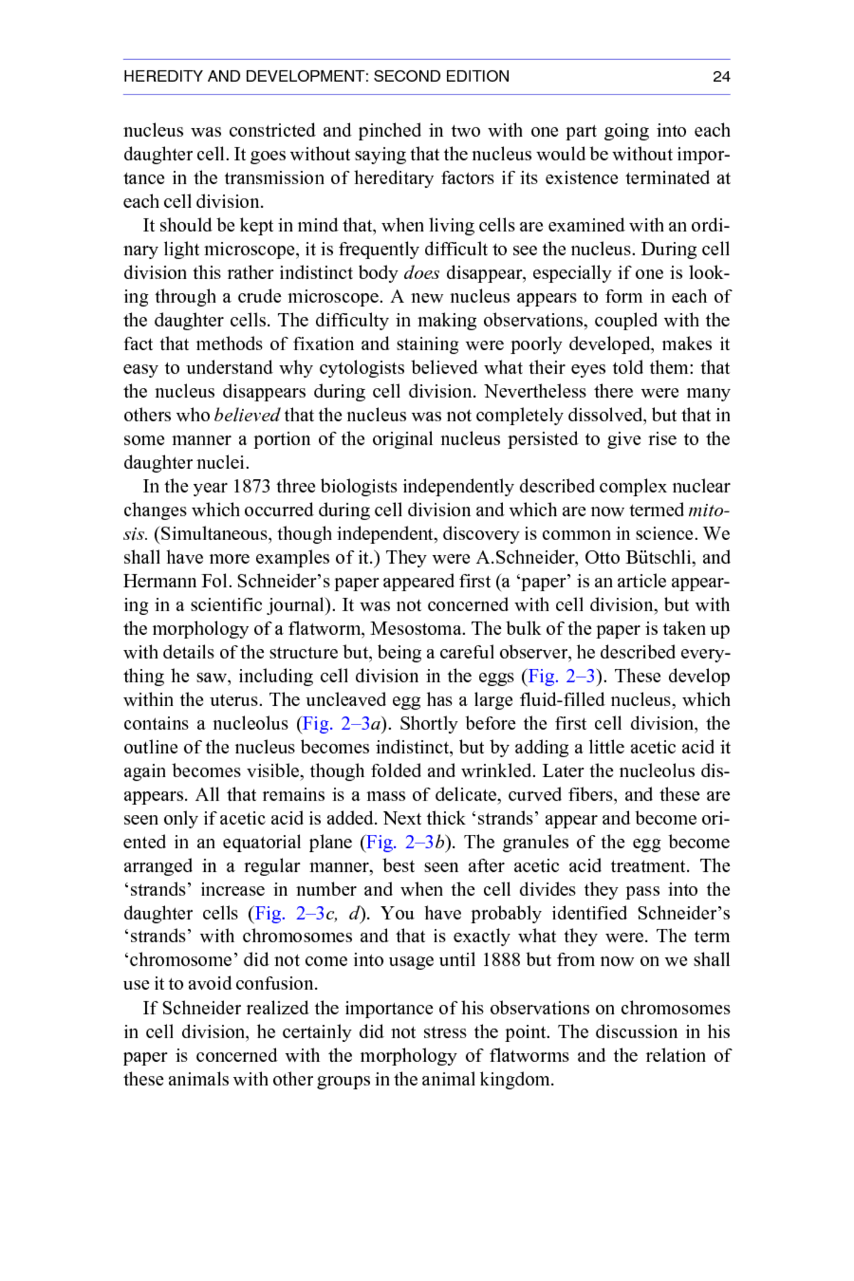
HEREDITY AND DEVELOPMENT: SECOND EDITION 24 nucleus was constricted and pinched in two with one part going into each daughter cell. It goes without saying that the nucleus would be without impor- tance in the transmission of hereditary factors if its existence terminated at each cell division. It should be kept in mind that, when living cells are examined with an ordi- nary light microscope, it is frequently difficult to see the nucleus. During cell division this rather indistinct body does disappear, especially if one is look- ing through a crude microscope. A new nucleus appears to form in each of the daughter cells. The difficulty in making observations, coupled with the fact that methods of fixation and staining were poorly developed, makes it easy to understand why cytologists believed what their eyes told them: that the nucleus disappears during cell division. Nevertheless there were many others who believed that the nucleus was not completely dissolved, but that in some manner a portion of the original nucleus persisted to give rise to the daughter nuclei. In the year 1873 three biologists independently described complex nuclear changes which occurred during cell division and which are now termed mito- sis. (Simultaneous, though independent, discovery is common in science. We shall have more examples of it.) They were A.Schneider, Otto Bütschli, and Hermann Fol. Schneiderâs paper appeared first (a âpaperâ is an article appear- ing in a scientific journal). It was not concerned with cell division, but with the morphology of a flatworm, Mesostoma. The bulk of the paper is taken up with details of the structure but, being a careful observer, he described every- thing he saw, including cell division in the eggs (Fig. 2â3). These develop within the uterus. The uncleaved egg has a large fluid-filled nucleus, which contains a nucleolus (Fig. 2â3a). Shortly before the first cell division, the outline of the nucleus becomes indistinct, but by adding a little acetic acid it again becomes visible, though folded and wrinkled. Later the nucleolus dis- appears. All that remains is a mass of delicate, curved fibers, and these are seen only if acetic acid is added. Next thick âstrandsâ appear and become ori- ented in an equatorial plane (Fig. 2â3b). The granules of the egg become arranged in a regular manner, best seen after acetic acid treatment. The âstrandsâ increase in number and when the cell divides they pass into the daughter cells (Fig. 2â3c, d). You have probably identified Schneiderâs âstrandsâ with chromosomes and that is exactly what they were. The term âchromosomeâ did not come into usage until 1888 but from now on we shall use it to avoid confusion. If Schneider realized the importance of his observations on chromosomes in cell division, he certainly did not stress the point. The discussion in his paper is concerned with the morphology of flatworms and the relation of these animals with other groups in the animal kingdom.
HEREDITY AND DEVELOPMENT: SECOND EDITION 25 2â3 Nuclear changes during cleavage in Mesostoma embryos. a is an uncleaved ovum. The large clear area is the ovum itself, which contains a nucleus and nucleolus. The surrounding structures are follicle cells, which the embryo uses for food. They are not shown in b, c, and d. The spiral structures are sperm. b, c, and d show the âstrands,â which we now realize are chromosomes, and their movements during cell division. (A.Schneider, âUntersuchungen über Plathelminthen.â Oberhessischen Gesellschaft für Natur- und Heilkunde. 14:69â140. 1873.) It remained for others to interpret and show the importance of the phenomena that Schneider had observed. Schneider was of the opinion that the nucleus persisted during division, though we must remember that he used acetic acid to establish this point. One could always question Schneiderâs interpretation, since the acetic acid treat- ment might have produced artifacts (abnormal structures) and the âstrandsâ could be so interpreted. Bütschli, in the same year, described cell division in a roundworm, Rhabdi- tis. He agreed with Schneider that the nucleus persisted during cell division. Fol, the third investigator to describe cell division in 1873, thought that the nucleus entirely disappeared during division and was re-formed in the daugh- ter cells. This view was shared by Flemming and Auerbach who published observations on cell division in 1874. It should be emphasized that these observers based their descriptions wholly or largely on what they observed in living eggs. The problem of cell division was immediately recognized as being of con- siderable importance, and numerous investigators followed Schneider, Bütschli, and Fol. A review article on cell division and related topics was published by Professor Mark of Harvard in 1881. He quoted 194 papers (by 86 authors) which appeared in the five years from 1874 through 1878. This period was one of more or less blind experimentation and exploration. The animal and plant kingdoms were combed for favorable material. Some of the investigators observed living cells, and others
HEREDITY AND DEVELOPMENT: SECOND EDITION 26 those that had been chemically treated. Interpretations of the observed phe- nomena were numerous and varied. Some order was brought out of chaos by Walther Flemming in 1878 (and more especially in his monograph of 1882). He was outstanding, first in selecting excellent material, namely, the epidermal cells of larval salaman- ders; second, in being careful to check in living cells all things that he observed in fixed and stained preparations; and third, in employing hitherto unsurpassed technical methods. Techniques and Instruments. Before Flemmingâs contribution is consid- ered in detail, we shall digress to discuss the development of techniques for preparing cells for microscopic observation. Many earlier workers used dyes in a more or less haphazard way, but in 1858 Gerlach described an adequate staining method. He found that the nuclei of preserved cells take up the dye from a dilute solution of carmine, while the rest of the cell remains unstained or becomes only slightly stained. This became a vastly improved method for observing nuclei, most of which, it must be remembered, are seen with great difficulty in the living state. Gerlach did not discover carmine; he merely perfected its use in cytology. This dye was well known to the Indians of Mex- ico long before the coming of the Spanish. They obtained it from the crushed and dried bodies of cochineal insects reared especially for this purpose. Later the commercial use of carmine spread to Europe. In all probability Gerlach tried it as a âhunch.â It happened to work. Another dye, hematoxylin, was first used successfully by Böhmer in 1865. Commercial preparations were available, derived from a tropical American tree known as logwood. This dye, like carmine, stains the nucleus. The first synthetic aniline dye was made by Perkin. The date of this discov- ery is generally given as 1856, when Perkin was a lad of 18 trying to synthe- size quinine. Many different aniline dyes were made later, and soon they became the principal ones used commercially. They were tried by cytologists from time to time, but it was not until the period of 1875â80 that their use was perfected. It was found that some aniline dyes, such as eosin, would stain parts of the cell not affected by carmine or hematoxylin. It was then possible to use the double-stain methods that are now standard. The nucleus could be stained deep blue with hematoxylin and the cytoplasm a pale pink with eosin. This gave a much improved picture of cell structure. Technical advances in still another field were providing an aid to cytolo- gists. Rapid improvement was being made in microscopes. In the 1870s Abbe, the greatest microscope designer of recent times, began his
HEREDITY AND DEVELOPMENT: SECOND EDITION 27 association with the Carl Zeiss optical works in Germany and increasingly fine instruments were turned out by this concern. In 1878 Abbeâs oil- immersion objective was first produced (one of the initial users was the famous bacteriologist Koch). The oil-immersion lens enabled one to obtain a good image of cell structures at magnifications as much as 2,500 diameters. Technical advances in this field continued with the invention of Abbeâs sub- stage condenser, and in 1886 Zeiss produced the apochromatic objective. This is the finest lens so far developed for the light microscope. These advances meant that cytology had reached a point in its develop- ment where a person like Flemming could make a culminating advance in our understanding of cell division. He certainly did not discover mitosis (nei- ther did any single person) but we owe to him more than to any other the con- cept of mitosis that we hold today. After Flemming, only details were added. Flemmingâs Description of Mitosis. It was well known to Flemming and his contemporaries that the structures observed in living cells might be quite different in appearance from those seen in preserved cells. In some types of living cells no nuclei could be seen, yet after staining typical nuclei were visi- ble. In a situation of this sort the question arises âAre nuclei present in all normal cells or, in some cells, can they be artifacts resulting from the treat- ment used in preparing the cells for study?â Flemming reasoned his answer this way. In some types of living cells, which we can call type 1, nuclei can be seen. When these cells are fixed and stained, a nucleus of characteristic shape and color appears. In other types of living cells, which we can call type 2, no nucleus can be seen. Nevertheless when type 2 cells are fixed and stained, a nucleus that in all respects is identical in appearance to the nuclei of fixed and stained cells of type 1, can be seen. Since the stained nuclei of cell types 1 and 2 have the same appearance, and the treatment is the same in both cases, the most reasonable hypothesis is that a nucleus is present, though invisible, in living cells of type 2. It seems most unlikely that cells of type 2 could be without a nucleus in the living state and that fixation and staining could produce an artifact that was identical to the nuclei of fixed and stained type 1 cells. Flemming made an attempt to apply this type of reasoning to all cell structures, and in every case he tried to use the living cell as the basis of reference. Structures that could never be seen in living cells and that made their appearance only after fixation and staining must be regarded as questionable. The Resting Stage. Flemmingsâ studies led to this concept of mitosis (Figs. 2â4 and 2â5). A resting stage cell is one not in mitosis. The
HEREDITY AND DEVELOPMENT: SECOND EDITION 28 2â4 Flemmingâs drawing of mitosis in fixed and stained cells of the salamander embryo. The figures are arranged in sequence beginning with a resting stage in a. (W.Flemming, Zellsubstanz, Kern und Zelltheilung, 1882.) a. Resting stage. The chromosomes are invisible. The nucleus con- tains chromatin and two nucleoli. The
HEREDITY AND DEVELOPMENT: SECOND EDITION 29 nucleus is spherical and generally occupies the central region of the cell. A nuclear membrane is present. In living resting stage cells the nucleus does not seem to have any internal structure. After fixation and staining, an irregular network of strands and granules, named chromatin, can be detected. In addi- tion, one or more large spherical granules, the nucleoli, are present. Chromo- somes cannot be seen in either the living or the preserved resting stage nucleus with the light microscope. Prophase. Changes in the nucleus are the first indications that mitosis is under way. Long, delicate threads, the chromosomes, make their appearance. At first they are not easy to see, but with the passage of time they become increasingly distinct. Mitosis is a continuous process, but for descriptive pur- poses it can be divided into a number of stages. When chromosomes first become visible we say that the prophase stage has begun. If prophase chro- mosomes are examined carefully, they are seen to be double structures, each chromosome being composed of two long strands, the chromatids, lying side by side. It should be emphasized that only in the very best preparations is it possible to see the chromatids. In most instances, and this is true today, only the entire chromosome is seen. Flemming was able to see the duplicate prophase chromosomes both in living and preserved salamander cells. Dur- ing prophase the nucleoli become smaller and eventually they disappear. Metaphase. Prophase ends and the next stage, metaphase, begins with the disappearance of the nuclear membrane. By this time the chromosomes have become very distinct. In stained preparations they are prominent cell struc- tures, grouped together in the center of the cell. Early in metaphase the spin- dle and asters become prominent. The spindle is given this name because of its shape. In the living cell the spindle appears as a transparent body. In fixed and stained cells there are one or more tiny nuclear membrane is present. b. Late resting stage. The chromosomes are forming. The nucleoli are disappearing. c. Prophase. The chromosomes have formed. No nucleoli. The nuclear mem- brane is still present. (The cytoplasm is not shown.) d. Metaphase. The nuclear membrane has disappeared. Two centrioles, each with a tiny aster, are shown. e. Metaphase. The centrioles and the astral rays surrounding them are distinct. The chromosomes are moving to the middle of the cell. f. Metaphase. The spindle has formed between the two centrioles. g. Metaphase. This is an exceptionally good preparation. Each chromosome is seen to be double, that is, each composed of two chromatids. hâj. Anaphase. The chromosomes are moving apart and one group is approaching each centriole. k. Telophase. The chromosomes have separated into two groups. The spindle has nearly disappeared and the astral rays are becoming indistinct. l. The cell is dividing. The chromosomes are being surrounded by a nuclear membrane and shortly each will be in the resting stage.
HEREDITY AND DEVELOPMENT: SECOND EDITION 30 2â5 Flemmingâs drawings of mitosis in living epidermal cells of a salamander larva. The draw- ings are arranged in sequence, beginning with a prophase in a and ending with the two daughter nuclei in l. The nuclear membrane, asters, spindle, and centrioles are not shown (W.Flemming, Zellsubstanz, Kern und Zelltheilung, 1882).
HEREDITY AND DEVELOPMENT: SECOND EDITION 31 granules, the centrioles, at each end. One can also see long strands, the spin- dle fibers, connecting the two centriole regions. At metaphase the chromo- somes become arranged in a plate perpendicular to the long axis of the spin- dle. The asters are observed in fixed and stained cells as a series of fibers radiating out from the centrioles. Anaphase. Metaphase ends and the next stage, anaphase, begins with the separation of the chromosomes into two groups. One group goes to each pole of the spindle. Flemming thought it possible that the double nature of the prophase chromosomes might be of significance in this respect. Could it be that each chromosome duplicates itself, forming two chromatids, and that at anaphase one chromatid goes to one pole and the other chromatid to the oppo- site pole of the spindle? (Flemmingâs belief was found to be true five years later by van Beneden.) Telophase. The two groups of chromosomes move to the poles of the spin- dle. When they arrive there, telophase, the last stage in mitosis, begins. The chromosomes become increasingly less distinct and the nuclear membrane is re-formed. The spindle and asters begin to disappear. The cell as a whole now divides into two daughter cells with the plane of division cutting across the spindle at the equator. As a result, each daughter cell contains a group of chromosomes. Eventually it becomes impossible to see the chromosomes; the cell has entered the resting stage once more. It should be emphasized that the term ârestingâ means only that the nucleus is not in mitosis. It does not signify a lack of metabolic activity. These nuclear changes, known as mitosis, were observed in so many dif- ferent kinds of animal cells that Flemming believed that they must be a uni- versal feature of living organisms. The nuclei of plants were found to behave in an almost identical manner. Figure 2â6 shows mitosis in a lily. The chro- mosome stages are identical with those in the salamander but the lily, like most plants, differs from animals in lacking centrioles and asters. Our general conclusions based on the work of cytologists up to 1882 are these: cells come from pre-existing cells, nuclei from pre-existing nuclei, and chromosomes from pre-existing chromosomes. At the time when some cytologists were studying the chromosomal events during mitosis, others were investigating fertilization and the formation of ova and sperm. These studies were to contribute greatly to the understanding of heredity, which was to come in the early years of the twentieth century.
HEREDITY AND DEVELOPMENT: SECOND EDITION 32 2â6 Mitosis in the lily (W.Flemming, Zellsubstanz, Kern und Zelltheilung, 1882). FERTILIZATION The elementary fact of fertilization, namely, that a sperm rather than the sem- inal fluid is required to initiate development of the ovum, was discovered by J.L.Prevost and J.B.Dumas in 1824. At this time the precise role of the sperm was not understood. In 1854, George Newport conducted experiments that suggested the sperm actually penetrates the ovum. A full understanding of this event had to wait until it was realized that both the ovum and the sperm are cells. Schwannâs belief that the ovum was a cell was not shared by many cytologists, but the work of Carl Gegenbauer in 1861 seemed to show that it was. Several years later it was also established that the sperm was a single cell. Inheritance, then, must be based on the transmission of cellsâan ovum from the mother and a sperm from the father. Fertilization in the Sea Urchin. In 1873â4 several investigators reported that two nuclei could be seen in the ovum soon after fertilization and
HEREDITY AND DEVELOPMENT: SECOND EDITION 33 before cell division had begun. It remained for Oskar Hertwig (1876) to demonstrate for the sea urchin that one of these nuclei was the nucleus of the ovum and the other was derived from the sperm. He found that these two nuclei approached each other, made contact, and in a slightly later stage only one nucleus was present (Fig. 2â7). In Hertwigâs opinion this single nucleus was the result of fusion of a maternal nucleus of the ovum and a paternal nucleus of the sperm. Almost immediately some other workers came to the same conclusion. Observations were made on eggs of many different species, and it was realized that the formation of the zygote nucleus through the fusion of a paternal pronucleus derived from the sperm and a maternal pronucleus from the ovum is a general phenomenon. It is necessary to emphasize that an attempt is being made only to trace the sequence of key ideas, observations, and experiments that led to an improved understanding of inheritance. This account is not âtrueâ history. Hertwigâs observations and interpretations were important steps in coming to this understanding. One must not assume, however, that all other scientists imme- diately recognized that this was so. Many doubted his observations and inter- pretations and it took several years before he was generally believed to have been correct. New discoveries are rarely accepted when they are proposed. They must be repeated by others before they are accepted as part of scientific knowledge. Two types of material proved of the greatest usefulness in studies of 2â7 Hertwigâs figures of sea-urchin embryos showing the nuclear events in fertilization. a shows an embryo 5 minutes after ova and sperm were mixed. The egg nucleus is the clear area on the left side of the embryo. The sperm nucleus is in the upper right portion of the embryo. b is an embryo 10 minutes after the ova and sperm were mixed. The two nuclei are in contact near the center of the embryo. c is an embryo 15 minutes after the ova and sperm were mixed. A single nucleus is present. This is the zygote nucleus that will undergo a series of mitotic divisions to form all of the nuclei of the individual (O.Hertwig, âBeiträge zur Kenntniss der Bildung, Befruchtung und Theilung des thierischen Eies,â Morph. Jahrb. 1:347â434. 1876).
HEREDITY AND DEVELOPMENT: SECOND EDITION 34 fertilization: the sea urchin (a marine animal related to the starfish) and Ascaris (a parasitic worm found in the intestine of man and other mammals). The sea urchin was especially suitable because it was easy to obtain the ova and sperm, because fertilization could be carried out under the controlled conditions of the laboratory, and because of the transparency of the ova and early embryos. The adults were collected in the ocean, usually by dredging, and in the laboratory both males and females could be stimulated to shed their gametes. The ova could be collected in one dish and the sperm in another. These would number in the millions. When the two were mixed, fertilization occurred in a matter of seconds. One of the most striking things about fertilization and early development in the sea urchin is the fact that events are synchronous in all the zygotes fer- tilized at one time. Thus, if one preserves embryos at successive five-minute intervals after fertilization, the sequence of nuclear events can be worked out with precision. Shortly after fertilization, the paternal pronucleus would be noticed close to the outer membrane of the ovum. At later times it would be found progressively closer to the maternal pronucleus, and eventually fused with it. Fertilization in Ascaris. As cytological material the sea urchin has one serious defect: its chromosomes are small and numerous. One can observe the general events in fertilization, but the details of chromosome movements and changes could not be determined with ease. On the other hand, the para- sitic worm Ascaris provides excellent material for studying the behavior of chromosomes since it has only four chromosomes and these are large and stain successfully. As a consequence the detailed nuclear events in fertiliza- tion were first observed in Ascaris. The process of fertilization in Ascaris was described by Edouard van Beneden (1846â1912) in 1883 and by others such as Theodor Boveri (1862â 1915) in 1888. Boveriâs figures, as reproduced in Figure 2â8, will be the basis of our account of fertilization. (For the present please ignore the legend for this figure, since it cannot be fully understood until the entire chapter has been read.) The first figure, a, shows a section of the entire ovum shortly after fertilization. The paternal pronucleus is in the lower right-hand quadrant. It contains two chromosomes. The structure forming a wrinkled cap immedi- ately above it is the acrosome, which is the portion of the sperm head com- posed of Golgi material. In the center of the ovum there is a dark granular area. This is the centrosome, which was formed by a part of the sperm lying immediately behind the sperm nucleus. There are two structures near the top of the figure. The one within the ovum is the maternal pronucleus. It contains two chromo-
HEREDITY AND DEVELOPMENT: SECOND EDITION 35 2â8 Fertilization in Ascaris. In a the sperm has entered the ovum and formed the paternal pronu- cleus. The maternal chromosomes have undergone the second meiotic division. This resulted in a maternal pronucleus with 2 chromosomes and a second polar body with 2 chromosomes. The dark structure in the center of the egg is the centrosome brought in by the sperm. In b the maternal and paternal pronuclei have enlarged and are approaching each other. In c the pronuclei are con- tinuing to enlarge. Notice the two granules between the pronuclei and in the centrosome sub- stance. These are the centrioles. In d it can be seen that each pronucleus has 2 chromosomes. The centrioles are moving apart and the surrounding centrosome substance is dividing into two por- tions. In e the centrioles and the associated centrosomes have nearly reached opposite sides of the egg. The 2 chromosomes of each pronucleus are more prominent than before. The second polar body is still attached to the top of the egg. In f the first mitotic division of the embryo has begun. Four chromosomes, the diploid number for this species, can be seen on the spindle. These were derived from the 2 pronuclei. At this division each of these 4 chromosomes will split and the two cells that result from the division will each receive 4 chromosomes (Th.Boveri, âDie Befruchtung und Teilung des Eies von Ascaris megalocephala,â Jenaische Zeit. 22:685â882. 1888).
HEREDITY AND DEVELOPMENT: SECOND EDITION 36 somes. The other structure, which is attached to the top of the ovum, is a polar body. It can be seen in b, c, and e as well. For the present we shall disre- gard it since it is concerned with meiosisâa subject to be considered in the last part of the chapter. In b the maternal and paternal pronuclei have moved somewhat closer and their chromosomes have become indistinct. In c the chromosomes in both pronuclei have become elongated and coiled. Two cen- trioles have appeared in the centrosome material. In d the centrosome itself has divided, half being centered around each centriole. The two centrioles, with their associated centrosomes, move farther apart in e. In f they are on opposite sides of the cell with a spindle between them and an aster radiating out from each. During this period considerable changes have been occurring in the pronuclei. In d the chromosomes have shortened and it can be seen that each pronucleus contains two. A further shortening of the chromosomes is apparent in e. During the interval between e and f the membranes around both the maternal and paternal pronuclei disappear and in f the four chromosomes have entered the spindle. The mitotic stage shown in f is an early metaphase. Somewhat later each of these four chromosomes will become double to make a total of eight, and at anaphase these will separate and four chromosomes will move to each pole of the spindle. The chromosome number of the zygote, therefore, is four. Half of this total is provided by the paternal pronucleus and half by the maternal pronucleus. The number of chromosomes in a pronucleus is spoken of as the haploid (or monoploid) number and the number in the zygote is the diploid number. It was clear from the work of van Beneden, Boveri, and others that each parent transmits an equal number of chromosomes to the zygote. So far as one could tell the chromosomes in the maternal pronucleus were morphologically the same as those in the paternal pronucleus. Further study revealed that throughout the animal kingdom similar events are observed with only a few exceptions. Fertilization involves the combina- tion of a haploid pronucleus derived from the sperm and a haploid pronucleus derived from the ovum. Their pooled chromosomes form the diploid number in the zygote. Since the increase in cell number during embryonic develop- ment involves mitosis, all the cells of the embryo and adult should be expected to contain the diploid number of chromosomes. Research has shown this to be true with only a few exceptions. THE FORMATION OF GAMETES An important problem was raised by these discoveries of the chromosomal events during fertilization: if the nuclei of embryonic and adult
HEREDITY AND DEVELOPMENT: SECOND EDITION 37 cells are diploid, how do the nuclei of ova and sperm become haploid? Ascaris provided excellent material for the study of this problem and the observations of van Beneden, Boveri, and Hertwig established the essential points during the 1880s, first solving the problem in the ovum and later in the sperm. They discovered that there are two unusual cell divisions during the formation of gametes. As a result of these divisions diploid cells have their chromosome numbers reduced to the haploid condition. These two divisions are highly modified mitotic divisions; they are known as the meiotic divi- sions. The process itself is meiosis. The relation between mitosis and meiosis can be brought out by a description of the chromosomal changes during the formation of the ovary and of mature ova. Mitosis in the Early Ovarian Cells. The ovary of Ascaris begins to form early in development. At first it consists of a few cells and in the course of time these divide to form the tremendous number comprising the ovary of the adult. This increase in the number of cells is brought about by mitosis. In mitosis each chromosome duplicates itself at every cell division so the num- ber of chromosomes remains constant from one cell generation to the next. So far as individual cells are concerned this is what occurs: the Ascaris nucleus contains four chromosomes as the diploid number; before every cell division there is a duplication of each of these four chromosomes to give a total of eight chromatids; at anaphase the chromatids are separated, four going to each daughter cell. This process is repeated with the result that all the cells of the ovary are diploid. Meiosis in the Female. Many of these diploid ovarian cells become enlarged and form ova. The ovum of Ascaris remains diploid until it has been released from the ovary and entered by a sperm. The ovum nucleus then undergoes a series of two meiotic divisions that leads to each of the resulting cells having the haploid number of chromosomes. The process of meiosis in the Ascaris ovum is shown in Figure 2â9, which is reproduced from the work of Boveri. The First Meiotic Division of the Ova. At the onset of meiosis each of the four long chromosomes (as shown in Figure 2â8f) becomes condensed to form a tiny sphere. Next the chromosomes come together in pairs, a process that is known as synapsis. The chromosomes do not fuse during synapsis, they merely come close to one another. Next, each chromosome becomes duplicate. Thus, each of the two pairs of synapsed chromosomes becomes a group of four, such a group being known as a tetrad. The first of Boveriâs figures, namely 2â9a, shows an ovum in this condition, which is the metaphase of the first meiotic division. In it we see the chromosomes grouped into two tetrads. In b the tetrads are being
HEREDITY AND DEVELOPMENT: SECOND EDITION 38 2â9 Meiosis in Ascaris eggs. a shows the upper portion of the egg and its nucleus. Previously the 4 chromosomes have undergone synapsis to form two pairs. Each chromosome then duplicated itself. The result is 2 tetrads, each composed of 4 chromatids. In this figure the tetrads are in the metaphase of the first meiotic division. b is the anaphase of the first meiotic division. Each tetrad has divided into 2 dyads. c the first meiotic division is complete. The first polar body has pinched off from the egg. It contains 2 dyads. The egg likewise contains two dyads. d the second meiotic division has begun and the 2 dyads are in the spindle. The first polar body with its chromosomes is beneath one of the egg membranes. It can be seen in all of the remaining figures except h. In e the dyads are rotating prior to their separation. f is a metaphase of the second meiotic division. g is the anaphase of the second meiotic division. h is the telophase of the second meiotic division. i the second meiotic division is complete. The second polar body has formed and it contains 2 chromosomes. The egg nucleus also contains 2 chromosomes (Th.Boveri, âDie Bildung der Rich- tungskörper bei Ascaris megalocephala und Ascaris lumbricoides,â Jenaische Zeit. 21:423â515. 1887). divided and in c they have separated completely. Half of each tetrad, or a dyad, goes to each pole of the spindle. It will be noticed that the spindle is not in the center of the cell but instead it is at the periphery. Inasmuch as the cell will divide across the equator of the spindle, the result will be two cells of very unequal sizes. The large cell resulting from the division is the ovum and the small cell is the first polar body. The chromosomes that enter the first polar body are morphologically and numerically equivalent to those that remain in the ovum. The Second Meiotic Division of the Ova. In d the first polar body is well separated from the ovum and the two dyads within the ovum are
HEREDITY AND DEVELOPMENT: SECOND EDITION 39 on the spindle of the second meiotic division. At this division the chromo- somes do not duplicate themselves. Consequently the dyads are divided and as a result two chromosomes go to each pole of the spindle. This second mei- otic division divides the cell unequally, as did the first, the result being a large ovum and a tiny second polar body. At the end of the second, and last, meiotic division there are only two chromosomes in the Ascaris ovum. The nuclear membrane forms around these two chromosomes, the haploid num- ber, and in this manner the maternal pronucleus is produced. The subsequent history of the maternal pronucleus has been discussed as an aspect of fertilization and Figure 2â8 should be re-studied (the maternal pronucleus in 2â9i is in the same stage as in 2â8a). Meiosis in the Male. The observation that the paternal pronucleus was haploid, yet the male diploid in its body cells suggested that a process similar to that just described must also occur in the male. A study of sperm formation in Ascaris showed this to be the case (Fig. 2â10). The last two cell divisions before a sperm forms are meiotic divisions. As in the egg, the four chromo- somes form two pairs and each chromosome duplicates itself. The result is two tetrads each composed of four chromatids. During the first meiotic divi- sion the tetrads are divided and half of each goes into each of the daughter cells. Not only is nuclear division equal but cell division is also equal, which is in contrast to the situation in the ova. At the next division the dyads are divided between the two daughter cells, which are again of equal size. Thus, from one cell with four chromosomes, and by means of two meiotic divi- sions, four cells each with two chromosomes are formed. Each of these four haploid cells develops without further division into a sperm cell. The essential difference between meiosis and mitosis is this: in mitosis there is one duplication of every chromosome for each cell division; in meio- sis there is only one duplication of every chromosome for the two meiotic divisions. As a consequence, in mitosis the chromosome number remains constant from one cell generation to the next; in meiosis the two meiotic divi- sions form cells with the haploid number of chromosomes. With full realization that the nuclear events associated with maturation and fertilization were important biological phenomena, cytologists examined many species of animals and plants. It was found that the reduction divisions leading to haploid pronuclei occur throughout the animal and plant king- doms. In short, another principle of almost universal application (a few excep- tions were found) had been discovered. The facts as outlined in this section were generally, though not universally, believed by 1890.
HEREDITY AND DEVELOPMENT: SECOND EDITION 40 2â10 Meiosis in Ascaris males. The diploid chromosome number in Ascaris is 4. The cells of the testis that will later form the sperm are diploid as shown in a. b shows a nucleus near the begin- ning of meiosis. The 4 chromosomes are undergoing synapsis. As meiosis continues each chro- mosome becomes shortened until it forms a tiny sphere. During this process each chromosome splits. As a result each
HEREDITY AND DEVELOPMENT: SECOND EDITION 41 A summary of meiosis and fertilization in Ascaris is given in Figure 2â11. These observations are interesting and important in their own right but it is not immediately obvious how they relate to the mechanism of inheritanceâ neither was it obvious to the early cytologists. The conceptual gap between observing an orderly sequence of chromosomal behavior and a general the- ory of inheritance is awesome. Few minds are capable of being the first to bridge a gap of this magnitude. As we have learned, Darwinâs theory of pangenesis was not generally accepted. Various alternatives were proposed, were subjected to the merci- less scrutiny of the scientific community, and judged inadequate. But at least some progress was made: by the end of the nineteenth century, it was gener- ally agreed that inheritance, however it was effected, was related to the nucleus. THE NUCLEUS AND HEREDITY The middle years of the 1880s witnessed several attempts to see if inheri- tance was controlled by some definite part of the cell. We might have expected this to be the case when we realize that cytologists, in a decade of uparalleled discovery, had worked out the essentials of mitosis, fertilization, and meiosis. Haeckelâs Hypothesis of the Nuclear Control of Inheritance. An effort to find a cytological basis for inheritance was made as early as 1866 by Ernst Haeckel (1834â1919), who postulated that the nucleus was responsible for the transmission of the inherited features of an organism. The data available to Haeckel in 1866 were not sufficient to test this hypothesis. As E.B.Wilson (1856â1939), the great American cytologist, was to remark some years later, it was a lucky guess. If a lucky guess of this sort had been made by some obscure scientist, it is probable that its influence on subsequent events would have been negligible. But Haeckel was a leader in the field of biology in his day. An idea of his, no matter how slight the factual basis, would have been noticed. It is conceivable, therefore, that Haeckelâs hypothesis of nuclear control of of the 2 pairs of synapsed chromosomes forms a tetrad, as shown in c. At the first meiotic division the 2 tetrads enter the spindle (d) and are divided, half of each tetrad (a dyad) going to each pole as shown in e. As a result of the first meiotic division 2 cells are formed (f, g). Each of these con- tains 2 dyads. In the second meiotic division (h, i, j, k) the dyads of the 2 cells are pulled apart. At the end of this division there are 4 cells (l, m, n, o). Each of these contains 2 chromosomes, the haploid number. There is no further division of these 4 cells and they develop directly into sperm (A.Brauer, âZur Kenntniss der Spermatogenese von Ascaris megalocephala,â Arch. Mikr. Anat. 42:153â213. 1893).
HEREDITY AND DEVELOPMENT: SECOND EDITION 42 2â11
HEREDITY AND DEVELOPMENT: SECOND EDITION 43
HEREDITY AND DEVELOPMENT: SECOND EDITION 44 inheritance helped to prepare others for thinking and experimenting along these lines. Nägeliâs Idioplasm Theory. In 1884 Carl Wilhelm von Nägeli (1817â 1891) suggested that a substance which he called the idioplasm was responsi- ble for inheritance. The idioplasm was thought to be an invisible chemical network that extended throughout the cell and from cell to cell. Nägeli did not observe the idioplasm in cells. He invented it to account for inheritance. He did not regard it as a highly stable material, but as one that might change during development, or as the result of nutrition or other external conditions. In any event it must return to the original condition in the embryo. Nägeli did considerable theorizing on the subject of inheritance, but his concept of pos- sible mechanisms was extremely vague. His hypothesis was nearly impossi- ble to test, and hence it could be of no real usefulness in directing efforts to profitable experimentation. Early Evidence for the Nuclear Control of Inheritance. In 1884â85 four German scientists, working independently, came to the conclusion that the physical basis of inheritance must lie in the chromosomes. They were Oskar Hertwig, Edouard Strasburger, Rudolf Kölliker, and August Weis- mann (1834â1914). The first three were primarily laboratory scientists. For at least a decade they had been leaders in the analysis of problems concerned with the nucleus. Weismann, on the contrary, is remembered largely for his theoretical work. These four men believed that the chromosomes were the physical basis of inheritance for the following reasons. 1. Even though inheritance was not well understood, it seemed that both parents have an equal share in transmitting their characteristics to the offspring. The soundest support for this belief came from work on plant hybrids. In the 1760s, Joseph Gottlieb Kölreuter crossed Nicotiana pan- iculata and Nicotiana rustica, two species of the tobacco genus that dif- fer in many ways. So far as he could tell, the hybrid offspring were the same whether the cross was paniculata âÃrustica â or rustica â Ãpan- iculata â. The obvious conclusion was that both parents contributed equally to the characteristics of the offspring. What is the physical basis of this equality? It was know, of course, that the only links between parent and offspring are the ovum and sperm. These two cells are about as different as any two cells could be. Usually the ovum has a mass thousands or millions of times the mass of the sperm. Ova usually contain a large quantity of cytoplasm, whereas sperm contain almost none. This would suggest that the cytoplasm was not the basis of inheritance because, if it were, it might be expected that the
HEREDITY AND DEVELOPMENT: SECOND EDITION 45 femaleâs contribution would be much greater than the maleâs. The only parts of the sperm and ova that seemed to these four scientists to be equivalent were the nuclei. The sperm pronucleus and the egg pronu- cleus were identical so far as one could tell. Perhaps this equivalence of structure was the basis of the equivalent importance of the two gametes in inheritance. Van Benedenâs description of the pronuclei in Ascaris, each with two chromosomes, seemed most suggestive. 2. During cell division, the cytoplasm and its formed structures seem to be divided passively. The chromosomes, on the other hand, go through a complicated mitosis which results in each of the daughter cells receiving exactly the same number of chromosomes. It seemed to Hertwig and the others that the significance of this complicated process might be that the nucleus was the basis of inheritance: why should the chromosomes, alone among the cell structures, be duplicated and then divided equally unless they were of great importance in inheritance? 3. The complex chromosomal changes during meiosis were understand- able in terms of keeping the chromosomes constant from generation to generation. There was no similar phenomenon for any other cell struc- ture. Since inheritance was an intergeneration phenomenon and the chromosomes seemed to be the only cell structures that were transmitted in an exact way from one generation to another, perhaps the chromo- somes were of importance in inheritance. 4. Finally, there was a more direct test of nuclear function in regenerating protozoa. The forms selected for this work were single-celled organisms with one nucleus. It was possible to cut the animals into two parts, one part containing cytoplasm and the other cytoplasm and the nucleus. Both parts healed. The part without a nucleus lived for some time, but it was unable to regenerate to form a whole animal, and it was incapable of reproduction. The part with the nucleus could regenerate a whole animal and could reproduce normally. These observations were suggestive, but they did not âproveâ that the nucleus was the physical basis of inheritance. The fact that chromosomes appeared to be the only cell structure that remained constant from cell to cell, and from generation to generation, could mean that inheritance was by way of the chromosomes. Many famous cytologists believed that a good working hypothesis was âThe nucleus is important in heredity.â In the next chapter, we shall learn that in the year 1900 the rediscovery of a scientific paper written much earlier by Mendel put the subject of inheritance in an entirely new light. It is of interest, therefore, to summarize the advances that those cytologists interested in heredity
HEREDITY AND DEVELOPMENT: SECOND EDITION 46 had made up to the year Mendelâs results became generally known. Such a summary was given retrospectively by E.B.Wilson in 1914: The work of cytology in its period of foundation laid a broad and substantial basis for our more general conceptions of heredity and its physical substratum. It demonstrated the basic fact that heredity is a consequence of the genetic con- tinuity of cells by division, and that the germ-cells are the vehicle of transmis- sion from one generation to another. It accumulated strong evidence that the cell-nucleus plays an important role in heredity. It made known the significant fact that in all the ordinary forms of cell-division the nucleus does not divide en masse but first resolves itself into a definite number of chromosomes; that these bodies, originally formed as long threads, split lengthwise so as to effect a meristic division of the entire nuclear substance. It proved that fertilization of the egg everywhere involves the union or close association of two nuclei, one of maternal and one of paternal origin. It established the fact, sometimes desig- nated as âVan Benedenâs lawâ in honor of its discoverer, that these primary germ-nuclei give rise to similar groups of chromosomes, each containing half the number found in the body-cells. It demonstrated that when new germ-cells are formed each again receives only half the number characteristic of the body- cells. It steadily accumulated evidence, especially through the admirable stud- ies of Boveri, that the chromosomes of successive generations of cells, though commonly lost to view in the resting nucleus, do not really lose their individual- ity, or that in some less obvious way they conform to the principle of genetic continuity. From these facts followed the far-reaching conclusion that the nuclei of the body-cells are diploid or duplex structures, descended equally from the original maternal and paternal chromosome-groups of the fertilized egg. Continually receiving confirmation by the labours of later years, this result gradually took a central place in cytology; and about it all more specific discov- eries relating to the chromosomes naturally group themselves. All this had been made known at a time when the experimental study of hered- ity was not yet sufficiently advanced for a full appreciation of its significance; but some very interesting theoretical suggestions had been offered by Roux, Weismann, de Vries, and other writers. While most of these hardly admitted of actual verification, two nevertheless proved to be of especial importance to later research. One was the pregnant suggestion of Roux (1883), that the forma- tion of chromosomes from long threads brings about an alignment in linear series of different materials or âqualities.â By longitudinal splitting of the threads all the âqualitiesâ are equally divided, or otherwise definitely dis- tributed, between the daughter-nuclei. The other was Weismannâs far-seeing prediction of the reduction division, that is to say, of a form of division involv- ing the separation of undivided whole chromosomes instead of the division- products of single chromosomes. This fruitful suggestion (1887) pointed out a way that was
HEREDITY AND DEVELOPMENT: SECOND EDITION 47 destined to lead years afterwards to the probable explanation of Mendelâs law of heredity. Such, in birdâs-eye view, were the most essential conclusions of our science down to the close of the nineteenth century.* Suggested Readings Readings in Heredity and Development contains selections from Grew (1682), Roget (1836), Virchow (1858), Weismann (1885, 1887, 1891), and Wilson (1900) as well as an extensive bibliography. The article by Coleman, listed below, provides an excellent introduction to the history of cytology as related to inheritance. BRADBURY, S. 1967. The Evolution of the Microscope. New York: Pergamon Press. COLEMAN, WILLIAM. 1965. âCell, nucleus, and inheritance: an historical study.â Pro- ceedings of the American Philosophical Society 109:124â58. HUGHES, ARTHUR. 1959. A History of Cytology. New York: Abelard-Schuman. MARK, E.L. 1881. âMaturation, fecundation, and segmentation of Limax campestris Bin- ney.â Bulletin Museum of Comparative Zoology 6:173â625. WILSON, E.B. 1914. Croonian Lecture: The bearing of cytological research on heredity.â Proceedings of the Royal Society of London. B. 88:333â52. Questions 1. Examine Figure 2â2 carefully. Do you detect any element of sameness among these diverse objects that suggests all should be put into the same class, that is, be identified as cells? Had Hooke seen these objects do you believe that he would have regarded them as equivalent to the struc- tures he observed in cork? 2. The nucleus of a cell does disappear before cell division. What could have been the reason, therefore, why some cytologists believed that the nucleus had an unbroken continuity from one cell generation to another? 3. In most instances it is far easier to see cell structures in fixed and stained cells than in living cells. That being the case, why did Flemming lay such stress on the importance of observing structures in living cells? 4. After studying Flemmingâs illustrations of chromosomes, would you have thought that all of the chromosomes of a cell are more or less alike, or that each is unique? 5. Would it be possible to work out the sequence of events in mitosis from a study of fixed and stained material only? Would it be necessary to assume any relation between a nucleus in the resting stage and a metaphase? 6. Prevost and Dumas performed experiments that led them to believe that the sperm and not the seminal fluid is the active agent in fertilization. Can you suggest how such experiments might have been done? * This quotation is from Wilsonâs Croonian Lecture, which is given in its entirety in Readings, Chapter 4.
HEREDITY AND DEVELOPMENT: SECOND EDITION 48 7. The meiotic mechanisms described for Ascaris are almost universal in animals and very similar in plants. Can you devise other ways of halving the number of chromosomes? 8. You probably already know something about DNA and its role in inheri- tance. Study Figures 2â4, 2â9, and 2â10 and try to image what is hap- pening to the DNA. 9. Haeckelâs hypothesis of nuclear control of inheritance was shown to be correctâa generation after he proposed it. Why cannot scientific ideas be accepted more promptly? 10. Compare Darwinâs Theory of Pangenesis with Nägeliâs Theory of Idioplasm. 11. In terms of what you know of cell biology, how would you evaluate these statements of Virchow (Readings, Chapter 2)? a. âEvery animal presents itself as a sum of vital unities, every one of which manifests all the characteristics of life.â b. After saying that cells are the basis for all the phenomena of life, he writes, âAccording to my ideas, this is the only possible starting point for all biological doctrines.â c. ââ¦the cell is really the ultimate morphological element in which there is any manifestation of life andâ¦we must not transfer the seat of real action beyond the cell.â 12. How could one prove the hypothesis âomnis cellula e cellulaâ? 13. Can you suggest why the phenomenon of mitosis was not discovered in the eighteenth century? 14. Do you believe that the chromosomes maintain their essential structure during the resting stage?
