
Radiation Physics Relevant to
Advanced Imaging Technology
X-ray backscatter advanced imaging technology (AIT) exploits the physical properties of X rays to create images using photons backscattered from a scanned passenger. This chapter summarizes the physics principles and the associated transfer of energy to the passenger that enable image formation. Additional information, including details of these processes, can be found in radiological physics and health physics texts.1
THE PHYSICS OF X-RAY ABSORPTION
X-ray backscatter AIT uses a narrow beam of X-ray photons with energies, hv, less than 100 keV.2 There are five basic interactions that can occur as X rays penetrate material. These are the photoelectric effect,3,4 Compton scattering, Thompson scattering, Rayleigh scattering, and pair production. Figure 2.1 shows the rela
_______________
1 See for example, H.E. Johns and J.R. Cunningham, Physics of Radiology, Fourth Edition, Charles C Thomas Publisher, Springfield, Ill., 1983.
2 An X-ray tube operating at 100 kV has a peak at a lower energy than 100 keV; for example, for 50 kV, the peak is at ~30 keV, as seen in Figure 2.2.
3 When a photoelectric interaction occurs, the energy of a photon is completely transferred to an atomic electron. The electron may thus gain sufficient kinetic energy to be ejected from the electron shell. The energy of the incoming photon must, however, be higher than the binding energy for the electron.
4Photoionization is the physical process in which an ion is formed from the interaction of a photon with an atom or a molecule.
tive interaction cross sections (i.e., attenuation coefficients) for photoelectric, Compton, and Rayleigh scattering in soft tissue as a function of photon energy. Pair production requires a minimum photon energy of 1.022 MeV and is not of concern here.
The photoelectric effect is the dominant interaction for X rays with energies below 30 keV. This reaction results in the disappearance of the photon. The result is the ejection of a bound electron, usually from an inner shell, with a kinetic energy of hv – EB, where EB is the original binding energy of the electron. The ejected electron loses its kinetic energy by exciting additional atoms. In some cases, it transfers sufficient energy to liberate an additional electron capable of creating additional ionization. This initiates a cascade process that ends only when all excitations have been thermalized and the system returns to a ground state, which may include chemical changes. Each ionized atom returns to its ground state by emitting one or more secondary photons and/or electrons, which add their contributions to the process. The result is a rapid dissipation of the energy of the incident X ray, diffused through a volume extending some 10 to 30 micrometers (µm) from the site of the initial absorption.
Because no scattered photon is generated, the photoelectric effect does not contribute photons to a backscatter image, but its contribution to energy absorbed in the material is significant. Furthermore, because the probability of a photoelectric interaction increases rapidly with the atomic number, Z, of the target atom, photons are more likely to be absorbed than scattered by high Z materials such as metals. This provides contrast in the backscatter image, with metallic objects appearing dark (indicating a lack of backscattered photons).
Compton scattering is the main interaction for energies above 30 keV. As seen in Figure 2.1, it is the dominant energy-loss mechanism in tissue for X rays of energy greater than about 30 keV. Here, the incident photon scatters off an electron in the material, transferring a fraction of its energy to the electron, thereby reducing its energy and changing direction. Energy conservation requires
where hv is the energy of the incident photon, hv′ is the energy of the photon after scattering, and Ee is the energy of the scattered electron.
By considering momentum conservation, it is possible to determine the properties of the scattered particles as a function of the scattering angle. If the photon scatters at an angle q from its trajectory prior to the interaction, the energy of the scattered photon is given by:
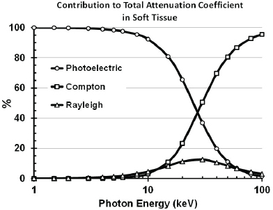
FIGURE 2.1 The relative interaction cross sections (i.e., attenuation coefficients) for photoelectric, Compton, and Rayleigh scattering in soft tissue as a function of photon energy. The cross section for Thompson scattering is too small to influence this figure. SOURCE: Courtesy of Tom Borak.
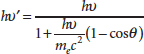
where mec2 is the rest mass energy of an electron. For example, a 50 keV photon scattered at θ = 180° emerges with an energy hv′ = 42 keV. The energy transferred to the electron is Ee = 8 keV.
The energy of the electron is dissipated in the material, whereas the scattered photon either escapes or interacts in the material. While it is unlikely that an incident photon will backscatter at exactly 180°, several Compton scattering events can occur, resulting in the photon emerging in a backward direction or terminating in a photoelectric event. Detector systems with large geometrical acceptance angles have a good probability of registering these scattered photons. An image is formed by associating all of the photons detected in a time interval with the location of the incident beam on the subject at that time. In effect, the output of the detectors is stored in a memory location synchronized with the position of the beam.
The third interaction, Thompson scattering, is the emission of radiation generated by the acceleration of point charges (electrons) by the electric field of the incident X ray. Because accelerated charges radiate, there is a finite probability that an X ray of the incident beam will be backscattered with no change in energy. This backscattered intensity can be calculated. It is independent of incident energy and equal to the intensity of the incident beam times the square of the ratio of the nuclear diameter (~2 × 10⁻¹⁵ m) to the distance between the scattering electron and the detector (~1 m). The resulting attenuation factor, ~10−30, effectively prevents Thompson-scattered electrons from contributing to the image, even when generous estimates of collection solid angles and the number of electrons in a material up to the X-ray absorption depth are assumed.
The final interaction, Rayleigh scattering, also involves acceleration of charges but differs from Thompson scattering in that the atoms are treated as objects with a nucleus and surrounding electrons and therefore have a finite size. This introduces a length scale and, as a result, a wavelength dependence, ~λ–4, for the scattering probability. This dependence strongly favors short wavelengths. Applied to visible light, it accounts for the blue color of the sky. The contribution of Rayleigh scattering of X rays is that of Thompson scattering multiplied by a resonance factor. The resulting cross sections are not negligible, but they are still small compared to the photoelectric and Compton cross sections, as seen in Figure 2.1. Hence, neither Thompson nor Rayleigh scattering make important contributions either to backscatter image formation or to the deposition of energy in the scanned object.
BEAM ATTENUATION AND DEPOSITED ENERGY
If it were possible to use monochromatic 50 keV photons for backscatter screening, it would be relatively easy to estimate the intensity of backscattered radiation reaching the detector and the energy deposited in the scanned person. The incident X-ray beam would attenuate exponentially in the person’s body, with an average penetration depth5 of the order of 1 cm, generating scattered photons and secondary electrons by processes described above. The resulting electrons would travel distances on the order of tens of micrometers, resulting in the energy being deposited relatively near the locations of the energy transfer events.
Practical X-ray sources do not produce monoenergetic beams. X-ray tubes use electrons produced at the negative terminal, the cathode, of a vacuum diode. These electrons are accelerated for AIT systems, usually to an energy of about 50 keV, then strike the positive terminal, the anode, which is typically made of a heavy, electron-
_______________
5 Penetration depth is a measure of how deep light or any electromagnetic radiation can penetrate into a material. It is defined as the depth at which the intensity of the radiation inside the material falls to 1/e (about 37%) of its original value.
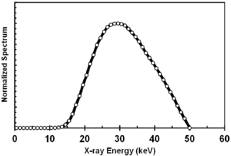
FIGURE 2.2 Typical energy spectrum of photons produced in an X-ray tube operating at 50 kV. SOURCE: Courtesy of Tom Borak.
dense metal such as tungsten. They lose energy either by interacting with the heavy nuclei to produce bremsstrahlung X rays or by interacting with the electrons of the anode to produce heat and characteristic X rays. The bremsstrahlung process produces a distribution of photon energies from zero to the electron energy itself. Because many electrons have slowed down before producing bremsstrahlung radiation, this results in a large number of photons at very low energies. The photons at very low energies are preferentially removed by absorption either in the tungsten anode or in the glass or metal body of the tube and any purposely inserted external filtration. The typical output spectrum peaks at around 30 keV, as shown in 6 However, the details of the spectrum depend on the atomic number of the anode material, the amount of anode material that the photons must traverse before exiting the tube (e.g., as a function of the exit angle), the atomic composition and thickness of the exit window, and other material (filters) in the beam path.
The average penetration depth of photons in a material such as the body of the person being scanned is the result of random absorption and scattering interactions. The number of unscattered photons per unit area typically decreases
_______________
6 Note that any characteristic radiation from the tungsten anode is only produced when the voltage is higher than 69.5 kV.
exponentially with depth. The rate of decrease depends on the probability of the various interactions. Compton scattering depends on the number density of electrons in the material, and the probability of Compton scattering is therefore a function of the average atomic number of the elements in the material. However, the photoelectric cross section also depends on the binding of the electrons and increases rapidly with increasing atomic number. Soft tissue (such as muscle and skin) consists mainly of hydrogen, oxygen, carbon, and nitrogen with a trace amount of phosphorus. The atomic numbers of these elements are all relatively low, so photon attenuation is easily evaluated. Photons with energies below about 10 keV that are incident on muscle are absorbed almost entirely in 1 cm. At 20 keV, about 30 percent of the incident photons penetrate 1 cm. At 50 keV, the fraction that penetrates 1 cm rises to 80 percent. Thus, the backscatter images represent the outer 1 cm or so of material. Bone contains calcium and has a significantly higher rate of photon attenuation but is often behind more than 1 cm of soft tissue, so it is not considered further.
To determine the amount and location of energy deposited in biological material—the likely initiator of biological effects of irradiation—one must take into account the scattered X rays and energetic electrons generated by Compton scattering as well as the electrons energized by the photoelectric process. At any location within the material, this depends on the branching ratio between (relative importance of) Compton scattering and the photoelectric effect. The scattered photons add to the photons that have not been scattered, and the excited electrons carry energy away from their point of creation. But electron ranges are typically a small fraction of the mean penetration depths of photons, so the location where the incoming energy is deposited is determined by the photons. For example, the mean free path of a 30 keV photon in water is 2.7 cm, three orders of magnitude larger than the 18 µm range of a 30 keV electron in the same material. However, electron transport may be significant near boundaries between materials—for example, tissue adjacent to bone. This will be discussed in more detail in Chapter 4.
The quantitative analysis of backscatter image formation and patterns of energy deposition in the person being scanned requires sophisticated computational techniques. This will be discussed in detail in Chapter 7.


