
7
Cancer
Chapter Overview
Based on new evidence and a review of prior studies, the current committee determined that epidemiologic results concerning an association between exposure to any one of the chemicals of interest (COIs; 2,4-dichlorophenoxyacetic acid (2,4-D), 2,4,5-trichlorophenoxyacetic acid (2,4,5-T), picloram, dimethylarsinic acid [DMA or cacodylic acid], and 2,3,7,8-tetrachlorodibenzo-p-dioxin [TCDD]) and monoclonal gammopathy of undetermined significance (MGUS) met the criteria for sufficient evidence of an association. No other changes in association level between the relevant exposures and other cancer types were made as either there were no published studies or the new evidence supported the findings of earlier updates. Thus, the current findings on cancer can be summarized as follows:
- There is sufficient evidence of an association between the COIs and soft tissue sarcomas, B-cell lymphomas (Hodgkin lymphoma, non-Hodgkin lymphomas, chronic lymphocytic leukemia, hairy-cell leukemia), and MGUS.
- There is limited or suggestive evidence of an association between the COIs and bladder cancer; laryngeal cancer; cancers of the lung, bronchus, or trachea; prostate cancer; multiple myeloma; and AL amyloidosis.
- There is inadequate or insufficient evidence to determine whether there is an association between the COIs and any other specific type of cancer.
Cancers are the second-leading cause of death in the United States (CDC, 2017a), with heart disease being the leading cause of death. However, among
men 55–79 years old, the group that includes most Vietnam veterans, the risk of dying from cancer exceeds the risk of dying from heart disease (CDC, 2017b). According to estimates from the National Cancer Institute (NCI), 1,735,350 new cases of cancer were expected to be diagnosed and 609,640 people of all ages were expected to die from cancer in the United States in 2018 (NCI, 2018a).
The objective of this chapter is to provide an assessment of whether the occurrence of cancers in Vietnam veterans may be associated with exposures to herbicides that they may have experienced during their military service. This chapter summarizes and presents conclusions about the strength of the evidence from epidemiologic studies regarding associations between exposure to the COIs and various cancer types. As described in Chapter 3, studies of exposure to polychlorinated biphenyls (PCBs) and other dioxin-like chemicals were also considered informative if their results were reported in terms of TCDD toxic equivalents (TEQs) or concentrations of specific congeners of dioxin-like chemicals. Studies that report TEQs based only on mono-ortho PCBs (which are PCBs 105, 114, 118, 123, 156, 157, 167, and 189) are considered even though their TEQs are several orders of magnitude lower than those of the non-ortho PCBs (77, 81, 126, and 169), based on the revised World Health Organization (WHO) toxicity equivalency factor (TEF) scheme of 2005 (La Rocca et al., 2008; van den Berg et al., 2006). The lower TEQs of the mono-ortho PCBs, however, may be counterbalanced by their abundance, which is generally many orders of magnitude higher than the non-ortho PCBs (H.-Y. Park et al., 2010).
A compendium of all of the studies reviewed by the various Veterans and Agent Orange (VAO) and VAO Update committees can be found at www.nap.edu/catalog/25137. In this update, if a new study reported on only a single type of cancer and did not revisit a previously studied population, then its design information is summarized here with its results; design information on studies that are updates of or new analyses on populations or cohorts that have been previously studied can be found in Chapter 5. Studies of childhood cancers in relation to parental exposure to the COIs are discussed in Chapter 8, which addresses possible adverse effects in the veterans’ offspring. Studies that consider only childhood exposure are not considered relevant to the committee’s charge.
In evaluating possible connections between herbicide exposure and the risk of cancer, the approach used to assess the exposure of study subjects is of critical importance in determining the overall relevance and usefulness of findings. There is great variation in the detail and the accuracy of exposure assessments among studies, which can distort the true relationship between exposure and disease. A few studies used biologic markers of exposure, such as the presence of a chemical in serum or tissues; others developed an index of exposure from employment or activity records; and still others used other surrogate measures of exposure, such as an individual’s presence in a locale when herbicides were used.
Each section on a specific cancer type opens with background information, including data on its incidence in the general U.S. population and its known
or suspected risk factors. Cancer incidence in the general U.S. population is included in the background material to provide a context for consideration of the cancer risk in Vietnam veterans; the numbers presented are estimates of incidence in the entire U.S. population, not predictions for the Vietnam veteran cohort. The data on the expected numbers of new cases and deaths for specific types of cancerin 2017 are based on estimates from NCI’s Surveillance, Epidemiology, and End Results (SEER) program or, if estimates for a particular cancer type were not available from NCI, on estimates by the American Cancer Society. Using the most recent SEER data available when this was written (SEER-18; 2000–2014), incidence data were derived for all races combined and separately for blacks and whites using age groups likely to include most Vietnam-era veterans. Incidence data are presented by sex, age, and race, all of which can have profound effects on risk (NCI, n.d.a). For example, the incidence of prostate cancer is about 2.6 times higher in men who are 70–74 years old as in men 60–64 years old and about 75% higher in blacks 60–64 years old than in whites in the same age group (NCI, 2015). Many other factors can influence cancer incidence, including screening methods, tobacco and alcohol use, diet, obesity, genetic predisposition, and medical history. Those factors can modify the risk of developing a given kind of cancer; they also need to be taken into account in epidemiologic studies of the possible contributions of the COIs.
Each section of this chapter pertaining to a specific type of cancer includes a summary of the findings described in the previous 10 VAO reports. That is followed by a discussion of the most recent scientific literature, and, when appropriate, the literature is discussed by exposure type (service in Vietnam, occupational exposure, or environmental exposure). A summary of biologic plausibility, which corresponds to the third element of the committee’s congressionally mandated Statement of Task, follows the description of newly identified epidemiologic studies. In fact, the degree of biologic plausibility itself influences whether the committee perceives positive findings to be indicative of an association or the product of statistical fluctuations (chance) or bias. Following a synthesis of the material reviewed, each section ends with the committee’s conclusion regarding the strength of the evidence from epidemiologic studies. The categories of association and the committee’s approach to categorizing the health outcomes are discussed in Chapter 3.
Chapter 4 contains information on the general biologic mechanisms by which exposure to TCDD and the other COIs contribute to malignant transformation. Toxicology studies use a variety of methods and animal or cellular models to derive results on the interactions of the COIs with the cellular machinery known to be important in the development of cancer at any site. When biologic plausibility is discussed in each section, this generic information is implicit, and only experimental data specific to carcinogenesis at the site in question are presented. There is increasing evidence that TCDD and the COIs may disturb cellular processes through epigenetic mechanisms, and reference to this evidence as it applies to specific cancers is included where it exists.
Considerable uncertainty remains about the magnitude of the risk posed by exposure to the COIs. Many of the veteran, occupational, and environmental studies reviewed by the committee did not fully control for important confounders. There is not enough information about the exposure experience of individual Vietnam veterans to permit combining exposure estimates for them with any potency estimates that might be derived from scientific research studies to quantify risk. The committee therefore cannot accurately estimate the risk to Vietnam veterans that is attributable to exposure to the COIs. The significant challenges in deriving useful quantitative estimates of the risks of various health outcomes in Vietnam veterans are explained in Chapter 2 of this report.
ORGANIZATION OF CANCER GROUPS
Consistent with the previous report in this series, the organization of cancer groups follows the major and minor categories of cause of death related to cancer sites established by the National Institute for Occupational Safety and Health (NIOSH; Robinson et al., 2006). For the present update, the committee gave more attention to the WHO’s classification of lymphohematopoietic neoplasms (WHO, 2008), which stresses partitioning of the disorders first by the lymphoid or myeloid lineage of the transformed cells, rather than categorizing into lymphomas and leukemias.
The system of organization used by the committee simplifies the process for locating a particular cancer type for readers. For any cancer type for which no epidemiologic research findings have been identified, the default category has always been “inadequate or insufficient evidence” of association with exposure to the COIs. A failure to review a specific cancer or other condition separately reflects the paucity of information concerning that cancer, so there is indeed inadequate or insufficient information to categorize an association with such a disease outcome.
BIOLOGIC PLAUSIBILITY
The studies considered by the committee that speak to the biologic plausibility of associations between human cancers and exposure to 2,4-D, 2,4,5-T, picloram, cacodylic acid, and TCDD have been performed primarily in laboratory animals (rats, mice, hamsters, and monkeys) or in cultured cells.
The animal studies examining the carcinogenicity of 2,4-D, 2,4,5-T, and picloram have, in general, produced negative results, although some bioassays used in those studies would not meet current standards. For example, there is a question of whether the highest doses (generally 30–50 mg/kg) used in some of the experiments reached a maximum tolerated dose or represented the doses that are capable of inducing carcinogenesis. Therefore, it is not possible to have absolute confidence that these chemicals have no carcinogenic potential at higher
doses. Additional evidence of a lack of carcinogenic potential comes from negative findings on the genotoxic effects of assays conducted primarily in vitro that indicate that 2,4-D and 2,4,5-T are genotoxic only at very high concentrations.
There is evidence in laboratory animals that cacodylic acid is carcinogenic, based on studies that have shown that it can induce neoplasms of the kidney (Yamamoto et al., 1995), bladder (Arnold et al., 2006; Cohen et al., 2007b; A. Wang et al., 2009; Wei et al., 2002; Yamamoto et al., 1995), liver, and thyroid gland (Yamamoto et al., 1995). Treatment with cacodylic acid induced the formation of neoplasms of the lung when administered to mouse strains that are genetically susceptible to developing those tumors (Hayashi et al., 1998; Yamanaka et al., 2009). Other studies have used the two-stage model of carcinogenesis in which animals are exposed first to a known genotoxic agent and then to a suspected tumor-promoting agent; with this model, cacodylic acid has been shown to act as a tumor promoter with respect to lung cancer (Yamanaka et al., 1996). These studies are further discussed in Chapter 4.
Collectively, the evidence obtained from studies of TCDD supports a connection between human exposure and cancers. The effects of TCDD on cellular function make carcinogenesis biologically plausible, and evidence from model systems indicate that TCDD can enhance carcinogenesis. This will be discussed in a generic sense below and more specifically in the biologic plausibility sections on individual cancers. Several reviews have affirmed the well-established mechanistic roles of the aryl hydrocarbon receptor (AHR) in TCDD-induced cancers (S. Ahmed et al., 2014; Androutsopoulos et al., 2009; Barouki and Coumoul, 2010; Dietrich and Kaina, 2010; Ide et al., 2017; Murray et al., 2014; Ray and Swanson, 2009; Rysavy et al., 2013; Tsay et al., 2013). The effect can be both cancer promoting—by activating oncogenes (Gardella et al., 2016) or blocking apoptosis (Bekki et al., 2015)—or protective, depending on the tissue type and timing of the exposure (Y. Li et al., 2014; Moore et al., 2016). The role of the AHR is further established by:
- its activation of several proteins of the P450 system of enzymes that play crucial roles in detoxification and drug metabolism (Al-Dhfyan et al., 2017a,b);
- activation of the paraoxanase antioxidant enzymes (Shen et al., 2016); and
- activation of the transforming growth factor (TGF)-β pathway (Silginer et al., 2016);
all of which are important in oncogenesis. TCDD can disrupt circadian rhythms via the AHR, and chronic disruption of circadian rhythms is associated with an increased incidence of cancer, suggesting a potential additional pathway by which TCDD increases cancer risk (C. Wang et al., 2014; C. X. Xu et al., 2013). TCDD increases the incidence or progression of human cancers through a variety of cellular mechanisms, and the biologic plausibility of an association between TCDD
exposure and cancer has been firmly established in a mechanistic sense. Data also indicate that AHR can play a protective role in cancer (reviewed in Kolluri et al., 2017; Murray et al., 2014), and its role as a therapeutic target for cancer therapy is being investigated. However, cancer therapies were considered beyond the scope of the committee’s charge, and not included in this report.
TCDD is considered a non-genotoxic carcinogen, as reviewed by Hernández et al. (2009), because it does not produce changes is DNA sequences. However, because of the oxidative stress it produces, TCDD does have some genotoxic potential. In vitro work with mouse hepatoma cells has shown that activation of the Ahr results in increased concentrations of 8-hydroxy-2-deoxyguanosine (8-OHdG), a product of DNA-base oxidation and a marker of DNA damage. The induction of cytochrome P4501A1 (CYP1A1) in these cells by TCDD or indolo (3,2-b) carbazole is associated with oxidative DNA damage (Park et al., 1996). In vivo experiments in mice corroborated those findings by showing that TCDD caused a sustained oxidative stress, as determined by measurements of urinary 8-OHdG (Shertzer et al., 2002), and that it involves AHR-dependent uncoupling of mitochondrial respiration (Senft et al., 2002). Mitochondrial reactive-oxygen production depends on the AHR. Electronics-dismantling workers who experienced complex exposures, including exposure to polychlorinated dibenzop-dioxins and dibenzofurans (PCDD/Fs), were shown to have increased concentrations of urinary 8-OHdG, indicative of oxidative stress and genotoxicity; however, this finding cannot be ascribed confidently to these compounds (Wen et al., 2008). Other than these observations of 8-OHdG formation and oxidative stress, there is little evidence that TCDD is genotoxic, and it appears likely that some of its mechanisms of action may involve epigenetic modifications of the DNA or chromatin (described in Chapter 4).
The ability to induce oxidative stress contributes to TCDD’s recognized activity as a potent tumor promoter and as a weak initiator in two-stage initiation–promotion models for ovarian cancer (Davis et al., 2000), liver cancer (Beebe et al., 1995), and skin cancers (Wyde et al., 2004). Work with a mouse lung cancer model suggests that in addition to increasing cell division, the tumor-promoting activities of TCDD include blocking apoptosis (R. J. Chen et al., 2014a). Early studies demonstrated that TCDD is two orders of magnitude more potent than the “classic” promoter tetradecanoyl phorbol acetate and that its skin-tumor promotion depends on AHR (Poland et al., 1982).
Laboratory animals exposed to TCDD show an increase in the incidence of several neoplasms, most notably of the liver, lungs, thyroid, and oral mucosa (Kociba et al., 1978; NTP, 2006). In long-term bioassays in both sexes of several strains of rats, mice, hamsters, and fish, TCDD increases the incidence of tumors, including those at sites distant from the site of treatment, at doses well below the maximum tolerated dose (Rysavy et al., 2013). TCDD exposure has also been shown to cause hyperplasia or metaplasia of epithelial tissues. In both laboratory animals and cultured cells, TCDD has been shown to exhibit a wide array of effects
on growth regulation, hormone systems, and other factors associated with the regulation of cellular processes that involve growth, maturation, and differentiation, in most cases via its interaction with AHR (Murray et al., 2014; Rysavy et al., 2013). In rat liver, TCDD downregulates reduced folate carrier (Rfc1) mRNA and protein, whose normal levels are essential in maintaining folate homeostasis (Halwachs et al., 2010). Reduced Rfc1 activity and a functional folate deficiency may contribute to the risk of carcinogenesis posed by TCDD exposure, perhaps via an epigenetic effect of interfering with DNA methylation levels (Davis and Uthus, 2004; Williams, 2012).
Tissue-specific protective cellular mechanisms may also be important to the response to TCDD and may complicate our understanding of its site-specific carcinogenic effects. For example, studies reviewed by this committee (and further described in specific cancer outcome sections below) include investigations of TCDD and other AHR ligands that found anti-proliferative and anti-metastatic activity in cell lines of different cancers, including breast, ovarian, and prostate (Hanieh, 2015; Hanieh et al., 2016; Ide et al., 2017; Y. Li et al., 2014).
In humans, the cancer-causing effects of TCDD (and the other COIs) have to be evaluated with respect to inherent genetic susceptibility or resistance, which can vary considerably across human beings. Several polymorphisms in the AHR gene have been identified in humans, although the functional significance remains uncertain. One genome-wide association study found a weak association with between the AHR locus and cutaneous squamous cell carcinoma (Chahal et al., 2016), and another study (Spink et al., 2015) found that the (GGGGC)n repeat polymorphism in the human AHR was overrepresented in a small sample size of lung cancers, compared to a neonatal population in New York thought to represent a the incidence in the general population. Variants of the DNA repair gene XRCC1 have been associated with urothelial cancer risk and this risk was increased with arsenic exposure (Chiang et al., 2014). Some of these genes exert their effect by modulating the cellular exposure to COIs, such as the effects of the CYP1A1, GSTM1, and p53 on polycyclic aromatic hydrocarbon (PAH) exposure (Gao et al., 2014). Genetic variants can also be associated with non-neoplastic health effects; for example, common polymorphisms in some cytochrome P450 genes (CYP1A1, CYP1B1, CYP17) are associated with benign prostatic hyperplasia related to organochlorine pesticide exposure (V. Kumar et al., 2014) and may have an impact on chronic kidney disease and dioxin levels in an endemic area of exposure (C. Y. Huang et al., 2016). Thus, identical exposures can have non-identical effects in different individuals, making it challenging to perform genotype/phenotype assessments of TCDD effects for specific cancers.
Several potential pathways for TCDD carcinogenesis have been proposed. TCDD may contribute to tumor progression via the inhibition of p53 tumor suppressor activity induced by genotoxic agents (Gardella et al., 2016). This inhibition may occur through AGR2 (Ambolet-Camoit et al., 2010) or through interaction with the AHR and FHL2 (four and a half LIM protein domains 2)
(Kollara and Brown, 2009). A study in fish by Calò et al. (2018) suggests that dioxin-like PCB 126 may promote the degradation of tumor suppressor p53 through the proteasome ubiquitin system. (In related work, Luecke-Johansson et al. [2017] explored the mechanism of activating AHR function as E3 ubiquitin ligase.) Borlak and Jenke (2008) demonstrated that AHR is a major regulator of c-Raf and proposed that there is cross-talk between AHR and the mitogen-activated protein kinase signaling pathway in chemically induced hepatocarcinogenesis. TCDD inhibits ultraviolet-C radiation-induced apoptosis in primary rat hepatocytes and in Huh-7 human hepatoma cells, supporting the hypothesis that TCDD acts as a tumor promoter by preventing exposed cells from undergoing apoptosis (Bekki et al., 2015; R. J. Chen et al., 2014b; Chopra et al., 2009). AHR activation by TCDD in human breast and endocervical carcinogenic cell lines induces sustained high concentrations of the cytokine interleukin (IL)-6. IL-6 has tumor-promoting effects in numerous tissues—including breast, prostate, and ovary—which opens the possibility that TCDD may promote carcinogenesis in these and possibly other tissues (Hollingshead et al., 2008). However, recent work in normal mammary cells indicates that AHR may function as inhibitors of mammary tumors (Hall et al., 2010; Hanieh, 2015; Hanieh et al., 2016; S. Zhang et al., 2009, 2012), supporting work indicating that TCDD’s effect is cell-type specific. More recent work has shown an interaction between the AHR and ADM (adrenomedullin) oncogene in cell lines and lung tissue (Portal-Nunez et al., 2012), and AHR repression experiments in gastric and head and neck cancers suggest that AHR expression leads to increased cancer cell growth and invasion (DiNatale et al., 2012; X. F. Yin et al., 2013). In cell culture studies of mechanisms of cancer progression and metastasis, TCDD exposure increased the epithelial-to-mesenchymal transition and the loosening of cell–cell contacts (Diry et al, 2006; Gao et al., 2016).
Genetic disturbances arising from confirmed exposure to herbicides were evaluated by analyzing sister-chromatid exchanges in lymphocytes from a group of 24 New Zealand Vietnam War veterans and 23 matched control volunteers (Rowland et al., 2007). The results showed a highly significant difference (p < 0.001) between the veterans and the control group in the mean frequency of sister-chromatid exchanges, which is thought to be an indicator of genetic damage. The distribution was skewed left, and the Vietnam veterans also had a much higher proportion of cells with sister-chromatid exchanges frequencies above the 95th percentile (≥17 sister chromatid exchanges per cell) than the controls (11.0% versus 0.07%). A study of sister-chromatid exchanges frequencies in blood samples taken from Vietnamese women from high- and moderate-TCDD-sprayed areas also showed increased sister-chromatid exchanges of 2.40 per cell and 2.19 per cell, respectively, compared with Vietnamese women from unexposed areas (1.48 per cell, p < 0.001) (Suzuki et al., 2014).
The weight of evidence that TCDD and dioxin-like PCBs make up a group of chemicals with carcinogenic potential includes unequivocal animal carcinogenesis
and biologic plausibility based on mechanistic mode-of-action data. Although the specific biological and genetic mechanisms by which dioxin causes cancer remain to be elaborated, the intracellular factors and mechanistic pathways involved in dioxin’s cancer-promoting activity all have parallels in animal and human studies. Nevertheless, the extrapolation of animal studies to humans should be viewed with caution since there are many biological differences among these species. The International Agency for Research on Cancer (IARC) has classified TCDD in group 1 as carcinogenic to humans. The strongest evidence for carcinogenicity was observed when all cancers sites were aggregated, but a positive association between TCDD exposure and soft-tissue sarcomas, non-Hodgkin lymphomas, and lung cancer has also been found (IARC, 2012b), which likely contributes to the strong association of all cancers combined. Risks for specific cancers in reports of TCDD-exposed workers and in the TCDD-exposed population in Seveso have been sporadic and inconsistent (J. Xu et al., 2016b), diluting the strength of the evidence for anything more than an aggregation of all cancers.
Thus, the toxicologic evidence indicates that a connection of TCDD and perhaps cacodylic acid with cancer in humans is, in general, biologically plausible. However, as discussed in the next section, whether such carcinogenic potential contributes to an individual type of cancer must be evaluated on a case-by-case basis. Experiments with 2,4-D, 2,4,5-T, and picloram in animals and cells have not provided a strong biologic basis for the presence or absence of carcinogenic effects for those COIs.
CURRENT VIEWS OF CANCER MECHANISMS
To address its charge, the committee weighed the scientific evidence linking the COIs to specific individual cancer sites. Before considering each site individually, it is important to address the concept that cancers share some characteristics among organ sites. All cancers share phenotypic characteristics: unregulated cell proliferation, increased cell survival, invasion outside normal tissue boundaries, and eventual metastasis. The current understanding of cancer development holds that a cell must acquire a series of specific genetic mutations that release it and its progeny from regulated growth in order to establish growth independence. These mutations can occur in a variety of genes that positively (oncogenes) or negatively (tumor suppressor genes) control cell growth, cell death (apoptosis), or the repair of genes when mutations do occur (Hanahan and Weinberg, 2000). Hanahan and Weinberg further add that for a tumor to survive, four other changes are necessary: changes in metabolism that give cells a selective growth advantage, evasion of the immune system, genetic instability leading to additional mutations, and local inflammation. In addition to mutational events, non-mutational or epigenetic events contribute to malignant transformation by altering the expression of genes that contribute to malignant transformation. However, some researchers have hypothesized that whatever the triggers, the earliest
cancer cells have recognizable somatic mutations and establish clonality (Burgio and Migliore, 2015). Also, angiogenesis, or the formation of new blood vessels, allows a developing malignancy to obtain nutrients and enable the cells of that malignancy to invade the local normal tissue. Recent work has drawn attention to the interaction of cancer cells and the tumor microenvironment. Derbal (2017) has described how dysregulation of cellular metabolism locks cancer cells into a “state of mutual dependence with the tumor microenvironment and deepens the tumor’s inflammation and immune-suppressive state,” therefore making it more difficult to treat.
Both genetic (mutational) and epigenetic (non-mutational) effects of carcinogenic agents can further contribute to and stimulate oncogenesis. Genotoxic damage by environmental exposures, such as the committee’s COIs, can affect tumor establishment through many non-carcinogenic processes, such as those that take place in the the metabolic and immune systems. As discussed above and in Chapter 4, 2,4-D, 2,4,5-T, and picloram have shown little evidence of genotoxicity in laboratory studies, except at very high doses, and little ability to induce carcinogenesis in laboratory animals. However, cacodylic acid and TCDD—acting more like promoters than genotoxic initiators—have been shown to induce tumors in laboratory animals. Extrapolating organ-specific results from animal experiments to humans is problematic because of important differences among species in the overall susceptibility of various organs to cancer development and in organ-specific responses to putative carcinogens. While experiments using animal models can be carefully designed to control for confounding risk factors, this is often not possible in human studies. Therefore, conclusions about the potential carcinogenicity of a chemical in humans rely heavily on the results of epidemiologic studies that examine evidence of an excess cancer risk for individual or multiple organ sites. As the evaluations of specific types of cancer in the remainder of this chapter indicate, the committee finds that TCDD appears to be a multisite carcinogen. That finding is in agreement with IARC, which has placed TCDD as a category 1 “known human carcinogen” (Baan et al., 2009; IARC, 2012b); with the U.S. Environmental Protection Agency (EPA), which has concluded that TCDD is “likely to be carcinogenic to humans” (EPA, 2004); and with the National Toxicology Program (NTP), which regards TCDD as “known to be a human carcinogen” (NTP, 2011). It is important to emphasize that the goals and methods of IARC and EPA in making their determinations were different from those of the present committee: Those organizations focus on anticipating hazards to minimize future exposure, whereas this committee focuses on risk after exposure. Furthermore, the recognition that TCDD and cacodylic acid are multisite carcinogens does not imply that they cause human cancer at every organ site.
The distinction between a general carcinogen and a site-specific carcinogen is more difficult to make because of the common practice of beginning analyses of epidemiologic cohorts with a category of “all malignant neoplasms,” a routine first screen for increased cancer activity in a study population without any
test of a biologically based hypothesis. When the distribution of cancers among anatomic sites is not provided in the report of a cohort study, a statistical test for an increase in all cancers is not meaningless, but it is usually less scientifically supportable than analyses based on specific sites for which more substantial biologically based hypotheses can often be developed. The size of a cohort and the length of the observation period often constrain the number of cancer cases that are observed and which specific cancers have enough observed cases to permit analysis. For instance, an analysis of the cumulative results on diabetes and cancers in the prospective Air Force Health Study (AFHS; Michalek and Pavuk, 2008) produced important information summarizing previous findings on diabetes, a fairly common condition, but the cancer analysis does not go beyond “all cancers.” The committee does not interpret the cancer findings as an indication that exposure to herbicides increases the risk of every variety of cancer, but rather as an indication that the agent is carcinogenic to humans. For example, the committee acknowledges that the results of the highly stratified analyses conducted in the AFHS found an increased incidence of certain cancers as well as all cancers combined in the Ranch Hand subjects. It views the result of all cancers combined as a conglomeration of information on individual malignancies. However, it also recognizes that melanoma and prostate cancer are two malignancies for which increased risk has been published (Akhtar et al., 2004; Pavuk et al., 2006), and therefore, that these conditions merit continued individual longitudinal analysis to resolve outstanding questions and to confirm the association with TCDD.
OVERVIEW OF STUDIES THAT REPORT MULTIPLE CANCER OUTCOMES
To avoid needless redundancy, the current committee made the decision to summarize those studies that reported separate results on five or more individual cancer outcomes here, at the beginning of the chapter. Two studies met this criterion: Collins et al. (2016) and Coggon et al. (2015). The discussion of the relevant study in each individual cancer section only includes the study population and specific effect estimates as well as any nuances of which the reader should be aware.
Collins et al. (2016) provides additional follow-up to a retrospective analysis of a cohort of 2,192 workers (only 5 of whom were female) exposed to dioxins during trichlorophenol (TCP) and pentachlorophenol (PCP) production at a Dow chemical manufacturing plant in Midland, Michigan (see Chapter 5). The U.S. population was used as the comparator for standardized mortality ratio (SMRs). Work history records were used to determine the length of exposure. Serum samples to measure levels of six types of dioxins were collected for 431 TCP and PCP workers. Historic concentrations for each dioxin congener were calculated from the median concentrations from the serum samples and the known half-lives associated with each congener. A job exposure matrix was created for both the TCP
and PCP production facilities based on the measured concentrations for workers in different jobs. A pharmacokinetic model was applied to job-specific concentrations based on the work history of each member of the study group to estimate their time-dependent serum concentration profiles for each dioxin congener (i.e., TCDD as well as Hexa-CDD, Hepta-CDD, and Octa-CDD). Complete vital status follow-up through December 2011 was achieved for the cohort, and there were 1,198 decedents through the entire study period (1979–2011); 1,615 TCP workers and 773 PCP workers (196 workers were exposed to both TCP and PCP and were included in both groups). SMRs were reported for more than 20 types of cancer and other health outcomes. Estimates were reported for all workers, TCP workers (196 of whom were also exposed to PCP), and PCP workers (196 who also had TCP exposure). This study is referred to throughout the chapter as the Dow Midland, Michigan, plant workers.
Coggon et al. (2015) extended the follow-up period of a large IARC-sponsored study and examined the carcinogenicity of phenoxy herbicides and their associations with, primarily, Hodgkin lymphoma (HL), STS, and chronic lymphocytic leukemia, but other types of cancers were also included as outcomes. The original IARC study, a nested case-control study within a large international cohort study included 36 subcohorts, 6 of which were made up of men who worked at 5 factories in the United Kingdom manufacturing or formulating a variety of phenoxy herbicides or else were contract workers spraying the compounds. The IARC study followed workers from 1947 through 1990/1991, and Coggon et al. (2015) extended the follow-up of the six UK cohorts to December 2012. Data were derived from individual employment and health care system records as well as from cancer registries and death records to detect additional cases. SMRs were reported, and the effect estimates were reported for all workers, workers exposed to herbicide levels above background, and workers exposed for more than 1 year at levels above background. The many results on specific cancer mortality in this group are referenced as the UK phenoxy herbicide manufacturers and sprayers.
STUDIES OF OVERALL CANCER MORTALITY OR INCIDENCE
The literature search for this update identified a number of publications on populations with relevant exposures that included risk estimates for overall mortality from any cancer (Cappelletti et al., 2016; Coggon et al., 2015; Collins et al., 2016; Kashima et al., 2015; S. A. Kim et al., 2015) or overall cancer incidence or prevalence (Ljunggren et al., 2014; Van Larebeke et al., 2015). However, grouping all cancer incidences or deaths is not informative for determining specific health effects that may be due to an exposure to the COIs versus those attributable to many other factors. This method also imposes the assumption of homogeneity of association across the combined deaths or cancer types. For example, Van Larebeke and colleagues (2015) assessed the prevalence of all cancer based on a self-reported affirmative response to the question, “Do you suffer or have
you suffered from one or another form of cancer?” The committee believed that this was not specific enough to be useful for the assessment of cancer and the potential effects from the COIs and excluded it from further review in the cancer chapter. Likewise, Ljunggren et al. (2014) assessed the distribution of dioxin-like chemicals in the lipoprotein fractions of cancer patients and controls but did not distinguish the specific types of cancer diagnoses.
The committee identified four studies that examined the association between the COIs and cancer mortality. Among the Dow Midland, Michigan, plant workers exposed to TCP or PCP or both, Collins et al. (2016) found that there was no difference in mortality for all cancer when the 1,615 TCP workers were compared with the standardized U.S. population (SMR = 0.98, 95% confidence interval [CI] 0.86–1.11). Similarly, there was no difference found for mortality from all cancer sites for the 773 PCP workers (SMR = 1.04, 95% CI 0.86–1.24). Additional results for site-specific cancer mortality are covered in each applicable section.
S. A. Kim et al. (2015) used serum concentrations of persistent organic pollutants, including dioxin-like and non-dioxin-like PCBs (n = 633) and organochlorine pesticides (n = 675) collected within the 1999–2004 National Health and Nutrition Examination Survey (NHANES) and adjusted for fat mass to make associations with overall mortality, mortality from all cancers combined, and mortality from cardiovascular diseases in people aged 70 years and older. Models were adjusted for age, sex, race, cigarette smoking, and physical activity. When fat mass was not included in the analysis, no association was found between any of the persistent organic pollutants and total mortality. When fat mass was included in the analysis, PCBs were inversely associated with total mortality in persons with high fat mass, but not in those with low fat mass. Organochlorine pesticides were found to be positively associated with total mortality for low fat mass, but the association was weaker with higher fat mass. Cancer mortality was highest among persons with fat mass less than the 25th percentile and who had the highest tertile concentration of PCB and organochlorine pesticides. None of the hazard ratios for cancer mortality were statistically significant. The analysis is limited by the low numbers of deaths in the follow-up period, which reduces the power to calculate cause-specific mortality. One possible explanation for the observed association may be that persistent organochlorine pesticides influence disease pathogenesis but not mortality, which may be influenced by a number of other factors.
The extended follow-up study of UK phenoxy herbicide manufacturers and sprayers found that of the total 4,093 deaths reported among this cohort of workers, 1,205 deaths were attributable to cancer (Coggon et al., 2015). However, neither overall mortality nor cancer-specific mortality were elevated among all workers (SMR = 1.0, 95% CI 0.97–1.03 and SMR = 0.99, 95% CI 0.94–1.05, respectively) or among workers potentially exposed to phenoxy herbicide levels above background (SMR = 1.02, 95% CI 0.99–1.06 and SMR = 1.02, 95% CI
0.96–1.09, respectively). Additional results for site-specific cancer mortality are covered in each applicable section.
Cappelletti et al. (2016) performed a retrospective study of 331 male electric arc foundry workers at a single plant in Trentino, Italy, to determine if they experienced excess mortality from all causes, all cancers, and specifically respiratory cancers, or if they experienced increased risk for other morbidities. An analysis of the dust emissions found that the dust contained metals (including iron, aluminum, zinc, manganese, lead, chromium, nickel, cadmium, mercury, and arsenic), PAHs, PCBs, and PCDD/Fs (reported as TEQs). Therefore, the authors could not determine which of the agents were associated with a specific outcome or to what extent. The men had worked at the factory for at least 1 year and, for the mortality analysis, were compared with the standardized general population of Region Trentino-Alto Adige (where the factory was located) because there were few non-exposed foundry workers and high attrition rates. Company and medical records were used to determine vital status; the cause of death was determined from death certificates or other registries. The workers were followed from March 19, 1979 (or their first day of employment), through December 31, 2009, or the date of death. No difference between exposed workers and the general population was found for all causes of mortality (SMR = 1.13, 95% CI 0.76–1.62, p = 0.53) or for all deaths from cancer (SMR = 1.36, 95% CI 0.75–2.29, p = 0.238). No differences in the mortality rates of all causes or all cancers were found when the cause of death was stratified by years of employment or time since first exposure. This study is most limited by the fact that foundry dust is a complex mixture, which makes it difficult to discern the impact of the specific contaminants of the foundry dust on the health outcomes of those exposed workers. Estimates were adjusted only for age group and not for other risk factors such as tobacco use, body mass index (BMI), or other jobs or activities that could result in similar exposures. Exposure to foundry dust by the general population, which was used for comparison, is not discussed, although the foundry appears to be in the local vicinity and emissions were reported to be present within a 2-kilometer radius.
The remainder of this chapter deals with the committee’s review of the evidence on each individual cancer site in accordance with its charge to evaluate the statistical association between exposure and cancer occurrence, the biologic plausibility and potential causal nature of the association, and the relevance to U.S. veterans of the Vietnam War. For each outcome, the relevant studies are presented for populations of Vietnam veterans and then for other exposed, non-veteran subjects (occupational cohort studies, environmental studies, and case-control studies).
A number of studies of populations that received potentially relevant exposures were identified in the literature search for this review but did not characterize exposure with sufficient specificity for their results to meet the committee’s criteria for inclusion in the evidentiary database (see Chapter 3). For instance, this rubric would apply to the occupational study conducted by Ruder et al.
(2014) in which 24,865 eligible workers from capacitor manufacturing, repair, and maintenance sites in the United States were exposed to arochlor 1254, 1242, and 1016, among others (mixed PCBs), and the authors sought to examine the relationship between PCB exposure and different causes of mortality. However, specific dioxin-like PCBs were not named, and no TEQs or other quantification of relevant exposures was presented. Similarly, the hospital-based case-control study by Niu et al. (2016) that examined hepatocellular carcinoma and risk factors including environmental exposures such as exposure to pesticides (not further defined) did not measure the levels of dioxins in serum samples and, as a result, lacked the necessary specificity to contribute to the weight of the evidence of an association between the COIs and hepatobiliary cancer; it was therefore excluded from further consideration. In previous updates as well as in the current update, numerous cancer studies have been identified that used case-control design and had exposure characterizations that were no more specific than job titles, farm residence, or herbicide exposure. The committee acknowledges that those studies were identified and presents briefly the reasons that they were not further considered and did not contribute to the evidentiary weight for an outcome under the heading of “Other Identified Studies.”
ORAL, NASAL, AND PHARYNGEAL CANCERS
Oral, nasal, and pharyngeal cancers develop in anatomical sites of the head and neck: the structures of the oral cavity (inside lining of the lips, cheeks, gums, tongue, and hard and soft palate: International Classification of Diseases, 9th Revision (ICD-9) codes 140–145; ICD-10 codes C00–C08), oropharynx (ICD-9 146; ICD-10 C09–C10), nasopharynx (ICD-9 147; ICD-10 C11), hypopharynx (ICD-9 148; ICD-10 C13), other buccal cavity and pharynx (ICD-9 149; ICD-10 C14), and nasal cavity and paranasal sinuses (ICD-9 160; ICD-10 C30–C31). The salivary glands may or may not be included. The oropharynx includes the soft palate, the tonsils, the side walls, and the posterior tongue. The nasopharynx is made up of the structures from the part of the throat that is behind the nose, whereas the hypopharynx consists of the area from the hyoid bone to the cricoid cartilage. The larynx refers to only the laryngeal structures and is covered separately. Although the above cancers are classified together in the same category, the epidemiological risk factors for cancers that occur in the oral cavity and oropharynx are different from the risk factors for cancer of the nasopharynx.
Tobacco and alcohol use are well-established risk factors that contribute synergistically to the incidence of oral cavity and oropharyngeal cancers and, to a certain degree, nasopharyngeal cancers. Infection with human papilloma virus (HPV), particularly HPV16, is a relatively newly recognized major risk factor for oropharygeal cancers (Gillison and Shah, 2001; Gillison et al., 2012; Hashibe et al., 2007, 2009; Kreimer et al., 2013; Marur et al., 2010; Michaud et al., 2014; Oliveira et al., 2012; Szentirmay et al., 2005). Some evidence has also been found
linking HPV to tonsillar and base-of-tongue cancers (Ramqvist et al., 2015). The risk factors for nasal cavity cancer include occupational exposure to nickel and chromium compounds (d’Errico et al., 2009; Feron et al., 2001; Grimsrud and Peto, 2006), wood dust (d’Errico et al., 2009), leather dust (Bonneterre et al., 2007), and high doses of formaldehyde (Nielsen and Wolkoff, 2010). Nasopharyngeal cancer is a very specific malignancy, and although alcohol, tobacco, and other environmental pollutants are risk factors, infection with the Epstein–Barr virus in combination with certain genetic predispositions and the consumption of poorly preserved food (Chang and Adami, 2006) constitutes the biggest attributable risk factor, especially in Africa, China, and other Asian countries.
Ecological studies in the United States have shown that between 2001 and 2010 the incidence of cancers of the oral cavity decreased (possibly because of the decreasing prevalence of smoking), whereas the incidence rates for oropharyngeal cancers increased annually by 2.9%, which has been attributed to HPV infection (Chaturvedi et al., 2011). In the United States in 2018 there were an estimated 51,540 new cases of and 10,030 deaths from oral cavity and pharyngeal cancers (NCI, n.d.b). Nasopharyngeal cancers occur very rarely in the United States (less than 1 case per 100,000 individuals); an estimated 3,200 cases were reported in 2015 (ACS, 2016). Most oral, nasal, and pharyngeal cancers are squamous-cell carcinomas. Nasopharyngeal carcinoma is the most common malignant epithelial tumor of the nasopharynx and can be further classified into one of three types: keratinizing squamous-cell carcinoma, nonkeratinizing carcinoma, and undifferentiated carcinoma.
The median age of diagnosis of oral cavity and pharynx cancers is 63 years, and 30.8% of new cases are diagnosed among people 55–64 years old, and 24.5% of cases are diagnosed among people 65–74 years old. Men of all races and ethnicities are at greater risk than women. Age-adjusted incidence rates were highest among white males and females and lowest among Hispanic men and women.
Conclusions from VAO and Previous Updates
The committee responsible for the original VAO report concluded that there was inadequate or insufficient information to determine whether there is an association between exposure to any of the COIs and oral cavity, nasal, and pharyngeal cancers. Additional information available to the committees responsible for Update 1996 through Update 2014 did not change that conclusion.
In Update 2006, a separate evaluation of tonsil cancer cases was performed. In the United States an estimated 70% of oral cavity and oropharyngeal cancers are caused by HPV infection. Therefore, if herbicide exposure had inhibitory or suppressive effects on cell-mediated or humoral-mediated immunity, then Vietnam veterans with HPV16 infection might be at increased risk for the development of these cancers through the persistence of a high-risk oncogenic virus. The Update 2006 committee concluded that, based on the three identified studies
that provided the number of tonsil cancer cases in their populations, there was not sufficient evidence to determine whether an association existed between exposure to the COIs and tonsil cancer. No new published studies have offered any important additional insight into this specific question. The present committee strongly reiterates the recommendation repeatedly made in Updates 2006, 2008, 2010, 2012, and 2014 that the Department of Veterans Affairs (VA) develop a strategy that uses existing databases to evaluate tonsil cancer in Vietnam-era veterans.
Subsequent committees reviewed studies of U.S. and international cohorts of Vietnam veterans. No statistically significant increase in oral cavity and pharyngeal cancers was found between deployed and nondeployed Vietnam-era Army Chemical Corps veterans (Cypel and Kang, 2010); such findings were consistent with a prior report on mortality through 1991 (Dalager and Kang, 1997). Among the cohort of 2,783 New Zealand veterans who served in Vietnam and were followed prospectively beginning in 1988 for cancer incidence and mortality, no statistically significant increased risk of head and neck cancers overall and specifically cancers of the oral cavity, pharynx, and larynx was observed compared with the general population of New Zealand. Based on 11 cases each, statistically significant increased risks of death from head and neck cancers and from cancers of the oral cavity, pharynx, and larynx were observed among the New Zealand Vietnam veteran cohort compared with the general New Zealand population (McBride et al., 2013). However, that study had several limitations, including the lack of observation until 15 years post-conflict and missing information on (and therefore the inability to adjust for) known confounding factors, including smoking, drinking habits, and HPV status, which limits the interpretation of the data. The Update 2014 committee concluded that the greater than two-fold excess risks of mortality from head and neck cancers as well as from cancers of the oral cavity, pharynx, and larynx cannot be completely attributed to confounding by smoking because excess risks were not found in this cohort for deaths from other smoking-related diseases such as lung cancer, chronic obstructive pulmonary disease, or coronary artery disease.
The Korean Veterans Health Study followed 185,265 male Vietnam veterans who were alive in 1992 for cancer incidence through 2003 (Yi, 2013; Yi and Ohrr, 2014) and for mortality through 2005 (Yi et al., 2014b) from cancers of the oral cavity, nasal cavity, and pharynx. For the internal comparison analysis of high- versus low-exposure categories derived from the exposure opportunity index (EOI) scores generated by the EOI model, Yi and Ohrr (2014) found a 2.54 increase in hazard ratio for cancers of the mouth and a relative risk of nearly 7.0 for salivary glands (though the estimate was very imprecise) as well as a nonstatistically significant increase in the risk of oropharyngeal cancer. No difference between the high- and low-exposure groups was found for tonsil cancer, and no differences in incidence were observed for the other head and neck cancers analyzed separately: lip, tongue, nasopharynx, hypopharynx, and nose and sinuses. Yi et al. (2014b) reported only on head and neck cancers as a group defined by
ICD-10 codes C00–C14 and found no association when comparing the high- versus low-exposure categories, nor in the analysis based on the logarithms of the individual EOI scores.
Several studies of occupational cohorts that reported on cancers of the oral cavity or pharynx were examined by previous committees, but the evidence was inconsistent. Specifically, studies of workers at Dow’s plant in Midland, Michigan, and in the NIOSH PCP cohort reported no increases in incidence (C. J. Burns et al., 2011) or mortality (Ruder and Yiin, 2011) from oral cavity and pharyngeal cancers. Likewise, McBride et al. (2009a) reported on mortality through 2004 in the New Zealand cohort of 1,599 workers who had been employed in manufacturing phenoxy herbicides from TCP; picloram was also produced in the plant. The researchers reported a non-significant excess in mortality from buccal cavity and pharyngeal cancers, but there were no deaths from nasopharyngeal cancers in either group. By contrast, Manuwald et al. (2012) reported a more than two-fold increase in mortality from cancers of the lip, oral cavity, or pharynx in a cohort of male and female chemical plant workers versus Hamburg’s general population. Squamous cell oral cancer risk was also found to be elevated, but the estimate was imprecise, in Sweedish workers who worked for the pulp industry and with wood or wood products and workers who were exposed to phenoxyacetic acids (Schildt et al., 1999).
Update of the Epidemiologic Literature
No new studies of Vietnam veterans or published environmental or case-control studies of exposure to the COIs and oral, nasal, or pharyngeal cancers were identified for the current update. Reviews of the relevant studies are presented in the earlier reports. Table 2, which can be found at www.nap.edu/catalog/25137, summarizes the results of studies related to oral, nasal, and pharyngeal cancer.
Occupational Studies
Cancers of the lip, tongue, and mouth were addressed by Coggon et al. (2015) in an extension of the follow-up of UK phenoxy herbicide manufacturers and sprayers. No deaths were due to cancer of the lip. Tongue cancer mortality was not statistically significant, and effect estimates were imprecise (wide CIs) for all workers (n = 8; SMR = 1.93, 95% CI 0.83–3.80), for workers exposed to herbicide levels above background (n = 5; SMR = 1.63, 95% CI 0.53–3.80), or for persons exposed for more than 1 year at levels above background (n = 3; SMR = 2.16, 95% CI 0.45–6.32). The estimates for mouth cancer showed a decreased risk that was likewise not statistically significant, with even more imprecise estimates for each of the groups: all workers (n = 2; SMR = 0.53, 95% CI 0.06–1.90), workers exposed to herbicide levels above background (n = 1; SMR
= 0.36, 95% CI 0.01–1.99), and workers exposed for more than 1 year at levels above background (n = 0; SMR = 0.00, 95% CI 0.00–2.89). A decreased risk of pharynx cancer was found, but the estimates were imprecise and not statistically significant across the three groups of workers: all workers (n = 4; SMR = 0.49, 95% CI 0.13–1.25), workers exposed to herbicide levels above background (n = 4; SMR = 0.66, 95% CI 0.18–1.68), and workers exposed for more than 1 year at levels above background (n = 0; SMR = 0.00, 95% CI 0.00–1.34). These data do not support an association between exposure to phenoxy herbicides and cancer of the lip, tongue, or mouth.
Other Identified Studies
Although Ruder et al. (2014) examined U.S. workers exposed to mixed PCBs and reported SMRs from all buccal cavity and pharynx neoplasms overall as well as those specifically for the tongue, pharynx, and other parts of buccal cavity, the authors did not state the specific dioxin-like PCBs at issue, and no TEQs or other quantification of relevant exposures were presented. Akahane et al. (2017) examined the prevalence of many long-term health effects, including tongue cancer, of people exposed to PCBs, dioxins (e.g., PCDD/Fs), and dioxin-like chemicals through the ingestion of contaminated rice bran oil (Yusho accident) compared with a group of age-, sex- and residential-area-matched individuals. Because TEQs or other quantification of relevant exposures were not presented, the study was not considered further.
Biologic Plausibility
Long-term animal studies have examined the effects of exposure to the COIs on tumor incidence (Charles et al., 1996; Stott et al., 1990; Walker et al., 2006; Wanibuchi et al., 2004). The National Institute of Environmental Health Sciences conducted a 2-year study of female Harlan Sprage Dawley rats treated with TCDD and other dioxin-like PCBs (Nyska et al., 2005; Yoshizawa et al., 2005a). Yoshizawa et al. (2005a) reported an increase in the incidence of gingival squamous-cell carcinoma in the rats treated orally (by gavage) with TCDD only at doses as low as 3 ng/kg and average severities increased with higher dosing levels. In the groups receiving 46 ng/kg or greater of TCDD, the incidence of oral squamous-cell carcinoma increased, and a statistically significant difference occurred in the highest dosed group (incidence rate: 19%) compared to the control group (2%). In the 100 ng/kg for 5 days/week for 104 weeks stop group, the incidence of oral gingival squamous hyperplasia was also increased significantly, and increased occurrence of squamous-cell carcinoma was observed (incidence rate 10% versus 2% among controls). When a mixture of TCDD, PCB 126, and 2,3,4,7,8-pentachlorodibenzofuran was administered, all doses (ranging from 6 ng/kg to 200 ng/kg) induced gingival squamous hyperplasia significantly with
no differences in severities, but the incidence of oral squamous-cell carcinoma, however, did not increase. A second publication from this study examined olfactory epithelial metaplasia and hyperplasia outcomes (Nyska et al., 2005). Squamous-cell carcinoma of the oral mucosa of the palate was increased. This study did not, however, find any pathologic effect of TCDD on nasal tissues (Nyska et al., 2005). Increased neoplasms of the oral mucosa had previously been observed and described as carcinomas of the hard palate and nasal turbinates (Kociba et al., 1978). Kociba et al. (1978) also reported a small increase in the incidence of tongue squamous-cell carcinoma.
DiNatale et al. (2012) used head and neck squamous-cell carcinoma cell lines to investigate mechanisms for tumor progression associated with AHR activation. This tumor type typically produces large amounts of cytokines, and its IL6 expression levels correlate with disease aggressiveness. In this model, AHR activation by TCDD enhances IL-6 production induced by another cytokine (IL-1β), so TCDD may play a role in head and neck squamous-cell oncogenesis. The potential impact of AHR activation on oral squamous cell carcinoma was recently described in a study by Stanford et al. (2016), which demonstrated that exposure to AHR ligands resulted in enhanced stem-cell-like properties of the human oral cells in culture and which used a novel orthotypic xenograft model to demonstrate the ability of AHR inhibitors to inhibit oral squamous-cell carcinoma progression.
Synthesis
Tonsil cancers, or more generally squamous-cell carcinomas of the oropharynx, remain of interest to Vietnam veterans and the committee, but no new information on them with respect to possible herbicide exposure was available for this update. Previous studies on Vietnam veterans from the Korean Veterans Health Study did not find an association between herbicide exposure and the risk of tonsillar cancers. Several previous studies have reported on oropharyngeal cancers broadly, but few have examined tonsil cancer as a distinct outcome.
The existing evidence from all published studies conducted among Vietnam veterans or various occupational cohorts reporting on the incidence of or mortality from cancers of the nose, oral cavity, or pharynx is largely inconclusive. Most of these studies have reported no association or else non-significant modest excesses in risk, while not characterizing exposure as specifically as needed for the committee’s decision making. The one new study that extended the follow-up period of men who worked at five factories in the United Kingdom manufacturing or formulating a variety of phenoxy herbicides or who were contract workers spraying the compounds also found no association with exposure to phenoxy herbicides and mortality from cancer of the lip, tongue, or mouth (Coggon et al., 2015).
The small numbers of oral, nasal, or pharyngeal cancer cases reported, in combination with a general lack of information on the smoking and drinking habits or HPV status of the tumors, limit the interpretation of the data. The other issue affecting the interpretation of the data is that this group of cancers is often grouped with respiratory cancers, most of which are cancers of the trachea, lung parenchyma, or bronchus. Because of the relatively small numbers of head and neck cancers, no meaningful conclusions can be drawn. Thus, in combination with the previously reviewed literature, the new information does not support an association between the cancers of oral cavity, nose, or pharynx with the herbicides sprayed in Vietnam.
Conclusion
Given the lack of new evidence, the committee concurs with the conclusion in Update 2014 and concludes that there is inadequate or insufficient evidence to determine whether there is an association between exposure to the COIs and oral, oropharyngeal, or nasopharynx cancers.
CANCERS OF THE DIGESTIVE ORGANS
Esophageal cancer (ICD-9 150; ICD-10 C15), stomach cancer (ICD-9 151; ICD-10 C16), colon cancer (ICD-9 153; ICD-10 C15), rectal cancer (ICD-9 154; ICD-10 C19–C21), hepatobiliary cancers (ICD-9 155; ICD-10 C22), and pancreatic cancer (ICD-9 157; ICD-10 C25) are the major cancers arising in the digestive organs. NCI estimated that 17,290 people would receive diagnoses of esophageal cancers in the United States in 2018 and that 15,850 people would die from esophageal cancers (NCI, n.d.c). The corresponding 2018 estimates of U.S. diagnoses and deaths for the other digestive organ cancers are: stomach cancer (incident diagnoses, 26,240; deaths, 10,800) (NCI, n.d.d), colon and rectal cancer (incident diagnoses, 140,250; deaths, 50,630) (NCI, n.d.e), pancreatic cancer (incident diagnoses, 55,440; deaths, 44,330) (NCI, n.d.f), and hepatobiliary cancers (incident diagnoses, 42,220; deaths, 30,200) (NCI, n.d.g), with other digestive cancers—for example, small intestine and anal cancers—adding an estimated 19,050 new diagnoses and 2,610 deaths (NCI, n.d.h, n.d.i). Collectively, tumors of the digestive organs were expected to account for 17.4% of new cancer diagnoses and 25% of cancer deaths in 2017.
The incidences of esophageal, stomach, colon, rectal, and pancreatic cancers increase with age. In general, the incidences are higher in men than in women and higher in blacks than in whites (NCI, 2018a). Risk factors for the cancers vary but always include a family history of the same form of cancer, some diseases of the affected organ, and diet. Tobacco use is a risk factor for pancreatic cancer and possibly for stomach cancer (Maisonneuve and Lowenfels, 2015; Stewart et al., 2008). An infection with the bacterium Helicobacter pylori increases the risk of
stomach and pancreatic cancers. Type 2 diabetes is associated with an increased risk of colorectal and pancreatic cancers (Berster and Göke, 2008).
Some studies of digestive cancers combine and report statistics on all digestive organ cancers rather than separating the data by types. For example, in a study of disease-related mortality through 2005 in Army Chemical Corps (ACC), veterans who handled or sprayed herbicides in Vietnam were compared with their non-Vietnam veteran peers or with U.S. men in general, with all gastrointestinal cancers reported collectively (Cypel and Kang, 2010). Adjusted estimates did not show any statistically significant excess in mortality from all cancers of the digestive tract in ACC Vietnam veterans compared with non-Vietnam veterans. Several other studies identified in Update 2014 also combined several digestive cancers for their analyses, making the results not particularly informative for individual cancers in the group (Boers et al., 2012; C. J. Burns et al., 2011; Manuwald et al., 2012).
Esophageal Cancer
Epithelial tumors of the esophagus (squamous-cell carcinomas and adenocarcinomas) are responsible for more than 95% of all esophageal cancers; 17,290 newly diagnosed cases and 15,850 deaths were estimated for 2018 in the United States (NCI, n.d.c). In the United States, adenocarcinoma of the esophagus has slowly replaced squamous-cell carcinoma as the most common type of esophageal malignancy; although squamous-cell carcinoma continues to be the most common form of esophageal cancer worldwide (Rubenstein and Shaheen, 2015). The incidence of esophageal cancer is higher among men than women, higher in black women than white women for all age groups from 60 to 74 years, but higher for white men than black men for all age groups from 65 to 74 years.1
Smoking and heavy alcohol ingestion are associated with the development of squamous-cell carcinoma (Dong and Thrift, 2017; Matejcic et al., 2017). For esophageal adenocarcinoma, smoking is an established risk factor, but alcohol consumption does not appear to be strongly associated (Dong and Thrift, 2017). Some data suggest that gastroesophageal reflux disease and Barrett esophagus are associated with an increased risk of esophageal adenocarcinoma. The rapid increase in obesity in the United States has been linked to increasing rates of gastroesophageal reflux disease, and the resulting rise in chronic inflammation has been hypothesized as explaining the link between gastroesophageal reflux disease and esophageal adenocarcinoma (Rubenstein and Shaheen, 2015).
___________________
1 As calculated on the site https://seer.cancer.gov/faststats/selections.php?#Output by choosing SEER 18 dataset, age-adjusted rates, esophageal cancer, and age ≥ 50 years. Data were displayed by choosing race and sex then separately by age.
Conclusions from VAO and Previous Updates
The committee responsible for VAO explicitly excluded esophageal cancer from the group of gastrointestinal tract tumors, and it was not separately evaluated or categorized with this group until Update 2004, when it was formally placed into the inadequate or insufficient category. No additional studies of esophageal cancer were reviewed until Update 2010.
Most evidence on the potential effects of the COIs and esophageal cancer had come from occupational cohorts until Update 2014, when three studies of international Vietnam veteran cohorts were published. The strongest evidence of an association between the COIs and esophageal cancer came from an occupational cohort of workers at a chemical plant in Hamburg, Germany, which reported a statistically significant increase in esophageal cancer mortality relative to men in the general population of Hamburg (SMR = 2.56, 95% CI 1.27–4.57) (Manuwald et al., 2012). Several papers on mortality in TCP and PCP workers employed by Dow Chemical Company in Midland, Michigan, from 1937 to 1980 have been reviewed. Collins et al. (2009b) followed 1,615 workers who worked at least 1 day in a department that had potential TCDD exposure; 5 esophageal cancer deaths were observed but no statistically significant associations were found. Among the 773 PCP workers who were exposed to chlorinated dioxins that did not include TCDD, there were two observed deaths from esophageal cancer (Collins et al. 2009c). In the Agricultural Health Study (AHS), Koutros et al. (2010a), found a statistically significant decrease in the incidence of esophageal cancer among the private applicators (52 cases, standardized incidence ratio [SIR] = 0.64, 95% CI 0.48–0.85) compared with the general population, which could indicate a healthy worker effect.
Other than the study of chemical plant workers in Hamburg (Manuwald et al., 2012), studies conducted outside the United States found no statistically significant associations between the COIs and esophageal cancer. McBride et al. (2009a) reported on a mortality follow-up of the workers in the Dow AgroSciences plant in New Plymouth, New Zealand, who were potentially exposed to TCDD; neither the SMR for esophageal cancer deaths in exposed workers nor the never-exposed group was statistically significant compared with the general New Zealand population. A follow-up analysis on cancer incidence in the men and women exposed to dioxin in the Seveso accident found no esophageal cancers in the high-exposure zone and no exposure-related pattern in the occurrence of esophageal cancer in the medium- and low-exposure areas (Pesatori et al., 2009).
Among studies of esophageal cancer in Vietnam veterans, two were related to incidence (Yi, 2013; Yi and Ohrr, 2014), and one examined cancer-specific mortality (Yi et al., 2014b) in the Korean Veterans Health Study, a large prospective cohort of 185,265 male Vietnam veterans alive in 1992, who were followed for cancer incidence through 2003 and for mortality through 2005. Comparing the
Vietnam veterans to the general Korean population, Yi (2013) reported a statistically significant decrease in the incidence of esophageal cancer (SIR = 0.70, 95% CI 0.64–0.85). However, in the internal comparison of those with high versus low EOI scores, Yi and Ohrr (2014) reported a statistically significant increased risk for esophageal cancer (hazards ratio [HR] = 1.36, 95% CI 1.00–1.85). This result was based on 184 incident esophageal cancers observed during followup, of which 113 cases were among veterans in the high-exposure category. Yi et al. (2014b) reported a non-statistically significant increase in mortality from esophageal cancer when comparing those in the higher exposure category (n = 98) with those with lower estimated exposure (n = 64). Similarly, mortality from esophageal cancer was not found to be associated with the individual, log-transformed EOI scores (Yi et al., 2014b). Information on smoking and alcohol consumption was not available, leading to concerns that some of the association could be due to confounding.
Update of the Epidemiologic Literature
No studies of Vietnam veterans or published environmental or case-control studies of exposure to the COIs and esophageal cancer were identified for the current update. Reviews of the relevant studies are presented in the earlier reports. Table 3, which can be found at www.nap.edu/catalog/25137, summarizes the results of studies related to esophageal cancer.
Occupational Studies Among the Dow Midland, Michigan, worker cohort that was compared with the standardized U.S. population, Collins et al. (2016) found no differences in mortality for esophageal cancer for the TCP workers (n = 8; SMR = 1.09, 95% CI 0.47–2.14) or the PCP workers (n = 5; SMR = 1.52, 95% CI 0.49–3.54).
Cancers of the digestive organs were addressed by Coggon et al. (2015) in an extension of the follow-up of UK phenoxy herbicide manufacturers and sprayers. No statistically significant associations between exposure to phenoxy acids and esophageal cancer were found for all groups of workers: all workers (n = 55; SMR = 0.99, 95% CI 0.74–1.28), workers exposed to herbicide levels above background (n = 46; SMR = 1.11, 95% CI 0.81–1.48), and workers exposed for more than 1 year at levels above background (n = 17; SMR = 0.89, 95% CI 0.52–1.43). These data do not support an association with phenoxy herbicides and cancer of the esophagus.
Other Identified Studies Two other studies of esophageal cancer were identified but were limited by a lack of exposure specificity (Ruder et al., 2014; Yildirim et al., 2014). A third study (Akahane et al., 2017) examined the prevalence of self-reported long-term health effects (including esophageal cancer) in people exposed to PCBs, dioxins (e.g., PCDD/Fs), and dioxin-like chemicals through the
ingestion of contaminated rice bran oil (Yusho accident) compared with an age-, sex- and residential-area-matched group. Because no TEQs or other quantification of relevant exposures was presented, the study was not considered further.
Biologic Plausibility
Long-term animal studies have examined the effect of exposure to the COIs on tumor incidence (Charles et al., 1996; Stott et al., 1990; Walker et al., 2006; Wanibuchi et al., 2004), and no increase in the incidence of esophageal cancer has been reported in laboratory animals after exposure. A previous biomarker study analyzed esophageal-cell samples from patients who had been exposed to indoor air pollution of different magnitudes and who did or did not have high-grade squamous-cell dysplasia or a family history of upper gastrointestinal-tract cancer (Roth et al., 2009). AHR expression was higher in patients who had a family history of upper gastrointestinal-tract cancer, but it was not associated with indoor air pollution, esophageal squamous-cell dysplasia category, age, sex, or smoking. These results might be interpreted to suggest that an enhanced expression of AHR in patients who had a family history of upper gastrointestinal-tract cancer may contribute to upper gastrointestinal-tract cancer risk associated with AHR ligands—such as PAHs, which are found in cigarette smoke—and with TCDD.
In a small series of studies, AHR expression was found to be higher in esophageal tumors than in corresponding normal mucosa and, somewhat surprisingly, played a role in the suppression of metastatic potential, in contrast to many other cancers (Safe et al., 2013). The significance of these observations and the mechanism underlying increased AHR expression was not determined (J. Zhang et al., 2012). No new mechanistic or biologic plausibility studies on esophageal cancer have been published since Update 2014.
Synthesis
In this update, two studies were reviewed that increased the follow-up period of workers exposed to dioxins (Collins et al., 2016) and phenoxy herbicides (Coggon et al., 2015) and examined the relationship of a variety of cancers, including esophageal, with mortality. Neither study provided additional evidence for a potential association between esophageal cancer overall and exposure to the COIs. Because the risk factors and etiologies for adenocarcinomas and squamous-cell carcinomas differ to some extent, it would have been more informative if the analyses were stratified by type. In combination with the studies reviewed previously, findings from the additional follow-up times do not provide adequate new evidence of the relationship between exposure to the COIs and esophageal cancer. No toxicologic studies provide evidence of the biologic plausibility of an association between the COIs and tumors of the esophagus.
Conclusion
On the basis of the evidence reviewed here and in previous VAO reports, the committee concludes that there is inadequate or insufficient evidence to determine whether there is an association between exposure to the COIs and esophageal cancer.
Stomach Cancer
The incidence of stomach cancer increases with age. NCI estimated that 26,240 people would receive a diagnosis of stomach cancer in the United States in 2018 and that 10,800 people would die from it. The incidence is almost twice as high in men than in women and is higher among all other race and ethnicity groups than in whites (NCI, n.d.d). Other risk factors include a family history of this cancer, some diseases of the stomach, and diet. Infection with Helicobacter pylori increases the risk of stomach cancer. Tobacco or alcohol use and the consumption of nitrite- and salt-preserved food may also increase the risk (Ang and Fock, 2014; Brenner et al., 2009; Key et al., 2004). The incidence rate of stomach cancer has been decreasing since 1975 when SEER began tracking it, and is estimated to be 7.2 per 100,000 men and women per year (NCI, n.d.d). Among men over age 65 years (the age group of Vietnam veterans), the age-adjusted modeled incidence rate of stomach cancer for all races combined was 36.7 per 100,000 for 2000–2014.2
Conclusions from VAO and Previous Updates
Stomach cancer was first considered independently in Update 2006, and that committee concluded that there was inadequate or insufficient evidence to determine whether there is an association between exposure to the COIs and stomach cancer. That conclusion has been maintained by the committees responsible for subsequent updates.
Case-control studies reviewed in previous updates examined agricultural exposures and stomach cancer. Both Ekström et al. (1999) and Mills and Yang (2007) found an association with herbicides and with phenoxy herbicides in particular. A study that compared mortality from stomach cancer among Iowa farmers versus other occupations found that the proportional mortality ratio of farmers was significantly higher (Burmeister et al., 1981, 1983). Occupational cohort studies reported little evidence of an exposure-related increase in stomach cancer. Updated mortality findings from Seveso concerning TCDD exposure
___________________
2 As calculated on the site https://seer.cancer.gov/faststats/selections.php?#Output by choosing SEER 18 dataset, age-adjusted rates, stomach cancer, all races, male, and over age 65.
(Consonni et al., 2008; Pesatori et al., 2009) found no evidence of an increase in stomach cancer.
Update 2014 reviewed cohort studies of Vietnam veterans from New Zealand and Korea that reported on stomach cancer. Stomach cancer mortality (Yi et al., 2014b) and incidence (Yi and Ohrr, 2014) were assessed among Korean veterans who had served in Vietnam between 1964 and 1973. Yi and Ohrr (2014) reported a modestly increased risk of incident stomach cancer in the internal comparison of the high- and low-exposure groups based on the EOI scores. Similarly, for stomach cancer mortality, Yi et al. (2014b) reported a similar modestly increased risk for the high- versus low-exposure groups and a positive association with the individual log-transformed EOI scores. Among 2,783 New Zealand Vietnam veterans who served in Vietnam between 1964 and 1975, McBride et al. (2013) reported that stomach cancer mortality was slightly elevated in the cohort, while stomach cancer incidence was slightly less than expected; however, neither estimate was statistically significant.
Update of the Epidemiologic Literature
No studies of Vietnam veterans or published environmental or case-control studies of exposure to the COIs and stomach cancer were identified for the current update. Reviews of the relevant studies are presented in the earlier reports. Table 4, which can be found at www.nap.edu/catalog/25137, summarizes the results of studies related to stomach cancer.
Occupational Studies Among the Dow Midland, Michigan, worker cohort that was compared with the standardized U.S. population, Collins et al. (2016) found no differences in mortality from stomach cancer for the TCP workers (n = 11; SMR = 1.58, 95% CI 0.79–2.83) or the PCP workers (n = 5; SMR = 1.30, 95% CI 0.42–3.04).
Cancers of the digestive organs were addressed by Coggon et al. (2015) in an extension of the follow-up of UK phenoxy herbicide manufacturers and sprayers. Mortality from stomach cancer was lower than expected for all groups of workers: all workers (n = 66; SMR = 0.78, 95% CI 0.61–1.00), workers exposed to herbicide levels above background (n = 43; SMR = 0.73, 95% CI 0.53–0.99), and workers exposed for more than 1 year at levels above background (n = 21; SMR = 0.75, 95% CI 0.46–1.15).
Other Identified Studies Two other studies of stomach and gastric cancer were identified but lacked sufficient exposure specificity to be included as contributing to the evidence base of the potential effects of the COIs (Ruder et al., 2014; Yildirim et al., 2014). A third study (Akahane et al., 2017) examined the prevalence of self-reported long-term health effects (including stomach cancer) in people exposed to PCBs, dioxins (e.g., PCDD/Fs), and dioxin-like chemicals
through the ingestion of contaminated rice bran oil (Yusho accident) compared with an age-, sex- and residential-area-matched group. Because no TEQs or other quantification of relevant exposures was presented, the study was not considered further.
Biologic Plausibility
Long-term animal studies have examined the effect of exposure to 2,4-D and TCDD on tumor incidence (Charles et al., 1996; Stott et al., 1990; Walker et al., 2006; Wanibuchi et al., 2004). No increase in the incidence of gastrointestinal cancers has been reported in laboratory animals. However, studies of laboratory animals have observed dose-dependent increases in the incidence of squamous-cell hyperplasia of the forestomach or fundus of the stomach after an administration of TCDD (Hebert et al., 1990; Walker et al., 2006). Similarly, in a long-term TCDD-treatment study in monkeys, hypertrophy, hyperplasia, and metaplasia were observed in the gastric epithelium (Allen et al., 1977). A transgenic mouse bearing a constitutively active form of the Ahr has been shown to develop stomach tumors (Andersson et al., 2002); the tumors are neither dysplastic nor metaplastic, but are indicative of both squamous-cell and intestinal-cell metaplasia (Andersson et al., 2005). The validity of the transgenic-animal model is indicated by the similarities in the phenotype of the transgenic animal (increased relative weight of the liver and heart, decreased weight of the thymus, and increased expression of Ahr target gene CYP1A1) and of animals treated with TCDD (Brunnberg et al., 2006). Recent cell culture work consistent with the in vivo studies showed that decreased AHR expression in two human gastric cancer cell lines was associated with decreased cell growth, migration, and invasion, all of which are hallmarks of malignant potential (X. F. Yin et al., 2013). In a biomarker study of cancer patients, AHR expression and nuclear translocation were significantly higher in stomach-cancer tissue than in precancerous tissue (Peng et al., 2009a). The results suggest that AHR plays an important role in stomach carcinogenesis. AHR activation in a stomach-cancer cell line has also been shown to enhance stomach-cancer cell invasiveness, potentially through a c-Jun-dependent induction of matrix metalloproteinase-9 (Peng et al., 2009b). No new mechanistic or biologic plausibility studies on gastrointestinal cancers have been published since Update 2014.
Synthesis
Studies that examined the association between the COIs and stomach cancer that were reviewed in previous VAO Updates reported mixed findings. Among the studies of Vietnam veterans, analyses from the AFHS did not report a statistically significant association of increasing serum levels of TCDD and stomach cancer (Pavuk et al., 2005). Other mortality studies of U.S. Vietnam veterans also did
not report an increased risk of death from stomach cancer. A modestly increased risk of stomach cancer was reported in Korean veterans, but there was inconsistent evidence in New Zealand Vietnam veterans. Whereas case-control studies of agricultural exposures reported evidence of an association with stomach cancer, studies of occupational cohorts—including the two reviewed above—found little evidence of an exposure-related increase in stomach cancer. Moreover, Collins et al. (2016) reported an increased but not statistically significant risk of stomach cancer among the U.S. TCP and PCP manufacturing workers, but Coggon et al. (2015) reported a statistically significant decreased risk of stomach cancer among the entire cohort and short-term UK phenoxy herbicide workers or sprayers.
There is some evidence of biologic plausibility in animal models, but overall the epidemiologic studies do not support an association between exposure to the COIs and stomach cancer.
Conclusion
Based on the evidence reviewed here and in previous VAO reports, the committee concludes that there is inadequate or insufficient evidence to determine whether there is an association between exposure to the COIs and stomach cancer.
Colorectal Cancers
Colorectal cancers include malignancies of the colon (ICD-9 153; ICD-10 C18) and of the rectum and anus (ICD-9 154; ICD-10 C19–C21); less prevalent tumors of the small intestine (ICD-9 152; ICD-10 C17) are often included. Colorectal cancers account for 8% of all incident cancers; NCI estimated that 140,250 people would receive diagnoses of colorectal cancer in the United States in 2018 and that 50,630 would die from it. Colon and rectum cancer is the second leading cause of cancer death in the United States (NCI, n.d.e).
The incidence of colorectal cancers increases with age; the median age of diagnosis is 67 years. Incidence is higher in men than in women, and highest in blacks and lowest in Hispanics and Asians/Pacific Islanders. Between 2000 and 2013, incidence rates in adults aged 50 years and older declined by 32%, with the drop largest for distal tumors in people aged 65 years and older. Over this same period, colorectal cancer incidence rates increased by 22% among adults aged less than 50 years, driven solely by tumors in the distal colon and rectum (Siegel et al., 2015). (Screening can affect the incidence, and screening is recommended for all persons over 50 years old.) Among men over age 65 years (the age group of Vietnam veterans), the age-adjusted modeled incidence rate of colorectal cancer for all races combined was 203.6 per 100,000 for 2000–2014.3 Other risk factors
___________________
3 As calculated on the site https://seer.cancer.gov/faststats/selections.php?#Output by choosing SEER 18 dataset, age-adjusted rates, colorectal cancer, all races, age ≥ 65 years, and male sex.
include a family history of this form of cancer, obesity, a lack of physical exercise, and diet (Johnson et al., 2013; Kamangar et al., 2006). Type 2 diabetes is associated with an increased risk of colorectal cancers (Berster and Göke, 2008). Additional risk factors that have been studied include having a personal history of chronic ulcerative colitis or Crohn disease for 8 years or longer, smoking cigarettes, and having three or more alcoholic drinks per day (NCI, 2018c).
Conclusions from VAO and Previous Updates
Until Update 2006, colorectal cancers were considered as a group with gastrointestinal-tract cancers. Beginning with Update 2006, colorectal cancers were considered independently, but each update committee has concluded that the available evidence has not been sufficient to reassign the association between exposure to the COIs and colorectal cancers from inadequate or insufficient evidence.
Studies of veterans from New Zealand and Korea who served in Vietnam and reported colorectal cancer outcomes, in general, found no statistically significant associations. McBride et al. (2013) reported on mortality among 2,783 male New Zealand veterans who had served in Vietnam between 1964 and 1975, were alive in 1988, and were followed through 2008. Based on 20 deaths from colorectal cancer, the effect estimate (SMR) was not statistically significantly elevated compared with the general New Zealand population, and the all-cause mortality estimate was statistically significantly lower in the cohort. Colorectal cancer incidence, based on 63 cases, was not statistically significantly different than the general population.
Among Korean veterans who served in Vietnam, Yi and Ohrr (2014) found a lower incidence of colon cancer among the more highly exposed compared with the less exposed as well as a small excess of rectal cancer, but neither was statistically significant. Regarding mortality from colorectal cancers, Yi et al. (2014b) reported no evidence of an increase in mortality from these cancers combined for high- versus low-exposure opportunity groups or in association with the individual log-transformed EOI scores.
Update of the Epidemiologic Literature
No studies of Vietnam veterans or published environmental or case-control studies of exposure to the COIs and colorectal cancer were identified for the current update. Reviews of the relevant studies are presented in the earlier reports. Table 5, which can be found at www.nap.edu/catalog/25137, summarizes the results of studies related to colon cancer.
Occupational Studies Among the Dow Midland, Michigan, worker cohort that was compared with the standardized U.S. population, Collins et al. (2016)
reported SMRs for colon and rectal cancers separately. Only 4 total deaths from rectum cancer were reported (3 among the TCP workers and 1 among the PCP workers), making the risk estimates unreliable. Compared with the standardized U.S. population, no statistically significant difference in mortality for colon cancer was found for the TCP workers (n = 22; SMR = 1.11, 95% CI 0.69–1.67) or the PCP workers (n = 12; SMR = 1.23, 95% CI 0.64–2.15).
Coggon et al. (2015) extended the follow-up of a cohort of UK phenoxy herbicide manufacturers and sprayers to examine the carcinogenicity of phenoxy herbicides. Mortality from colon and rectal cancers was reported separately. No difference in colon cancer mortality was found for all workers (n = 77; SMR = 0.97, 95% CI 0.77–1.22), for workers exposed to herbicide levels above background (n = 50; SMR = 0.87, 95% CI 0.65–1.15), or for persons exposed for more than 1 year at levels above background (n = 23; SMR = 0.86, 95% CI 0.54–1.29), although the all effect estimates indicated decreased risk. The estimates for rectal cancer were similarly not statistically significant and lower than expected for each of the groups: all workers (n = 39; SMR = 0.76, 95% CI 0.54–1.04), workers exposed to herbicide levels above background (n = 31; SMR = 0.84, 95% CI 0.57–1.19), and workers exposed for more than 1 year at levels above background (n = 14; SMR = 0.81, 95% CI 0.44–1.36).
Other Identified Studies Three other studies of colon and rectal cancers were identified. One lacked sufficient exposure specificity to be included as contributing to the evidence base of the potential effect of the COIs (Ruder et al., 2014). The second study examined disparities in colorectal cancer incidence in communities around Ontario, Canada, but did not collect information on the COIs (Sitharan et al., 2014). A third study (Akahane et al., 2017) examined the prevalence of self-reported long-term health effects (including bowel cancer) in people exposed to PCBs, dioxins (e.g., PCDD/Fs), and dioxin-like chemicals through ingestion of contaminated rice bran oil (Yusho accident) compared with an age-, sex- and residential-area-matched group. Because no TEQs or other quantification of relevant exposures was presented, the study was not considered further.
Biologic Plausibility
Long-term animal studies examining the effect of exposure to the COIs on tumor incidence (Charles et al., 1996; Stott et al., 1990; Walker et al., 2006; Wanibuchi et al., 2004) have reported no increase in the incidence of colorectal cancers. Xie et al. (2012) reported that AHR activation by TCDD induces robust proliferation in two human colon-cancer cell lines through Src-mediated epidermal growth factor receptor activation. That novel finding suggests that TCDD and other AHR ligands may contribute to an increased proliferation of colonic cells, but more studies are needed to understand the relation of increased proliferation of these cells to colorectal cancers, if any, and the potential role of AHR
activation in colorectal and intestinal carcinogenesis. Yin et al. (2016a) found keratinocyte growth factor (KGF), AHR, and CYP1A1 are overexpressed in colorectal cancer tissues. KGF promoted colon cancer cell growth in vitro and also upregulated and activated AHR. Furthermore, KGF promoted cell proliferation through the AHR–cyclin D1 pathway in colon cancer cells.
In another study, Yin et al. (2016b) found that an endogenous AHR agonist (6-formylindolo [3, 2-b] carbazole, or FICZ) inhibited LoVo colon cancer cell proliferation by inducing cell cycle arrest. Ahr activation by TCDD was anti-inflammatory in a mouse model of colon cancer by inducing acetylcholinesterase-targeting micro RNA-132, which suppressed production of TNF-α, IL-1β and IL-6 (Alzahrani et al., 2017). These studies support a plausible mechanism by which TCDD may influence colorectal cancers.
Synthesis
Epidemiologic findings for colorectal cancers have not been particularly suggestive of an association with exposure to the COIs. None of the studies of U.S. Vietnam veterans found an elevated risk of colorectal cancer, and similar results have been reported for Vietnam veterans from Australia, Korea, and New Zealand. In line with previous follow-up studies of the U.S. Dow chemical plant workers, Collins et al. (2016) found few deaths from rectal cancer, and colon cancer was not found to be elevated in either TCP or PCP workers compared with the standardized U.S. population. Coggon et al. (2015) reported decreased, but not statistically significant risks among the UK factory workers or sprayers of phenoxy herbicides. Limited evidence is available on the biologic plausibility of an association between exposure to any of the COIs and tumors of the colon or rectum. Overall, the available evidence does not support an association between the COIs and colorectal cancers.
Conclusion
Based on the evidence reviewed here and in previous VAO reports, the committee concludes that there is inadequate or insufficient evidence to determine whether there is an association between exposure to the COIs and colorectal cancers.
Hepatobiliary Cancers
Hepatobiliary cancers include cancers of the liver (ICD-9 155.0, 155.2; ICD-10 C22) and the intrahepatic bile duct (ICD-9 155.1; ICD-10 C22.1). NCI estimated that 42,220 men and women would receive diagnoses of liver cancer or intrahepatic bile duct cancer in the United States in 2018 and that 30,200 people would die from these cancers (NCI, n.d.g). Gallbladder cancer and extrahepatic
bile duct cancer (ICD-9 156; ICD-10 C23–C24) are uncommon and, when they are addressed, are often grouped with liver cancer.
In the United States, liver cancers account for 2.4% of new cancer cases and 4% of cancer deaths. Misclassification of metastatic cancers as primary liver cancer can lead to an overestimation of the number of deaths attributable to liver cancer (Chuang et al., 2009). Liver cancer is the second most common cause of death from cancer worldwide and it is estimated that it will be responsible for nearly 782,000 deaths in 2018 (Globocan, 2018). Liver cancers are most common and are among the leading causes of death in less developed countries and regions, especially those in Northern Africa, Micronesia, and Eastern and Southeastern Asia (Globocan, 2018). Known risk factors for liver cancer include chronic infection with the hepatitis B or hepatitis C virus and exposure to the carcinogens aflatoxin and vinyl chloride. Alcohol cirrhosis and obesity-associated metabolic syndrome may also contribute to the risk of liver cancer (Chuang et al., 2009; Farazi et al., 2006). In the general population, the incidence of liver and intrahepatic bile duct cancers is higher in men than in women and higher in blacks than in whites but highest among Asian/Pacific Islanders and American Indians/Alaska Natives (NCI, n.d.g). Among males of all races aged 65 years and older, the age-adjusted modeled incidence rate of hepatobiliary cancers was 53.3 per 100,000 for 2000–2014.4
Conclusions from VAO and Previous Updates
The committee responsible for VAO concluded that there was inadequate or insufficient information to determine whether there is an association between exposure to the COIs and hepatobiliary cancers. Additional information available to the committees responsible for subsequent updates did not change that conclusion.
The committee for Update 2014 reviewed two studies of Korean veterans who served in Vietnam and were part of the Korean Veterans Health Study. When compared to the general Korean population, there was no evidence of increased liver cancer risk (Yi, 2013). In the internal comparison of the high- versus low-exposure-opportunity group, Yi and Ohrr (2014) reported a nonstatistically significant elevation in liver cancer for the high-exposure group. The risk of liver cancer mortality was slightly increased in the internal comparison and from the analysis of the individual EOI scores (Yi et al., 2014b). The committee for Update 2014 also reviewed an occupational study of 3,529 employees of a Chinese automobile foundry that reported a statistically significantly elevated risk of liver cancer mortality, based on 32 cancer deaths (L. Wang et al., 2013). However, these studies did not change the committee’s conclusion that there is
___________________
4 As calculated on the site https://seer.cancer.gov/faststats/selections.php?#Output by choosing SEER 18 dataset, age-adjusted rates, hepatobiliary cancer, all races, age ≥ 65 years, and male sex.
inadequate or insufficient evidence to determine whether there is an association between exposure to the COIs and hepatobiliary cancers.
Update of the Epidemiologic Literature
No environmental or case-control studies of the COIs and hepatobiliary cancers have been identified since Update 2014. Reviews of the relevant studies are presented in the earlier reports. Table 6, which can be found at www.nap.edu/catalog/25137, summarizes the results of studies related to hepatobiliary cancer.
Vietnam-Veteran Studies Krishnamurthy et al. (2016) conducted a retrospective study in the Dayton, Ohio, Veterans Administration Medical Center to investigate all patients carrying dual diagnoses of both cirrhosis and hepatitis C virus (verified by viral genotype) at this facility from January 2000 to December 2013. Of the 509 patients with both diagnoses, 119 did not have genotype results and were excluded from the analysis. A total of 390 patients confirmed to have both cirrhosis and hepatitis C virus were identified, and 311 had follow-up information; there were 79 confirmed hepatocellular carcinomas among the 390 patients with dual diagnoses. Exposure to Agent Orange was determined by self-report. Effect estimates were adjusted for smoking, alcohol “addiction” (ill defined), and race. Alcohol addiction and African American race were associated with hepatocellular carcinoma among the patient sample (OR = 2.17, 95% CI 1.07–4.43 and OR = 2.07, 95% CI 1.22–3.51, respectively). The association between self-reported exposure to Agent Orange and hepatocellular carcinoma was not statistically significant (OR = 1.76, 95% CI 0.85–3.64).
Occupational Studies Among the Dow Midland, Michigan, worker cohort that was compared with the standardized U.S. population, only four deaths from hepatobiliary cancers were reported during the entire follow-up period (Collins et al., 2016). All four deaths occurred in the TCP workers, and although the estimate showed decreased risk, it was not statistically significantly different from the comparison population (SMR = 0.62, 95% CI 0.17–1.58).
Cancers of the digestive organs were addressed by Coggon et al. (2015) in an extension of the follow-up of UK phenoxy herbicide manufacturers and sprayers. Mortality from liver and gall bladder cancers was reported separately, but the effect estimates from gall bladder cancer (two deaths among all workers) are unreliable because of the small number of cases. The risk for liver cancer mortality was not different for any of the groups of workers: all workers (n = 20; SMR = 1.24, 95% CI 0.76–1.91), workers exposed to herbicide levels above background (n = 14; SMR = 1.14, 95% CI 0.62–1.91), or for workers exposed for more than 1 year at levels above background (n = 4; SMR = 0.72, 95% CI 0.20–1.85).
Other Identified Studies Four other studies of hepatobiliary cancers were identified but lacked sufficient exposure specificity (Niu et al., 2016; Ruder et al., 2014; VoPham et al., 2015) and either did not collect serum samples to measure levels of dioxins (Ruder et al., 2014; VoPham et al., 2015) or did not measure the dioxin levels in collected samples (Niu et al., 2016). Therefore, these studies were not considered as contributing to the evidence base of the potential effect of the COIs. A fourth study (Akahane et al., 2017) examined the prevalence of self-reported long-term health effects (including liver and gallbladder cancers) in people exposed to PCBs, dioxins (e.g., PCDD/Fs), and dioxin-like chemicals through the ingestion of contaminated rice bran oil (Yusho accident), compared with an age-, sex- and residential-area-matched group. Because no TEQs or other quantification of relevant exposures was presented, the study was not considered further.
Biologic Plausibility
Long-term animal studies have examined the effect of exposure to the COIs on tumor incidence (J. F. Brown et al., 2007; Charles et al., 1996; Stott et al., 1990; Walker et al., 2006; Wanibuchi et al., 2004). Studies performed in laboratory animals have consistently demonstrated that long-term exposure to TCDD results in the formation of liver adenomas and carcinomas (Knerr and Schrenk, 2006; Walker et al., 2006). Furthermore, TCDD increases the growth of hepatic tumors that are initiated by treatment with a complete carcinogen. Pathologic liver changes have been observed after exposure to TCDD, including nodular hyperplasia and massive inflammatory cell infiltration (Kociba et al., 1978; NTP, 2006; Walker et al., 2006; Yoshizawa et al., 2007); inflammation can be important for the development and progression of many cancers, including liver cancers (Mantovani et al., 2008). In monkeys treated with TCDD, hyperplasia and an increase in cells that stain for alpha-smooth muscle actin have been observed (Korenaga et al., 2007). Positive staining for alpha-smooth muscle actin is thought to indicate a process (the epithelial–mesenchymal transition) that is associated with the progression of malignant tumors (Weinberg, 2008). Zucchini-Pascal et al. (2012) showed that TCDD exposure induced an epithelial-to-mesenchymal transition in primary cultured human hepatocytes.
Bile duct hyperplasia (but not tumors) has been reported in rodents following chronic treatment with TCDD (Knerr and Schrenk, 2006; Walker et al., 2006; Yoshizawa et al., 2007). Similarly, monkeys treated with TCDD developed metaplasia, hyperplasia, and hypertrophy of the bile duct (Allen et al., 1977). Hollingshead et al. (2008) showed that TCDD-activated AHR in human breast and endocervical cell lines induces sustained high concentrations of the IL-6 cytokine, which has tumor-promoting effects in numerous tissues, including cholangiocytes; thus, TCDD might promote carcinogenesis in biliary tissue.
TCDD may contribute to tumor progression by inhibiting p53 regulation (phosphorylation and acetylation) triggered by genotoxicants through the increased
expression of the metastasis marker AGR2 (Ambolet-Camoit et al., 2010) and a functional interaction between the AHR and FHL2 (Kollara and Brown, 2009). AHR has also been shown to be a regulator of c-Raf and proposed cross-talk between AHR and the mitogen-activated protein kinase signaling pathway in chemically induced hepatocarcinogenesis (Borlak and Jenke, 2008). TCDD inhibits ultraviolet-C radiation-induced apoptosis in primary rat hepatocytes and Huh-7 human hepatoma cells, which supports the hypothesis that TCDD acts as a tumor promoter by preventing damaged cells from undergoing apoptosis (Chopra et al., 2009). TCDD inhibited the proliferation of isolated mouse oval cells, which are liver precursor cells, via an Ahr-dependent pathway, suggesting that these cells are not the precursor for TCDD-induced tumors in the mouse (Faust et al., 2013a).
Elyakim et al. (2010) found that human microRNA miR-191 was upregulated in hepatocellular carcinoma and that miR-191 was also upregulated after TCDD treatment, which may contribute to the mechanism of the carcinogenic activity of TCDD. Ovando et al. (2010) used toxicogenomics to identify genomic responses that may contribute to the development of hepatotoxicity in rats treated chronically with the AHR ligands, TCDD, or PCB 126. The researchers identified, respectively, 24, 17, and 7 genes that were differentially expressed in the livers of rats exposed to those Ahr ligands and in human cholangiocarcinoma, human hepatocellular adenoma, and rat hepatocellular adenoma. These findings may help elucidate the mechanisms by which dioxin-like chemicals induce their hepatotoxic and carcinogenic effects.
In rodents, TCDD may promote hepatocarcinogenesis through cytotoxicity, chronic inflammation, and liver regeneration and through hyperplastic and hypertrophic growth due to the sustained activation of Ahr (Köhle and Bock, 2007; Köhle et al., 2008). For example, TCDD exposure has been reported to increase liver fibrosis in mice via an Ahr-dependent pathway (Andreola et al., 2004). Kennedy et al. (2014) used transgenic mouse strains to measure dioxin-induced liver cancers in a model in which TCDD was used as a tumor promoter. One set of experiments showed that the number of TCDD-induced liver tumors was significantly higher in mice that expressed Ahr with high-binding affinity to TCDD than in an isogenic strain that expressed a low-binding-affinity Ahr. A second set of experiments showed that the genetic ablation of inflammatory cytokines significantly reduced TCDD-induced liver tumors. Likewise, the genetic ablation of AHR reduced TCDD-induction of the inflammatory cytokines (Pierre et al., 2014). Species differences governing AHR activation are demonstrated by the divergence in the transcriptomic responses to TCDD in mouse, rat, and human liver (Boutros et al., 2008, 2009; Carlson et al., 2009; S. E. Kim et al., 2009), but it should be noted that the in vitro human hepatocyte studies may not reflect the in vivo response of human liver to TCDD. In vitro studies with transformed cell lines and primary hepatocytes cannot replicate the complexity of a tissue response that is important in eliciting the toxic responses
observed in vivo (Dere et al., 2006). Finally, AHR expression was shown to be significantly elevated in human liver cancers, although the absolute level of increase is only about 30% to 40%, but the biological significance of this observation is not known (Z. Liu et al., 2013).
In a study of gene-expression changes in adult female primary human and rat hepatocytes exposed to TCDD in vitro, Black et al. (2012) used whole-genome microarrays to show that TCDD produced different gene-expression profiles in rat and human hepatocytes, both on an ortholog basis (conserved genes in different species) and on a pathway basis. For commonly affected orthologs or signaling pathways, the human hepatocytes were about one-fifteenth as sensitive as rat hepatocytes. Such findings are consistent with epidemiologic studies that show humans to be less sensitive to TCDD-induced hepatotoxicity. A study of gene-expression changes in cultured rat liver cells (the WB-F344 cell line) showed that AHR agonist PCB 126 identified hundreds of dysregulated genes that increased in number as a function of time after exposure from 6 to 72 hours; these included the Wnt and TGF-β signaling pathways, which are involved in tumorogenesis (Faust et al., 2013b). Peyre et al. (2014) studied TCDD activity in human HepG2 hepatocarcinoma cells and demonstrated pro-carcinogenic activity (anti-apoptosis and induction of epithelial to mesenchymal transition) via dysregulation of the TGF-β pathway. Reyes-Reyes et al. (2016) described another mechanism of cancer progression in HepG2 cells involving TGF-β; the long interspersed nuclear element-1 (L1) damages DNA and was activated in HepG2 cells via TGF-β signaling in a study of AHR activation by benzo[a]pyrene. L. T. Wang et al. (2017) found a high correlation between histone deacetylase 8 (HDAC8) and AHR expression as mRNA and protein levels. Expression of both HDAC8 and AHR was significantly upregulated in hepatocellular carcinoma cell lines and tumor tissues compared with normal hepatocytes; inhibition of HDAC8 inhibited hepatoma cell proliferation and transformation.
The chronic exposure of rats to TCDD was associated with fatty liver degeneration and necrosis (X. Chen et al., 2012). Another group reported that the hepatotoxic effects of TCDD were exacerbated in mice that had glutathione deficiency (Y. J. Chen et al., 2012). The combined exposure to PCBs and TCDD induced significant hepatotoxicity in rats (C. Lu et al., 2010). Brown et al. (2007) carefully evaluated the mechanisms by which dioxin-like PCBs caused liver cancer in rats suggesting a process involving the net activity of multiple mixed-function oxidases, redox cycling quinones, and reactive oxygen species.
Cacodylic acid (DMAIII and DMAV) is carcinogenic and has been shown to induce renal cancer. In F344/DuCrj rats treated with a mixture of carcinogens for 4 weeks, a subsequent exposure to DMA (not indicated whether this was DMAIII or DMAV) via their drinking water for 24 weeks caused tumor promotion in the liver, kidney, urinary bladder, and thyroid gland but inhibited induction of tumors of the nasal passages (S. Yamamoto et al., 1995). More recent studies have also
found that oral exposure of adult mice to 200 ppm DMAV in addition to fetal arsenic exposure can act as a promoter of renal and hepatocellular carcinoma, markedly increasing tumor incidence beyond that produced by fetal arsenic exposure alone (Tokar et al., 2012).
Synthesis
Epidemiologic findings for hepatobiliary cancers have not suggested an association with exposure to the COIs. Since the previous update, one new study of U.S. Vietnam veterans who attended a VA Medical Center examined hepatocellular carcinoma comorbid with dual diagnoses of cirrhosis and hepatitis C virus and exposure to Agent Orange (determined by self-report) (Krishnamurthy et al., 2016). Although the risk estimate was elevated, no statistically significant association was found between exposure to Agent Orange and hepatocellular carcinoma. Such null findings are consistent with studies of other cohorts of U.S. Vietnam veterans as well as those from Australia and Korea. Two occupational cohort studies that extended the long-term follow-up period of their cohorts were reviewed. Similar to findings from previous follow-ups, few deaths from hepatobiliary cancers were reported among the U.S. Dow chemical plant workers, and the risk estimate was lower for the workers than in the general U.S. population (Collins et al., 2016). Among the UK factory workers or sprayers of phenoxy herbicides that was followed since 1947, Coggon et al. (2015) also reported a decreased, but not statistically significant risk of hepatobiliary cancer.
Although one new study (Krishnamurthy et al., 2016) reported modest evidence of excess liver cancer among Vietnam veterans using VA medical center services, the weak design, nonspecific exposure, and confounding remain a concern for interpreting its results. The lack of evidence of association between exposure and hepatobiliary cancers in the well-designed and exposure-characterized occupational studies does not support an association. Despite the evidence of TCDD’s activity as a hepatocarcinogen in animals, the evidence from epidemiologic studies remains inadequate to link the COIs with hepatobiliary cancers, which has a relatively low incidence in Western populations. Overall, the available evidence does not support an association between the COIs and hepatobiliary cancers.
Conclusion
On the basis of the evidence reviewed here and in previous VAO reports, the committee concludes that there is inadequate or insufficient evidence to determine whether there is an association between exposure to the COIs and hepatobiliary cancers.
Pancreatic Cancer
The incidence of pancreatic cancer (ICD-9 157; ICD-10 C25) increases with age, and the median age of diagnosis is 70 years. NCI estimated that there would be 55,440 new diagnoses of pancreatic cancer in the United States in 2018, and that 44,330 people would die from it (NCI, n.d.f). The incidence is higher in men than in women. The age-adjusted modeled incidence rate of pancreatic cancer for men 50–64 years old of all races combined was 23.1 per 100,000 in 2014 and increased to 62.5 for 65- to 74-year-olds and 100.2 for men over 75 years.5 Blacks have the highest incidence rates for both men and women, and American Indians/Alaska Natives and Asians/Pacific Islanders have the lowest incidence rates. Risk factors include chronic pancreatitis (Yadav and Lowenfels, 2013), family history, diet, tobacco use, obesity, and type 2 diabetes (Huxley et al., 2005).
Conclusions from VAO and Previous Updates
Like other digestive organ cancers, pancreatic cancer was considered independently for the first time in Update 2006. In reviewing the existing evidence concerning an association between herbicide exposure and pancreatic cancer, the committee for Update 2006 noted a report of increased rates of pancreatic cancer in U.S. female Vietnam nurse veterans (Dalager et al., 1995a) but concluded that it alone did not constitute limited or suggestive evidence of an association. That increase persisted in the follow-up study of the American female veterans cohort (Cypel and Kang, 2008), but committees for subsequent updates have concurred with the decision of the committee for Update 2006, which concluded that there is inadequate or insufficient evidence of an association of exposure to any of the COIs and pancreatic cancer.
Four studies of Vietnam veterans were reviewed in Update 2014: a follow-up study of U.S. women veterans (H. K. Kang et al., 2014a), the cohort from New Zealand (McBride et al., 2013), and two studies of Korean veterans (Yi, 2013; Yi and Ohrr, 2014). Pancreatic cancer incidence was lower among the New Zealand and Korean veterans than in their respective general populations, but the difference was not statistically significant. Among the mortality analyses, deaths from pancreatic cancer were lower, but not statistically significantly so, among the New Zealand cohort veterans compared with the standardized general population of New Zealand. The risk of death from pancreatic cancer was higher among the other Vietnam veteran cohorts that were followed, but again the differences were not statistically significant. Among the U.S. women veterans, deployed women were compared with Vietnam-era women who remained in the United States, and no statistically significant differences in pancreatic cancer mortality were
___________________
5 As calculated on the site https://seer.cancer.gov/faststats/selections.php?#Output by using the SEER 18 dataset and choosing age-adjusted rates, pancreatic cancer, all races, age ≥ 65 years, and stratifying the results by sex.
observed overall or when the analyses were limited to deployed and nondeployed nurses. Among Korean veterans, neither pancreatic cancer incidence nor risk of mortality from pancreatic cancers in association with herbicide exposure from either the internal comparison of the high- and low-exposure-opportunity groups or the analysis of the individual EOI scores was statistically significant. However, none of the veteran studies controlled for smoking status, a known risk factor of pancreatic cancer.
Update of the Epidemiologic Literature
No studies of Vietnam veterans or environmental studies of the COIs and pancreatic cancer have been identified since Update 2014. Reviews of the relevant studies are presented in the earlier reports. Table 7, which can be found at www.nap.edu/catalog/25137, summarizes the results of studies related to pancreatic cancer.
Occupational Studies Two occupational cohort studies were identified since Update 2014 that examined the relationship between phenoxy herbicides and pancreatic cancer. Among the Dow Midland, Michigan, worker cohort that was compared with the standardized U.S. population, no differences in mortality for pancreatic cancer were found for the TCP workers (n = 7; SMR = 0.55, 95% CI 0.22–1.14) or the PCP workers (n = 6; SMR = 1.00, 95% CI 0.37–2.18) (Collins et al., 2016).
Mortality from pancreatic cancers was one of the outcomes addressed by Coggon et al. (2015) in an extension of the follow-up of UK phenoxy herbicide manufacturers and sprayers. No difference in risk of mortality from pancreatic cancer was found for any of the groups of workers: all workers (n = 54; SMR = 1.04, 95% CI 0.78–1.35), workers exposed to herbicide levels above background (n = 39; SMR = 1.03, 95% CI 0.73–1.40), or workers exposed for more than 1 year at levels above background (n = 13; SMR = 0.74, 95% CI 0.39–1.26).
Case-Control Studies One population-based case-control study of occupational exposure to pesticides, including phenoxy herbicides, and pancreatic cancer has been published since Update 2014. Fritschi et al. (2015) recruited 504 confirmed pancreatic cancer cases and 643 controls between January 2007 and June 2011 as part of the Queensland [Australia] Pancreatic Cancer Study. All participants were Queensland residents and at least 18 years old. Controls were randomly selected from the Australian electoral roll and frequency matched to cases by sex and 5-year age group at diagnosis. Participants completed face-to-face or telephone interviews that collected information about sociodemographic and lifestyle factors (including detailed smoking behaviors and history), medical history, history of cancer in first-degree relatives, and detailed lifetime job histories (including job title, industry, location, main tasks, and ages at start and finish). Additional questions were asked for people who reported ever working in
industries that are associated with nitrosamine exposure and the occupational or direct use of pesticides on animals or crops. An occupational hygienist, who was blinded to case status, reviewed the job history information to assess the likelihood of exposure to N-nitrosamines and pesticides and estimated level and frequency of such exposures. Pesticide assessments were made for organochlorine insecticides, organophosphate insecticides, phenoxy herbicides, other herbicides, fumigants, fungicides, and other pesticides. Risk estimates were adjusted for age and sex. No statistically significant associations were found with exposure to any of the individual pesticide groups. Specifically examining ever versus never exposure to phenoxy herbicides (19 cases and 24 controls reported ever exposure), no statistically significant association with pancreatic cancer was found (OR = 0.93, 95% CI 0.50–1.74). Among non-smokers the odds of pancreatic cancer with exposure to pesticides was 1.00 (95% CI 0.48–2.06), and among ever smokers it was 0.82 (95% CI 0.51–1.32).
Other Identified Studies Two other studies of pancreatic cancer were identified. The first lacked sufficient exposure specificity to be included as contributing to the evidence base of the potential effect of the COIs (Ruder et al., 2014). The second study (Akahane et al., 2017) examined the prevalence of self-reported long-term health effects (including pancreatic cancer) in people exposed to PCBs, dioxins (e.g., PCDD/Fs), and dioxin-like chemicals through the ingestion of contaminated rice bran oil (Yusho accident) compared with an age-, sex- and residential-area-matched group. Because no TEQs or other quantification of relevant exposures was presented, the study was not considered further.
Biologic Plausibility
Long-term animal studies have examined the effect on tumor incidence of exposure to each of the COIs: 2,4-D and 2,4,5-T (Charles et al., 1996), TCDD (Walker et al., 2006), picloram (Stott et al., 1990), and DMA (Wanibuchi et al., 1996, 2004). No increase in the incidence of pancreatic cancer in laboratory animals after the administration of cacodylic acid, 2,4-D, or picloram has been reported. A 2-year study of female rats reported increased incidences of pancreatic adenomas and carcinomas after treatment at the highest dose of TCDD (100 ng/kg per day) (Nyska et al., 2004). Other studies have observed chronic active inflammation, acinar-cell vacuolation, and an increase in the proliferation of the acinar cells surrounding the vacuolated cells (Yoshizawa et al., 2005b). As previously discussed, chronic inflammation and hyperproliferation are closely linked to the formation and progression of cancers, including cancers of the pancreas (Hahn and Weinberg, 2002; Mantovani et al., 2008). Also, metaplastic changes in the pancreatic ducts were observed in female monkeys treated with TCDD (Allen et al., 1977). No new mechanistic or biologic plausibility studies on pancreatic cancer have been published since Update 2014.
Synthesis
Although prior studies of Vietnam veterans from the United States and Australia had reported an excess of pancreatic cancer, studies of New Zealand and Korean veterans who served in Vietnam did not report an association between service and pancreatic cancer, although none of them controlled for smoking. In the current update, two studies of occupational cohorts that produced phenoxy herbicides or were exposed to TCDD did not find elevated risks of pancreatic cancer mortality and often did not report consistent directions of any effect. The population-based case-control study of Fritschi et al. (2015) explored exposure to organochlorine insecticides and herbicides and also found no evidence for an association of exposure to these compounds with pancreatic cancer. Limited evidence is available on the biologic plausibility of an association between exposure to any of the COIs and tumors of the pancreas; long-term animal studies of exposure to each of the COIs have not found increased incidence of pancreatic cancer. Overall, the existing evidence does not support a conclusion that exposures to the COIs are associated with the occurrence of pancreatic cancer.
Conclusion
On the basis of the evidence reviewed here and in previous VAO reports, the committee concludes that there is inadequate or insufficient evidence to determine whether there is an association between exposure to the COIs and pancreatic cancer.
Other Digestive Cancers
Findings on cancers of the small intestine (ICD-9 152; ICD-10 C17), ill-defined intestine, rectosigmoid junction, gallbladder (ICD-9 156; ICD-10 C23), retroperitoneum (ICD-9 158; ICD-10 C48), and other unspecified digestive organs (ICD-9 159; ICD-10 C26.8, C26.9, C48.8) are considered in this category.
Some studies have reported the risk of small intestine cancer separately, and others have grouped it under “other digestive cancers.” Epidemiologic findings for cancer of the small intestine and the COIs have not been encountered in any of the VAO updates except in analyses of the Korean Veterans Health Study, reviewed by the Update 2014 committee. In a mortality analysis, Yi et al. (2014b) found a statistically significant elevated risk for the internal comparison but not for the analysis of the individual EOI scores of mortality from cancer of the small intestine. Yi and Ohrr (2014) found a statistically significant increase in incidence of cancers of the small intestine when comparing the high- versus low-exposure opportunity groups, but the estimate was imprecise.
Since Update 2014, the committee has identified three studies, one that reported on mortality from small intestine cancer specifically, one that reported
mortality from “other digestive sites” that would include the small intestine and others as defined above, and a third that presented “malignant neoplasms of the peritoneum and other and unspecified of digestive organs” as a single group.
Among the Dow Midland, Michigan, worker cohort that used the standardized U.S. population for comparison, only four deaths were reported from other digestive cancers during the follow-up period (Collins et al., 2016). The small number of deaths resulted in a slightly elevated but imprecise risk estimate for all reported categories of workers: all TCP/PCP workers (SMR = 1.37, 95% CI 0.37–3.51), TCP workers (n = 3; SMR = 1.91, 95% CI 0.52–4.89), and PCP workers (n = 1; SMR = 0.94, 95% CI 0.02–5.25).
In an extension of the follow-up of UK phenoxy herbicide manufacturers and sprayers examining the carcinogenicity of phenoxy herbicides, Coggon et al. (2015) reported that there were four deaths due to cancers of the small intestine over the entire follow-up period. The risk estimates were not statistically significant and were imprecise for all three groups of workers presented: all workers (SMR = 1.67, 95% CI 0.65–4.28), workers exposed to phenoxy herbicides above background (n = 4; SMR = 2.26, 95% CI 0.62–5.80), and workers exposed to phenoxy herbicides above background levels for 1 year or longer (n = 2; SMR = 2.48, 95% CI 0.30–8.96).
A third identified occupational study (Ruder et al., 2014) reported SMRs from many types of malignant neoplasms in the digestive organs using a category of “malignant neoplasms of the peritoneum and other and unspecified of digestive organs” but lacked the necessary exposure specificity to be considered further.
Given the small number of studies and, in some cases, non-specific categorization of the outcomes, these data do not allow the committee to reach any definitive conclusions regarding the association of exposure to the COIs and other digestive cancers, including cancers of the small intestine.
LARYNGEAL CANCER
The larynx is a part of the throat between the base of the tongue and the trachea, and it contains the vocal cords, epiglottis, supraglottis, glottis, and sub-glottis. NCI estimated that in the United States in 2018, 13,150 people would receive a new diagnosis of and 3,710 men and women would die from laryngeal cancer (ICD-9 161; ICD-10 C32) (NCI, n.d.j). It is the 20th most common cancer diagnosis. The incidence of laryngeal cancer increases with age; the age-adjusted modeled incidence rate of laryngeal cancer for men 65 years and older (the age of Vietnam veterans) for all races combined was 25.1 per 100,000 for 2000–2014.6 It is more common in men than in women and in blacks than in whites. Exposure to tobacco smoke, paint fumes, metalworking fluids, and asbestos have been associated with laryngeal cancer, as has alcohol and occupational exposures
___________________
6 As calculated on the site https://seer.cancer.gov/faststats/selections.php?#Output by using SEER 18 dataset, choosing age-adjusted rates, laryngeal cancer, all races, male sex, and age ≥ 65 years.
to wood dust and employment in the petroleum, plastics, and textile industries (ACS, 2012a; IOM, 2006a).
Conclusions from VAO and Previous Updates
The original VAO committee reviewed five studies that presented data on laryngeal cancers separately and concluded that “although the numbers are too small to draw strong conclusions, the consistency of a mild increase in relative risk is suggestive of an association for laryngeal cancer.” The weight of evidence regarding laryngeal cancer has increased since the original VAO committee review, particularly with the addition of epidemiological studies of workers employed in manufacturing herbicides potentially contaminated with TCDD. An elevated rate of laryngeal cancer deaths in workers who were exposed to any phenoxyacetic acid herbicide or chlorophenol was found (SMR = 1.6, 95% CI 1.0–2.5, based on 21 deaths), especially in workers who were exposed to TCDD or higher-chlorinated dioxins (SMR = 1.7, 95% CI 1.0–2.8, based on 15 deaths) (Kogevinas et al., 1997). Ongoing updates have continued to indicate an increase in laryngeal cancer in the occupational cohorts making up this IARC cohort. An environmental study of residents of Chapaevsk, Russia, which was heavily contaminated by many industrial pollutants, including dioxin, showed an association with laryngeal cancer in men (relative risk [RR] = 2.3, 95% CI 1.2–3.8) (Revich et al., 2001).
Among the studies of Vietnam veterans, a positive association was found in the study of veterans in Australia that compared mortality from laryngeal cancer with that in the general population (ADVA, 2005a) but not in the study that compared Australian veterans who served in Vietnam with non-deployed soldiers (ADVA, 2005c). In contrast, Watanabe and Kang (1996) found a statistically significant 40% excess of mortality from laryngeal cancer in U.S. Army personnel deployed to the Vietnam theater. The AFHS did not have sufficient power to detect whether an association existed. The New Zealand cohort of 2,783 Vietnam veterans reported a total of five incident cases and two deaths from larynx cancers, but the study was insufficiently powered to provide reliable estimates (McBride et al., 2013). The Korean Veterans Health Study identified a large number of incident cases (n = 157) and deaths (n = 82) from larynx cancer during a 20-year follow-up (Yi, 2013; Yi and Ohrr, 2014; Yi et al., 2014a,b). Despite the large sample size, the modestly increased risks of both incidence and mortality from larynx cancer were not statistically significant. Although additional studies of laryngeal cancers and exposure to the COIs have been reviewed, the update committees have continued to conclude that there is limited or suggestive evidence of an association between exposure to at least one COI and laryngeal cancer.
Update of the Epidemiologic Literature
No studies of Vietnam veterans and laryngeal cancer have been published since Update 2014. Furthermore, no environmental or case-control studies of the COIs and laryngeal cancer have been identified since Update 2012. Reviews of the relevant studies are presented in the earlier reports. Table 8, which can be found at www.nap.edu/catalog/25137, summarizes the results of studies related to larangeal cancer.
Occupational Studies
Coggon et al. (2015) reported on mortality from laryngeal cancer as part of their follow-up study that examined the carcinogenicity of phenoxy herbicides among UK phenoxy herbicide manufacturers and sprayers. Fewer deaths than expected from laryngeal cancer were found for all groups of workers, but none of the estimates were statistically significantly different from the comparison population: all workers (n = 7; SMR = 0.66, 95% CI 0.26–1.35), workers exposed to herbicide levels above background (n = 7; SMR = 0.91, 95% CI 0.36–1.87), and workers exposed for more than 1 year at levels above background (n = 3; SMR = 0.84, 95% CI 0.17–2.44). These data do not support an association with phenoxy herbicides and cancer of the larynx.
Other Identified Studies
One other study of laryngeal cancer was identified. However, it lacked sufficient exposure specificity to be included as contributing to the evidence base of the potential effect of the COIs (Ruder et al., 2014).
Biologic Plausibility
Long-term animal studies have examined the effect of exposure to the COIs on tumor incidence (Charles et al., 1996; Stott et al., 1990; Walker et al., 2006; Wanibuchi et al., 2004). No increase in the incidence of laryngeal cancer in laboratory animals after the administration of any of the COIs has been reported.
Synthesis
Overall, most reports reviewed in previous updates suggested an increased risk of laryngeal cancer, although the individual studies often were based on small numbers of cases and did not control for smoking. In addition, there is evidence of an excess risk of laryngeal cancer among those who experienced chloracne—a marker of high exposure to dioxins. The literature provides a reasonable level of consistency regarding evidence of a moderate increase in the relative risk of
laryngeal cancer. In larger occupational studies with good exposure characterizations that focus on the COIs, the associations are generally strong for laryngeal cancer; however, in the extended follow-up time of the UK phenoxy herbicide workers and sprayers, Coggon et al. (2015) found decreased risk estimates of mortality from laryngeal cancer for all categories of exposure in workers. Studies of Vietnam veterans have provided modest, generally not statistically significant, associations.
Conclusion
Based on one additional study reviewed for the current update, the committee concurs with prior VAO committees and concludes that there is limited or suggestive evidence of an association between exposure to at least one COI and laryngeal cancer.
LUNG CANCER
Lung cancer (carcinoma of the lung or bronchus, ICD-9 162.2–162.9; ICD-10 C34) is the second most common diagnosed cancer and the leading cause of cancer death (accounting for about 26% of all cancer deaths) in the United States. An estimated 234,030 people will receive diagnoses of lung cancer in the United States in 2018, and 154,050 people will die from it (NCI, n.d.k). The principal types of lung neoplasms are identified collectively as bronchogenic carcinoma and carcinoma of the lung. Cancer of the trachea (ICD-9 162.0) is often grouped with cancers of the lung and bronchus under ICD-9 162.2, but it is a rare cancer. The lung is also a common site of metastatic tumors from other organ sites, but only studies of primary cancer sites are reviewed.
The incidence of lung cancer increases with age, and the median age of diagnosis is 70 years. The age-adjusted modeled incidence rate of lung and bronchus cancers for men 50–64 years old of all races combined was 83.3 per 100,000 in 2014 and increased to 298.6 for 65–74 year olds and 447.5 for men over 75 years. The increased incidence rate with age is similar for women, though not as high as it is for men of the same age groups.7 The incidence of lung cancer is consistently higher in black men than in white men, but slightly lower in black women compared with white women. It is lowest among Hispanic men and women (NCI, n.d.k).
Smoking is a major risk factor for lung cancer and increases the risk of all histologic types of this disease, but the associations with squamous-cell and small-cell carcinomas are the strongest. CDC’s 2014 Surgeon General report estimated that 82% of lung cancer deaths are attributable to cigarette smoking (CDC,
___________________
7 As calculated on the site https://seer.cancer.gov/faststats/selections.php?#Output by using SEER 18 dataset and choosing age-adjusted rates, lung cancer, all races, age ≥ 65 years, and male sex.
2014a). Other risk factors include exposure to asbestos, uranium, vinyl chloride, nickel chromates, coal products, mustard gas, chloromethyl ethers, gasoline, diesel exhaust, and inorganic arsenic. The risk posed by arsenic does not imply that cacodylic acid, which is a metabolite of inorganic arsenic, can be assumed to be a risk factor for lung cancer. Important environmental risk factors include exposure to secondary tobacco smoke and radon (Lantz et al., 2013; NRC, 1999; Samet et al., 2009).
Conclusions from VAO and Previous Updates
The committee responsible for VAO concluded that there was limited or suggestive evidence of an association between exposure to at least one COI and lung cancer. Additional information available to the committees responsible for subsequent Updates did not change that conclusion.
The most compelling evidence of an association with lung cancer has come from studies of heavily exposed occupational cohorts, including British 2-methyl-4-chlorophenoxyacetic acid (MCPA) production workers (Coggon et al., 1986), German production workers (Becher et al., 1996), a BASF cohort (Ott and Zober, 1996), a NIOSH cohort (Fingerhut et al., 1991; Steenland et al., 1999), and Danish production workers (Lynge, 1993). The methodologically sound AHS did not show any increased risk of lung cancer. Although there was substantial 2,4-D exposure in the AHS cohort (Blair et al., 2005b), the dioxin exposure of the contemporary farmers was probably negligible. In large part, the environmental studies have not been supportive of an association, although in the cancer-incidence update from Seveso (Pesatori et al., 2009), the highest risks of lung cancer occurred in the most exposed.
In studies of U.S. veterans, a significantly increased risk of lung-cancer risk was found in ACC veterans who used herbicides in Vietnam (Cypel and Kang, 2010; Dalager and Kang, 1997), and an increased risk of lung cancer was associated with increased serum TCDD concentrations in AFHS Ranch Hand veterans (Pavuk et al., 2005). The Australian cohort studies of Vietnam veterans (ADVA, 2005a,b,c), which presumably cover a large proportion of exposed soldiers, showed higher than expected incidence of and mortality from lung cancer. The main limitations of the Australian and American ACC studies are that there was no assessment of exposure and that some potential confounding variables, notably smoking, could not be accounted for. Additionally, the Korean Veterans Health Study (Yi, 2013; Yi and Ohrr, 2014; Yi et al., 2014b) found modestly elevated, but not statistically significant, relative risks of both lung cancer incidence and mortality compared with the general population and high- versus low-exposure groups. The results were not adjusted for smoking, but earlier self-reported information from a large portion of the cohort indicated that smoking behavior did not appear related to the extent of a veteran’s exposure to herbicides.
Despite evidence of an association with lung cancer incidence and mortality in the male Vietnam veteran studies, data from the U.S. veteran women showed no excess lung cancer mortality in comparison to the U.S. cohort of non-deployed women or those from the U.S. general population. Similar results were observed also among male Vietnam veterans in New Zealand. Despite their limitations, these studies of Vietnam veterans are largely suggestive of modest associations between herbicide exposure and lung cancer incidence and mortality.
Update of the Epidemiologic Literature
No new studies of Vietnam veterans (U.S. or international) and lung cancer have been identified since Update 2014. No environmental or case-control studies of the COIs and lung cancer have been identified since Update 2012. Reviews of the relevant studies are presented in the earlier reports. Table 9, which can be found at www.nap.edu/catalog/25137, summarizes the results of studies related to lung, bronchus, or trachea cancer.
Occupational Studies
Among the Dow Midland, Michigan, worker cohort that was compared with the standardized U.S. population, two estimates were presented––all cancers of the respiratory system combined (ICD-10 C30–C39) and also a subgroup of bronchus, trachea, and lung cancers (ICD-10 C33–C34) (Collins et al., 2016). For all cancers of the respiratory system combined, no differences in mortality were found for the TCP workers (n = 77; SMR = 0.88, 95% CI 0.69–1.10) or the PCP workers (n = 42; SMR = 1.05, 95% CI 0.76–1.42). Likewise, after restricting the analysis to the subgroup of bronchus, trachea, and lung cancers, no differences in mortality was found for the TCP workers (n = 72; SMR = 0.86, 95% CI 0.67–1.08) or the PCP workers (n = 39; SMR = 1.02, 95% CI 0.73–1.40).
A recent analysis of lung cancer incidence was conducted using data collected from the U.S. AHS (Bonner et al., 2017). The sample included 57,310 pesticide applicators from Iowa and North Carolina who were enrolled in the study between 1993 and 1997; vital status was updated through 2011. Exposure was assessed by extensive questionnaire, allowing for estimates of intensity and duration of exposure, and the information was updated from 1999 to 2005. In the 43 pesticides chosen for assessment of risk, there was considerable variation in the risk estimates associated with exposure estimates, with dicamba exposure estimated to be inversely related to lung cancer risk when modeled both as quartiles of lifetime days of exposure and as quartiles of intensity-weighted lifetime days of exposure and compared with the non-exposed group (p trend = 0.007 and 0.001, respectively). Similar results were found for 5-year and 15-year lagged lifetime-days of dicamba exposure presented as quartiles and again compared with no exposure (p trend = 0.001 and < 0.001, respectively). Robust
data collection allowed for an adjustment of confounders and common risk factors, including lag time from first exposure. However, the committee notes that the authors did not control for multiple comparisons (although there is some disagreement in the literature concerning such methods), the number of lung cancer cases is small, and the exposure data are based only on recall.
In an extension of the follow-up of UK phenoxy herbicide manufacturers and sprayers to examine the carcinogenicity of phenoxy herbicides, Coggon et al. (2015) reported deaths from lung cancer. Their analyses were not adjusted for smoking status, but mortality from lung cancer was not elevated for any of the groups of workers: all workers (n = 392; SMR = 1.01, 95% CI 0.91–1.11), workers exposed to herbicide levels above background (n = 298; SMR = 1.07, 95% CI = 0.95–1.20), or workers exposed for more than 1 year at levels above background (n = 138; SMR = 1.05, 95% CI 0.88–1.24).
Cappelletti et al. (2016) performed a retrospective study of 331 male electric arc foundry workers at a single plant in Trentino, Italy, to determine if they experienced excess mortality from all causes, all cancers, and specifically respiratory cancers or if they experienced increased risk for other morbidities. Their analysis of the dust emissions found that the dust contained metals (including iron, aluminum, zinc, manganese, lead, chromium, nickel, cadmium, mercury, and arsenic), PAHs, PCBs, and PCDD/Fs (reported as TEQs). Therefore, the authors could not determine which of the agents were associated with a specific outcome or to what extent. The men had worked at the factory for at least 1 year and, for the mortality analysis, were compared with the standardized general population of Region Trentino-Alto Adige (where the factory was located) because there were few nonexposed foundry workers and high attrition rates. Company and medical records were used to determine vital status; the cause of death was determined from death certificates or other registries. The workers were followed from March 19, 1979 (or their first day of employment), through December 31, 2009, or date of death. Compared with the general population, workers exposed for more than 1 year were at increased risk of mortality from malignant tumors of the larynx, trachea, bronchi, and lungs (reported as a group) (n = 8; SMR = 3.35, 95% CI 1.45–6.60, p = 0.01). When workers were stratified by the number of years of exposure, there were three individuals or fewer in each of the strata, resulting in large and imprecise effect estimates, but none was statistically significant. This study is most limited by the fact that foundry dust is a complex mixture, which results in an inability to discern the impact of the specific contaminants of the foundry dust on the health outcomes of those exposed workers. Estimates were only adjusted for age group and not adjusted for other risk factors such as tobacco use, BMI, or other jobs or activities that could result in similar exposures. Exposure to foundry dust by the general population that was used for comparison is not discussed, although the foundry appears to be in the local vicinity and emissions from it were reported to be present in a 2-kilometer radius of it.
Other Identified Studies
Two other studies of respiratory cancers were identified. The first lacked sufficient exposure specificity to be included as contributing to the evidence base of the potential effect of the COIs (Ruder et al., 2014). The second study (Akahane et al., 2017) examined the prevalence of self-reported long-term health effects (including lung cancer) in people exposed to PCBs, dioxins (e.g., PCDD/Fs), and dioxin-like chemicals through the ingestion of contaminated rice bran oil (Yusho accident) compared with an age-, sex- and residential-area-matched group. Because no TEQs or other quantification of relevant exposures was presented, the study was not considered further.
Biologic Plausibility
Long-term animal studies have examined the effect on tumor incidence of exposure to each of the COIs: 2,4-D and 2,4,5-T (Charles et al., 1996), TCDD (Walker et al., 2006), picloram (Stott et al., 1990), and DMA (Wanibuchi et al., 1996, 2004). As noted in previous VAO reports, there is evidence of an increased incidence of squamous-cell carcinoma of the lung in male and female rats exposed to TCDD at high concentrations (Kociba et al., 1978; Van Miller et al., 1977). A significant increase in neoplastic and non-neoplastic lung lesions was found in female rats exposed to TCDD for 2 years (Kociba et al., 1978; NTP, 1982a,b, 2006; Walker et al., 2006, 2007). The most common non-neoplastic lesions were bronchiolar metaplasia and squamous metaplasia of the alveolar epithelium. Cystic keratinizing epithelioma was the most commonly observed neoplasm. The lung was also identified as a target organ in an NTP tumor-promotion study, which examined the presence of tumors after 60 weeks of exposure to TCDD in ovariectomized female Sprague Dawley rats initiated with a single dose of diethyl-N-nitrosamine (Beebe et al., 1995; Tritscher et al., 2000). Those studies reported increased incidences of alveolar epithelial hyperplasia and alveolar–bronchiolar metaplasia—results that were similar to those found in the earlier NTP studies (Tritscher et al., 2000). A study with female mice in the lung-cancer-sensitive A/J strain background showed that estrogen exposure increased lung tumor incidence significantly in ovariectomized mice treated with a chemical carcinogen, 4-(methylnitrosamino)-1-(3-pyridyl)-1-butanone (NNK), known to induce these tumors. However, TCDD exposure did not increase lung tumor formation further in these ovariectomized and estrogen-treated mice (R. J. Chen et al., 2014a). TCDD by itself had little lung tumor–promoting activity in intact female A/J mice, but it exhibited a significant synergistic effect when combined with a low dose of NNK. Cell-culture experiments suggested that the TCDD effect was via the inhibition of apoptosis (R. J. Chen et al., 2014b). The AHR has been implicated in both the chemical induction of lung tumors (Tsay et
al., 2013) and the inhibition of their metastasis (Zhang et al., 2012) but has not been linked specifically at this time to TCDD or the other COIs.
Cacodylic acid (DMAIII and DMAV) is carcinogenic, but results from studies of DMA exposure and lung cancer in laboratory animals have not been consistent. In the mouse lung, cacodylic acid (DMAV) was been shown to act as a tumor initiator (Yamanaka et al., 1996, 2009) and as a tumor promoter (Mizoi et al., 2005). DMAV can also act as a complete carcinogen, inducing lung tumors in susceptible strains of mice, including those with deficient DNA-repair activity (Hayashi et al., 1998; Kinoshita et al., 2007). However, a 2-year study of F344 rats exposed to cacodylic acid at 0–100 ppm and B6C3F1 mice exposed at 0–500 ppm failed to detect lung neoplasms at any dose (Arnold et al., 2006). 2,4-D causes lung damage, and a recent report provided evidence that this effect occurs via disruption of the microtubule network (Ganguli et al., 2014).
Synthesis
Epidemiologic findings for the incidence and mortality of lung, bronchus, and trachea cancers have been suggestive of an association with exposure to the COIs. The several toxicologic studies of mechanistic activity provide further support for the conclusion that the evidence of an association is limited or suggestive.
Since Update 2014, four occupational studies have been published. Three of them extended the follow-up period of their respective, well-characterized cohorts. Consistent with the prior follow-ups of the Dow Midland, Michigan, plant workers, TCP workers had a slightly decreased risk of all respiratory system cancers combined and also when restricted to only lung, bronchus, and trachea cancers, but neither estimate was statistically significant. PCP workers had slight but not statistically significant increased risks of all respiratory system cancers combined as well as for lung, bronchus, and trachea cancers (Collins et al., 2016). Among the UK factory workers or sprayers of phenoxy herbicides who have been followed since 1947, Coggon et al. (2015) reported a slightly increased, but not statistically significant, risk of lung cancer mortality for all groups of workers; however, the analyses were not adjusted for smoking status. Although it was a relatively small cohort of male steel workers, Cappelletti et al. (2016) found that workers exposed for more than 1 year were at increased risk of mortality from malignant tumors of the larynx, trachea, bronchi, and lungs compared with the general population of the area. However, neither smoking nor residential proximity to the plant was considered in the analysis.
An analysis of lung cancer incidence in the U.S. AHS found that dicamba exposure, measured as both lifetime days and years of lag time, showed an inverse relationship with lung cancer (Bonner et al., 2017). However, the analysis is limited by the small number of lung cancer cases and the fact that exposure data are based only on recall.
Conclusion
On the basis of the evidence reviewed here and in previous VAO reports, the committee concludes that there is limited or suggestive evidence of an association between exposure to at least one COI and carcinomas of the lung, bronchus, and trachea.
BONE AND JOINT CANCERS
Primary bone and joint cancers (ICD-9 170; ICD-10 C40–C41) are relatively rare, with an estimated 3,450 new diagnoses and 1,590 expected deaths in the United States in 2018 (NCI, n.d.l). Primary bone and joint cancers refer to malignancies that originate in the bone joint; cancers that metastasize from another site are excluded from the discussion. Bone and joint cancers are most commonly diagnosed in persons less than 20 years of age, and these cancers are rare in adults, including those in the youngest age group of Vietnam veterans (62–70 years). Risk factors for bone and joint cancers in adults are gender, ethnicity, genetic and familial factors, exposure to ionizing radiation in treatment for other cancers, and a history of some non-cancer bone diseases, including Paget disease (Chung and Van Hul, 2012; Ottaviani and Jaffe, 2009).
Conclusions from VAO and Previous Updates
The committee responsible for VAO concluded that there was inadequate or insufficient information to determine whether there is an association between exposure to the COIs and bone and joint cancers. Very few studies of bone and joint cancers were conducted in veteran populations, and none found statistically significant associations. Additional information available to the update committees, in general, did not have adequate power or detailed information concerning exposures to the COIs to change that conclusion.
Update of the Epidemiologic Literature
Two studies that examined the prevalence of osteosarcoma (Akahane et al., 2017) and the mortality outcomes of bone and joint cancers (Ruder et al., 2014) have been identified since Update 2014. The first study examined the prevalence of a variety of self-reported conditions, including osteosarcoma, in Yusho patients (known to be exposed to PCBs, PCDD/Fs, and dioxin-like chemicals) and an age-, sex-, and residence-matched comparison group. Because TEQs or other quantification of relevant exposures were not presented, the study was not considered further. The study by Ruder et al. (2014) lacked sufficient exposure specificity to be included as contributing to the evidence base of the potential effect of the COIs. No environmental or case-control studies of exposure to the COIs and bone and joint cancers have
been published since Update 2012. Reviews of the relevant studies are presented in the earlier reports. Table 10, which can be found at www.nap.edu/catalog/25137, summarizes the results of studies related to bone and joint cancers.
Biologic Plausibility
No animal studies have reported an increased incidence of bone and joint cancers after exposure to the COIs.
Synthesis
There is a paucity of literature on bone and joint cancers to explain the biological plausibility or population-level risk from exposure to the COIs. This is not surprising, given the low frequency of these tumors in the adult population. The newly identified studies (Akahane et al., 2017; Ruder et al., 2014) either lacked exposure specificity or a quantification of exposure and, therefore, were not considered further by the committee.
Conclusion
Given the absence of new evidence for the current update, the committee concurs with prior VAO committees and concludes that there is inadequate or insufficient evidence to determine whether there is an association between exposure to at least one COI and bone and joint cancers.
SOFT-TISSUE SARCOMAS
Soft-tissue sarcomas (STSs) (ICD-9 164.1, 171; ICD-10 C38.0, C47, and C49) arise within organs and in the tissue between organs, such as fat, muscle, nerves, fibrous tissues, blood vessels, or deep skin tissues, and can be found in any part of the body. There are more than 50 types of STSs, but the three most common types are undifferentiated pleomorphic sarcoma (malignant fibrous histiocytoma), leiomyosarcoma, and liposarcoma (ACS, 2018a). Because of the diverse histological features of STS, accurate diagnosis and classification can be difficult. The American Cancer Society estimated that 13,040 new diagnoses of STS and 5,150 deaths were expected in the United States in 2018 (ACS, 2018a).
The incidence of STS increases with age. The age-adjusted modeled incidence rate of STS for men 65 years and older for all races combined was 15.4 per 100,000 for 2000–2014 and increases to 19.9 for men over 75 years. The increase
in incidence rate with age is similar for women, but it is not as high as it is for men of the same age groups.8
Among the risk factors for STS are exposure to ionizing radiation during treatment for other cancers, some inherited genetic conditions (including neurofibromatosis and Li-Fraumeni syndrome), chronic conditions including lymph-edema and immune deficiency (Kaposi sarcoma), and exposure to chemicals such as phenoxyacetic acids (herbicides), and woods preservatives that contained chlorophenols (Cormier and Pollock, 2004).
Conclusions from VAO and Previous Updates
The committee responsible for VAO concluded that there was sufficient epidemiologic data to support an association between exposure to the COIs and STS. Additional information available to the committees responsible for subsequent updates has reaffirmed that conclusion.
The available epidemiologic evidence suggests that phenoxy herbicides rather than TCDD may be associated with developing STS. Some of the strongest evidence of an association between STS and exposure to phenoxy herbicides comes from a series of case-control studies conducted in Sweden (Eriksson et al., 1979, 1981, 1990; Hardell and Eriksson, 1988; Hardell and Sandström, 1979; Wingren et al., 1990). The studies, involving a total of 506 cases, show an association between STS and exposure to phenoxy herbicides, chlorophenols, or both. The VAO committee concluded that although those studies have been criticized, there is insufficient justification to discount the consistent pattern of increased risks in these well-designed and conducted studies. Furthermore, a reanalysis of the data by Hardell (1981) to evaluate the potential influence of recall bias and interviewer bias confirmed the original results. Other studies among male Danish gardeners (Hansen et al., 2007) and forestry workers (Reif et al., 1989) also supported the association with STS. Case-control studies evaluating the association between phenoxy herbicide and chlorophenol exposure and STS incidence and mortality in New Zealand found the risk of STS to be elevated but not statistically significant so (A. H. Smith et al., 1983, 1984; Smith and Pearce, 1986). Other international case-control studies of STS and the COIs have been conducted in England (Balarajan and Acheson, 1984), northern Italy (Vineis et al., 1986), and Australia (Smith and Christophers, 1992).
Additional support for an association between exposure to the COIs and STS comes from a NIOSH study showing increased risk for STS in those production workers who were most highly exposed to TCDD (Fingerhut et al., 1991). Similarly, an increased risk of death 10–19 years after first exposure was seen in the
___________________
8 As calculated on the site https://seer.cancer.gov/faststats/selections.php?#Output by using the SEER 18 dataset and choosing age-adjusted rates, soft-tissue sarcoma, all races, age ≥ 65 years, and male sex, and using the modeled incidence rate.
IARC cohort (Kogevinas et al., 1992; Saracci et al., 1991) according to a fairly crude exposure classification. An updated and expanded study of the IARC cohort by Kogevinas et al. (1997) found risk of STS was not statistically significantly increased when follow-up was extended to 1992. The NIOSH and IARC cohorts are among the largest and the most highly exposed occupational cohorts. Smaller studies of workers that are included in the multinational IARC cohort—Danish herbicide manufacturers (Lynge et al., 1985, 1993) and Dow production workers in Midland, Michigan (Collins et al., 2009b), and in New Zealand ('t Mannetje et al., 2005)—showed an increased risk of STS, but the results were often not statistically significant, possibly because of the small samples (related to the relative rarity of STS in the population). Several studies have reported on STS in relation to living near waste incinerators that release dioxin as a contaminant; each of those studies found a statistically significant excess of STS, but none showed any objective evidence of human exposure. No cases of STS have been reported in zones A (highest exposure) and B (intermediate exposure) in the Seveso cohort (Consonni et al., 2008); the incidence of STS was slightly, but not significantly, increased in Zone R (lowest exposure) (Pesatori et al., 2009).
Case-control studies have also been conducted in various U.S. populations looking for associations of herbicides with STS and other lymphohematopoietic cancers (Hoar et al., 1986; Woods and Polissar, 1989; Woods et al., 1987) but no statistically significant associations were reported for STS. Case-control studies conducted among international populations have, for the most part, reported null associations.
Studies of Vietnam veterans have not found significant increases in STS. No increase was seen in Ranch Hand veterans (AFHS, 1996, 2000; Michalek et al., 1990) or in VA studies of U.S. Vietnam veterans (Breslin et al., 1986, 1988; Bullman et al., 1990; Watanabe and Kang, 1995; Watanabe et al., 1991). A nonstatistically significant increase in mortality from STS was seen in state studies of veterans in Massachusetts, Michigan, and New York. A slight increase in the incidence of STS was seen in Australian Air Force veterans compared with the general Australian population but not in Army or Navy personnel (ADVA, 2005a), and no increase in mortality was seen in Australian veterans who served in any of the military branches (ADVA, 2005b). No differences in incidence of or mortality from connective and soft-tissue cancers was found in the New Zealand veteran cohort (McBride et al., 2013), nor among the Korean veteran cohort (Yi and Ohrr, 2014).
Update of the Epidemiologic Literature
No studies of Vietnam veterans or environmental studies of the COIs and STS have been identified since Update 2014. Reviews of the relevant studies are presented in the earlier reports. Table 11, which can be found at www.nap.edu/catalog/25137, summarizes the results of studies related to STS.
Occupational Studies
Among the Dow Midland, Michigan, worker cohort that was compared with the standardized U.S. population (Collins et al., 2016), few deaths from STSs were reported (four among TCP workers and one among PCP workers), and risk estimates for the total worker population were imprecise (SMR = 2.31, 95% CI 0.63–5.91). When examined by the specific dioxin congener and exposure level, an increased risk for STS was found with increasing exposure level of TCDD, but the test for trend was not statistically significant (p = 0.32). None of the individual exposure-level risk estimates reached statistical significance, and all estimates were imprecise. The one caveat the authors present is that they knew that one of the four deaths at the highest exposure level of TCDD was determined to be misclassified and, in fact, was not an STS. Because this determination was discovered during the pathology review of an earlier study and the rest of the cases in this study were not reviewed, this finding was ignored, and the death was counted as an STS death. The authors concluded that any increased risk of STS with TCDD should be interpreted with caution, given the small numbers of cases of STS and the uncertainty of the diagnosis.
In an extension of the follow-up of UK phenoxy herbicide manufacturers and sprayers to examine the carcinogenicity of phenoxy herbicides and their association primarily with HL, STS, and chronic lymphocytic leukemia, Coggon et al. (2015) reported four cases of STS among all the workers. The highest risk estimate for STS was in workers with exposure above background and for more than 1 year (n = 3; SMR = 2.05, 95% CI 0.42–5.98), while the effect for all workers regardless of the extent and duration of exposure was less than 1.0 (n = 4; SMR = 0.92, 95% CI 0.25–2.36). Neither estimate was statistically significant, and both were imprecise. A nested case-control analysis was performed within the cohort that showed a protective effect for workers exposed to above background levels for less than 1 year (OR = 0.77, 95% CI 0.17–3.50) but an increased risk for workers exposed above background for more than 1 year (OR = 1.30, 95% CI 0.30–5.62), but again both estimates were imprecise. Stratifying the workers with above-background levels into low- and high-exposure groups resulted in decreased and imprecise risk estimates (OR = 0.79, 95% CI 0.16–3.89 and OR = 0.95, 95% CI 0.19–4.88, respectively). No clear link between phenoxy herbicides and STS was observed in this study. The authors concluded that if there is an association, it is very small, and it is unlikely that phenoxy herbicides were responsible for the increased risk of STS.
Case-Control Studies
In a Finnish study, Tuomisto et al. (2017) compared the odds ratios for STS calculated from survey data versus tissue TEQs for dioxin congeners. Previously, the authors had performed a prospective study in which patients undergoing
STS surgery or appendectomy (no cancer) consented to have a subcutaneous fat sample taken at the time of surgery. Fat samples were tested for 17 PCDD/F congeners. All participants filled out a survey regarding exposure history to wood preservatives, fungicides and herbicides, and insecticides. The data were collected prospectively, and this analysis included 87 STS patients age-matched to 308 controls who underwent an appendectomy. When exposure information from questionnaires was used to compute effect estimates, wood preservatives (n = 8; OR = 6.7, 95% CI 1.4–33), fungicides and herbicides (n = 7; OR = 16.0, 95% CI 1.9–138), and reported exposure to any of the chemical groups (n =15; OR = 7.0, 95% CI 2.2–22) were statistically significant but not precise. However, when the exposure assessment was based on the actual levels of PCDD/Fs in the same patients, none of the classes of chemicals were statistically significantly associated with STS. One reason for the differences between the survey results and the objective measures in the fat samples may be that cancer patients are more prone to recall bias than controls. A second factor for the difference in results between questionnaires and tissue samples may be that the risk for STS is not the contaminant dioxin, but rather the primary chemical exposure. Dioxin is poorly absorbed and, in order to accumulate a high level, an individual is also likely to have accumulated a high level of the primary chemical, which perhaps is more likely to be the real carcinogen. The authors concluded that the number of cases of STS was so low that it is unlikely that chlorophenols or phenoxy herbicides were major risk factors for STS. While this study contradicts the earlier literature supporting an association between exposure to the COIs and STS (Eriksson et al., 1981, 1990; Hardell and Eriksson, 1988; Hardell and Sandström, 1979), it has too few cases of STS to reverse the previous VAO committees’ conclusions. This study does, however, demonstrate the strength of using tissue levels versus questionnaires to overcome recall bias among cancer patients.
Other Identified Studies
One other study that reported on mortality of STSs was identified, but it lacked sufficient exposure specificity to be included as contributing to the evidence base of the potential effect of the COIs (Ruder et al., 2014).
Biologic Plausibility
In a 2-year study, dermal application of TCDD to Swiss-Webster mice led to an increase in fibrosarcomas in females but not in males (NTP, 1982b). There is some concern that the increase in fibrosarcomas may be associated with the treatment protocol rather than with TCDD. The NTP gavage study (NTP, 1982a) also found an increased incidence of fibrosarcomas in male and female rats and in female mice. No new mechanistic or biologic plausibility studies on STS have been identified by VAO committees.
Synthesis
Previous committees have concluded that the occupational, environmental, and Vietnam-veteran studies showed sufficient evidence to link herbicide exposure to STS. Mechanistic data from animal studies continue to be sparse and not compelling. Two new occupational studies that extended the follow-up period of established cohorts (Coggon et al., 2015; Collins et al., 2016) reported small numbers of STS cases and imprecise risk estimates, which do not provide enough evidence to refute the previous conclusion that there are sufficient data to link STS with one of the COIs. The findings from Tuomisto et al. (2017), which show sizable differences in risk estimates when using questionnaires versus tissue levels to determine exposure to the COIs, raise important methodological issues for future studies.
STS is a rare tumor that is frequently misclassified. In a small study, even a single death can have a large impact on effect estimates. The findings from the three studies reviewed for the current update are not as supportive of an association between STS and the COIs. This may be due to flaws in design in earlier studies, or it may reflect the possibility that the risk for this malignancy occurs within a shorter latency period than other malignancies. Therefore, the conclusion of sufficient evidence of an association between exposure to at least one of the COIs and STS remains unchanged.
Conclusion
On the basis of the evidence reviewed here and in previous VAO reports, the committee concludes that there is sufficient evidence of an association between exposure to at least one of the COIs and STS.
SKIN CANCERS
Skin cancers are generally divided into two broad categories: malignant melanoma (or simply melanoma) and non-melanoma skin cancers. The two most common non-melanoma skin cancers are squamous-cell carcinomas, which are derived from the squamous epithelium, and basal-cell carcinomas, which are derived from stem cells. Melanomas are derived from melanocytes. Non-melanoma skin cancers have a far higher incidence than melanoma but are less likely to metastasize and are more easily cured with primary resection.
The committee responsible for Update 1998 first chose to address melanoma studies separately from those of non-melanoma skin cancers. Some researchers report results by combining all types of skin cancers without specifying type. Although there is a general supposition that high mortality figures refer predominantly to melanoma and high-incidence figures refer to non-melanoma skin cancers, the committee believes that combined information is not interpretable,
and therefore, it is interpreting data only when the results specify melanoma or non-melanoma skin cancers.
Melanoma
Melanoma is the fifth most commonly diagnosed cancer; with an estimated 91,270 new diagnoses (ICD-9 172; ICD-10 C43) expected in the United States in 2018 accounting for 5.3% of all new cancer diagnoses. It is estimated that 9,320 people will die from melanoma in 2018 (NCI, n.d.m). Because non-melanoma skin cancers are not required to be reported to registries, the estimated number of cases is not as precise as those of other cancers. Although melanoma accounts for less than 5% of skin cancer cases, it is responsible for about 75% of skin cancer deaths (Siegel et al., 2017). The incidence of melanoma is higher in men than in women and increases with age. The age-adjusted modeled incidence rate of melanoma for men 50–64 years old of all races combined was 47.5 per 100,000 for 2000–2014 and increased to 118.3 for 65–74 year olds and 184.7 for men over 75 years. The increasing incidence rate with age is similar for women, though not as high as it is for men of the same age groups.9
Melanoma occurs more frequently in fair-skinned people than in dark-skinned people; the risk in whites is roughly 20 times higher than it is in blacks. Other risk factors include the presence of large numbers of moles (> 50) or dysplastic nevi (also called atypical moles), having a suppressed immune system, and excessive exposure to ultraviolet (UV) radiation, typically from the sun but also from artificial sources, such as tanning beds. A family history of the disease has been identified as a risk factor, but it is unclear whether that is attributable to genetic factors or to similarities in skin type and sun-exposure patterns (Rastrelli et al., 2014). There is a genetic predisposition to melanoma, known as BK Mole syndrome (also called familial atypical multiple mole melanoma syndrome), which is characterized by a mutation in the CDKN2A gene. In addition to the dermal forms of melanoma, these tumors occur much more infrequently in various tissues of the eye.
Conclusions from VAO and Previous Update
The committee responsible for VAO concluded that there was inadequate or insufficient information to determine whether there is an association between exposure to the COIs and skin cancers. The Update 1998 committee considered the literature on melanoma separately from that of non-melanoma skin cancers and found that there was inadequate or insufficient information to determine
___________________
9 Modeled incidence rate as calculated on the site https://seer.cancer.gov/faststats/selections.php?#Output using SEER 18 dataset and by choosing age-adjusted rates, melanoma of the skin, all races, age ≥ 50 years, and male sex.
whether there is an association between the COIs and melanoma. Additional evidence reviewed by update committees has not changed the conclusion.
Few studies of melanoma among veteran populations have been conducted. In a comparison of cause-specific mortality between the deployed and the nondeployed veterans who served in the U.S. ACC, a moderate but not statistically significant increase in the risk of malignant skin cancer was observed in the deployed cohort (Cypel and Kang, 2010). Few cases of melanoma were reported among the AFHS participants, and when stratified by serum TCDD exposure level, the risk estimates were elevated but imprecise and not statistically significant (Pavuk et al., 2005). An analysis using quartiles of years of service in Southeast Asia also failed to find an association with melanoma among Ranch Hands (Akhtar et al., 2004). Data from the final AFHS examination cycle indicate that many more melanoma cases were diagnosed in the comparison veterans than in the Ranch Hand subjects. Consequently, the committee responsible for Update 2006 (the report in which these studies were reviewed) recommended that a uniform TCDD-based analysis be performed on the on the most recent melanoma data for all subjects in the AFHS (Pavuk, 2005). These data would possibly support or contradict the suggestive findings of the Ranch Hand cohort (Akhtar, 2004). Such a comprehensive analysis of the most current melanoma data from the AFHS has not yet been published.
In a study of 2,783 New Zealand veterans who served in Vietnam, both melanoma mortality and incidence risk estimates were less than 1.0 but neither was statistically significant (McBride et al., 2013). Likewise, findings from the Korean Veterans Health Study showed that the incidence of melanoma in high-exposed veterans was lower, but not to the point of statistical significance, than in the low-exposed group, based on the calculated EOI scores (Yi and Ohrr, 2014). No statistically significant difference was observed for melanoma mortality between high- and low-exposed groups.
Studies of mortality from melanoma related to occupational exposures have also been reviewed. In TCP workers in New Zealand (McBride et al., 2009a) and in the Dow cohort in Midland, Michigan (Collins et al., 2009b), no evidence of an association between the COIs and melanoma was found. In evaluating the use of specific pesticides and melanoma in the AHS, Dennis et al. (2010) found that only an exposure to arsenic-based pesticides, among the COIs, was correlated with any increase in risk, which was weak and not statistically significant. Updates of cancer incidence in the Seveso cohort for the period 1977–1996 (Pesatori et al., 2009) continued to provide evidence that melanoma is associated with exposure to TCDD.
Update of the Epidemiologic Literature
No new studies of Vietnam veterans (U.S. or international) and melanoma or environmental studies of melanoma and the COIs have been identified since Update 2014. Reviews of the relevant studies are presented in the earlier reports.
Table 12, which can be found at www.nap.edu/catalog/25137, summarizes the results of studies related to melanoma.
Occupational Studies Among the Dow Midland, Michigan, worker cohort that was compared with the standardized U.S. population, Collins et al. (2016) found only two deaths from malignant melanoma during the follow-up period. Although the calculated estimate showed decreased risk, because of the few cases it is unreliable and imprecise (SMR = 0.36, 95% CI 0.04–1.30), and no conclusions should be made based on this finding.
In an extension of the follow-up of UK phenoxy herbicide manufacturers and sprayers to examine the carcinogenicity of phenoxy herbicides, Coggon et al. (2015) reported a total of seven deaths from melanoma among all the workers. Melanoma mortality was lower than expected for each of the three groups of workers presented, but no estimates were statistically significant: all workers (n = 7; SMR = 0.66, 95% CI 0.26–1.35), workers exposed to herbicide levels above background (n = 6; SMR = 0.73, 95% CI 0.27–1.59), or workers exposed for more than 1 year at levels above background (n = 2; SMR = 0.55, 95% CI 0.07–2.00). This study does not support an association between pheonxy herbicide exposure and melanoma.
Other Identified Studies Three other studies were identified that reported on the outcomes of melanoma or skin cancer. The first was a mortality study of an occupational cohort of capacitor manufacturers exposed to mixed PCBs as well as several other chemicals and metals; this study lacked sufficient exposure specificity to be included as contributing to the evidence base of the potential effect of the COIs and melanoma (Ruder et al., 2014). The second study was a well-designed case-control study to examine the association of pesticide use and the risk of cutaneous melanoma in Brazil (Segatto et al., 2015). Although a validated structured questionnaire was used to collect detailed information on exposure factors (including domestic and occupational exposures to pesticides and herbicides, frequency, types, and commercial names), only 3.5% of pesticide exposures were to organochlorines, which were not specified further, and a small number of participants (7 cases and 3 controls) reported exposure to herbicides, for which an effect estimate was not provided. A third study (Akahane et al., 2017) examined the prevalence of self-reported long-term health effects, including skin cancer without further differentiation, in people exposed to PCBs, dioxins (e.g., PCDD/Fs), and dioxin-like chemicals through ingestion of contaminated rice bran oil (Yusho accident) compared with an age-, sex- and residential-area-matched group. Because no TEQs or other quantification of relevant exposures was presented, the study was not considered further.
Biologic Plausibility
TCDD and related herbicides have not been found to cause melanoma in animal models. IARC considers DMA to be a Group 2B compound indicating that it is possibly carcinogenic to humans (IARC, 2012a). In general, rodents, which are used in most toxicology studies, are not a good model for studying melanoma. As discussed elsewhere in this chapter, TCDD and DMA are known tumor promoters and could act as a promoter for skin cancer initiators, such as UV radiation (Morikawa et al., 2005). Ikuta et al. (2009) examined the physiologic role of the AHR in human skin and theorized that over-activation can lead to skin cancers, but they provided no evidence that melanoma incidence is increased after TCDD exposure. Ahr has been shown to mediate UVB-induced skin tanning in a murine model through an action on melanocytes (Jux et al., 2011), which suggests that TCDD may affect skin pigmentation and potentiate other pathways in the developmet of melanoma. Studies of human cells have also confirmed a role of the AHR in the regulation of keratinocytes and melanocytes. Kalmes et al. (2011) showed that AHR signaling in immortalized HaCaT cells is associated with cell-cycle progression. Borland et al. (2014) found that peroxisome proliferator-activated receptor beta/delta is involved in AHR-mediated tumorigenic activity in keratinocytes. In human melanocytes, Luecke et al. (2010) demonstrated that TCDD exposure induced tyrosinase and tyrosinase-related protein 2 gene expression—an indication that AHR signaling after TCDD exposure modulates melanogenesis. O’Donnell et al. (2012) further showed that the activity of AHR was associated with the proliferation of melanoma cells. Finally, a study of a Han Chinese population (X. W. Wang et al., 2012) has shown that normal genetic variants of AHR are associated with the occurrence of vitiligo. Studies reviewed here strongly suggest that AHR is associated with melanocyte function and number in humans.
Synthesis
No association between the COIs and melanoma was observed in occupational studies that extended the follow-up period for their cohorts of interest (Coggon et al., 2015; Collins et al., 2016). For both studies, the total number of melanoma deaths in each cohort was less than five, leading to imprecise effect estimates. Mechanistic data from animal studies are sparse and have not found associations between the COIs and melanoma. As such, the committee maintains the previous conclusion of inadequate or insufficient evidence to determine whether there is an association between exposure to any one of the COIs and melanoma.
Conclusion
On the basis of the evidence reviewed here and in previous VAO reports, the committee concludes that there is inadequate or insufficient evidence to determine whether there is an association between exposure to the COIs and melanoma (dermal or ocular).
Basal-Cell and Squamous-Cell Cancers (Non-Melanoma Skin Cancers)
Basal- and squamous-cell skin cancers are the most common type of cancer, with an estimated 5.4 million basal and squamous cell skin cancers diagnosed each year (occurring in about 3.3 million Americans since some people have more than one). About 80% of these are basal-cell cancers. The incidence of these cancers has been increasing for several years, and some possible explanations for the increase are better skin cancer detection, people getting more sun exposure, and people living longer (ACS, 2018b).
Excessive exposure to UV radiation is the most important risk factor for nonmelanoma skin cancers (ICD-9 173; ICD-10 C44); radiation exposure, HPV, immune suppression, and a family history of non-melanoma skin cancers have also been identified as potential risk factors (Dubas and Ingraffea, 2013). Although exposure to inorganic arsenic is recognized as a risk factor for non-melanoma skin cancers (Bailey et al., 2010; Gilbert-Diamond et al., 2013), this does not imply that exposure to cacodylic acid (DMA), which is a metabolite of inorganic arsenic, can be assumed to be a risk factor. The relevance of aresenicals and DMA to the committee’s charge are discussed in Chapter 4.
Conclusions from VAO and Previous Updates
Until Update 1998, all skin cancers were considered together, and the evidence of an association between the COIs and skin cancers was determined to be inadequate or insufficient. Update 1998 and all subsequent updates through Update 2014 concluded that there was inadequate or insufficient information, based on new evidence of studies of veterans, occupational cohorts, and case-control studies, to determine whether there is an association between exposure to the COIs and basal-cell or squamous-cell cancers.
Update of the Epidemiologic Literature
Since Update 2014, no additional environmental or case-control studies of non-melanoma skin cancers and exposure to the COIs have been published. Reviews of the relevant studies are presented in the earlier reports. Table 13, which can be found at www.nap.edu/catalog/25137, summarizes the results of studies related to non-melanoma skin cancers.
Vietnam-Veteran Studies Nosrati et al. (2014) performed a retrospective medical chart review of 1,499 non-melanoma skin lesions from 1,024 patients seen at a VA medical center from 2003 to 2013 in order to determine the rates of spontaneous regression of residual tumor left behind after biopsy. In patients with positive margins, the absence of tumor tissue in the re-resection sample implies spontaneous regression. The authors hypothesized that Agent Orange–induced tumors would behave more aggressively and be less likely to undergo spontaneous regression. Agent Orange exposure was determined by participation in the Agent Orange Registry (n = 100). The overall regression rates for all non-melanoma subtypes was 43% for the Agent Orange exposed and 41% for unexposed; there was no difference between groups (p = 0.28).
Occupational Studies In an extension of the follow-up of UK phenoxy herbicide manufacturers and sprayers to examine the carcinogenicity of phenoxy herbicides, Coggon et al. (2015) reported four deaths from “other skin” (not melanoma) cancers among all the workers (SMR = 1.15, 95% CI 0.31–2.93). Two deaths were reported among workers exposed to herbicide levels above background (SMR = 0.81, 95% CI 0.10–2.91), and no deaths were reported among workers exposed for more than 1 year at levels above background. Because of the small number of deaths, the effect estimates are imprecise, which limits their interpretation. This study does not support an association between pheonxy herbicide exposure and non-melanoma/other skin cancer.
Other Identified Studies One other study that reported on the mortality of nonmelanoma skin cancers was identified, but it lacked sufficient exposure specificity to be included as contributing to the evidence base of the potential effect of the COIs (Ruder et al., 2014).
Biologic Plausibility
There are no new studies on animal models of skin cancers that are relevant to this update. TCDD and DMA have been shown to produce non-melanoma skin cancers in animal models (Morikawa et al., 2000; Wyde et al., 2004). As discussed elsewhere in this chapter, TCDD and DMA are known tumor promoters and each could act as a promoter for skin cancer initiators, such as UV radiation.
Synthesis
The two new studies of non-melanoma skin cancer do not support any change in the conclusion of inadequate or insufficient evidence to determine whether an association with exposure to at least one of the COIs exists. In the study of veterans seen at a VA medical center, exposure was based on self-report and participation in the Agent Orange Registry (Nosrati et al., 2014). Tumors
among the Agent Orange–exposed participants were not more aggressive or less likely to undergo spontaneous regression than those in the unexposed group. In a study extending the follow-up period of UK men who worked in factories manufacturing or formulating a variety of phenoxy herbicides or who were contract workers spraying the compounds, Coggon et al. (2015) reported small numbers of non-melanoma skin cancer deaths (n = 4) among all workers, yielding unstable and imprecise risk estimates that do not support an association between pheonxy herbicide exposure and non-melanoma skin cancer. Although TCDD has been previously shown to produce non-melanoma skin cancers in animal models (Wyde et al., 2004), no new studies on animal models of skin cancers have been published.
Conclusion
On the basis of the evidence reviewed here and in previous VAO reports, the committee concludes that there is inadequate or insufficient evidence to determine whether there is an association between exposure to the COIs and basal-cell or squamous-cell skin cancers.
BREAST CANCER
Breast cancer (ICD-9 174 for females, ICD-9 175 for males; ICD-10 C50 for both males and females) is the second most common type of cancer (after non-melanoma skin cancers) in women in the United States (ACS, 2018c). For 2018, NCI estimated that in the United States, there would be 266,120 incident cases of breast cancer and 40,920 deaths from it (NCI, n.d.n). Overall, those numbers represent 15% of all new cancer diagnoses and 6.8% of all cancer deaths. Breast cancer in men is much less common; the American Cancer Society estimated that in 2018 in the United States, there would be 2,550 incident cases of and 480 deaths from breast cancer in men (ACS, 2018d).
Breast cancer incidence generally increases with age; the median age of diagnosis is 62 years for females. In the age groups of most Vietnam veterans, the incidence in men is higher in blacks than in whites, while in women the incidence is generally higher in whites than blacks (NCI, n.d.n). The age-adjusted modeled incidence rate of breast cancers for women 50–64 years old of all races combined was 265.5 per 100,000 for 2000–2014 and increased to 448.6 for 65- to 74-year-olds and drops to 412.1 for women over 75 years.10 The roughly 5,000–8,000 female Vietnam veterans who were potentially exposed to herbicides in Vietnam would now be menopausal. Given the high incidence of breast cancer in older and
___________________
10 As calculated on the site https://seer.cancer.gov/faststats/selections.php?#Output by using the SEER 18 dataset, and choosing age-adjusted rates, breast cancer, female, all races, and the appropriate age range.
postmenopausal women in general, it is expected on the basis of demographics alone that the breast cancer burden in female Vietnam veterans will be increasing in the near future.
The age-adjusted modeled incidence of breast cancers for men 65 years and older for all races combined was 6.4 per 100,000 for 2000–2014.11 Breast cancer incidence in men has increased over the past 30 years (Kami ska et al., 2015). However, as the majority of breast cancer epidemiologic studies involve women, although instances of male breast cancer are noted below when they have been reported, the committee’s conclusions are based on the studies in women.
Established risk factors for women other than age include a personal or family history of breast cancer, alcohol consumption, and some characteristics of reproductive history, specifically early menarche, late onset of menopause, and either no pregnancies or a first full-term pregnancy after the age of 30 years (Kami ska et al., 2015). In a meta-analysis of studies on alcohol consumption and female breast cancer, Corrao et al. (2004) reported that, in comparison to those who never drank, light drinkers (≤ 1 drink/day or 12.5 g/day) had an elevated pooled relative risk (RR = 1.25, 95% CI 1.20–1.29), and the risk was markedly increased (RR = 1.55, 95% CI 1.44–1.67) for heavy drinkers (≥ 4 drinks/day or 50 g/day). Other post-menopausal lifestyle risk factors for breast cancer include high BMI/obesity and physical inactivity. In addition, breast cancer risk is increased by the prolonged use of hormone-replacement therapy, particularly preparations that combine estrogen and progestins, whereas estrogen-only therapy (only applied in women without a uterus) slightly decreased the risk (Anderson et al., 2004; Chlebowski et al., 2003). The potential of other personal behavioral and environmental factors (including the use of exogenous hormones) to affect breast cancer risk is being studied extensively.
Conclusions from VAO and Previous Updates
The committee responsible for VAO concluded that there was inadequate or insufficient information to determine whether there is an association between exposure to the COIs and breast cancer. The additional information available to the committees through Update 2014 did not change that conclusion. Several studies with positive, but mostly not statistically significant, findings in different populations and with different COI exposures have since been reviewed (for example, updates from the Seveso Women’s Health Study, women employed at an insecticide/herbicide plant in Hamburg, Dow’s Michigan plant cohort, the NIOSH PCP cohort). However, due to the null findings on mortality from breast cancer in the important cohorts of female Vietnam-era veterans (Cypel and Kang,
___________________
11 As calculated on the site https://seer.cancer.gov/faststats/selections.php?#Output by choosing incidence by cancer site for the SEER 18 dataset and using age-adjusted rates, breast cancer, all races, male sex, and selecting 65 years and older.
2008; Dalager et al., 1995a; H. K. Kang et al., 2000b, 2014a; Thomas et al., 1991) as well as other U.S. and foreign cohorts of male Vietnam veterans (ADVA, 2005b,c; CDVA, 1997; Yi and Ohrr, 2014) and the inconsistent findings of risk in studies of the incidence of breast cancer in several occupational cohorts and the Seveso study, the committees have maintained that breast cancer should remain in the category of inadequate or insufficient evidence to determine whether there is an association.
Update of the Epidemiologic Literature
No studies of breast cancer among Vietnam veterans or occupational cohorts of exposure to the COIs and breast cancer have been published since Update 2014. Reviews of the relevant studies are presented in the earlier reports. Table 14, which can be found at www.nap.edu/catalog/25137, summarizes the results of studies related to breast cancer.
Environmental Studies
Morgan et al. (2017) used 1999–2004 NHANES data to examine the association between breast cancer and environmental exposure to PCBs, bisphenol A (BPA), and phthalates. The study sample included women, ages 20–85 years, who participated in NHANES by completing questionnaires and for whom data were available on serum concentrations of PCBs and urinary phthalate and BPA. Cases were defined as women who self-reported breast cancer, and controls were chosen as those who had specifically responded that they did not have breast cancer. The women were grouped by age for analysis (20–59 years, 60–74 years, and 75 years and above). Serum levels of PCBs 74, 99, 118, 138, 153, and 180 were measured, as were the sums of dioxin-like PCBs (PCBs 74 and 118) and non-dioxin-like PCBs for 2,007 women. Urinary BPA and seven urinary phthalate metabolites were also analyzed (but the information is not presented, as these chemicals are not related to the COIs). Concentrations of all individual PCBs and sums of PCBs increased with age. When cases were compared with controls, the age-adjusted geometric means for individual PCB concentrations were significantly higher in cases for the non-dioxin-like PCBs only. When stratified by age groups, non-dioxin-like PCBs were significantly higher in cases versus controls only for the 20- to 59-year age group. When examined by ethnic background, the women with breast cancer who identified race as “other” had significantly higher levels of all PCBs (including the dioxin-like PCBs) except PCB 180 in comparison to non-Hispanic whites with breast cancer, with a geometric mean (standard deviation) for PCB 74 of 27.4 (1.05) versus 13.5 (1.07), p < 0.05; and for PCB 118 of 32.8 (1.01) versus 16.4 (1.21), p < 0.05. To further examine the relationship of PCB concentration and breast cancer, cases and controls were divided into two groups, PCB levels ranging from below the limits of detection
to the 50th percentile (reference) and those above the 50th percentile (no cases had PCB concentrations below the limits of detection). The geometric means of the PCB concentrations were significantly higher in cases versus controls for all PCBs, but when this model was adjusted for age, BMI, race/ethnicity, lactation, and age of menarche, the concentrations of non-dioxin-like PCBs 138 and 180 were imprecise, but significantly associated with breast cancer. For comparisons of the dioxin-like PCBs above the 50th percentile versus the reference, after full adjustment in the model, neither PCB 74 (OR = 2.64, 95% CI 0.59–12.0) or PCB 118 (OR = 2.01, 95% CI 0.48–8.44) or their sum (OR = 1.58, 95% CI 0.29–8.53) was statistically significantly associated with breast cancer. The authors then performed an analysis using the same reference group of women with PCB levels from below the limits of detection to the 50th percentile (reference) and comparing them with two groups of higher PCB exposure: those between 50th and 75th percentile and those above the 75th percentile. This model included adjuments for age, BMI, and race/ethnicity, and only non-dioxin-like PCB 138 was significantly associated with breast cancer in the 50th to 75th percentile (OR = 2.93, 95% CI 1.04–8.26) and the above 75th percentile groups (OR = 3.43, 95% CI 1.13-10.4). For the dioxin-like PCBs, neither the 50th to 75th nor the above 75th percentiles were statistically significant for PCB 74 (OR = 1.79, 95% CI 0.20–10.8 and OR = 1.65, 95% CI 0.24–11.4, respectively) or PCB 118 (OR = 1.27, 95% CI 0.21–7.79 and OR = 1.45, 95% CI 0.26–7.92, respectively). Likewise when the dioxin-like PCBs were summed and entered into the fully adjusted model, no statistically significant associations were found. Thus, in adjusted models that account for known risk factors for breast cancer, dioxin-like PCB were not associated with increased risk.
Danjou et al. (2015) reported findings from a prospective study of dioxin exposure through dietary intake and risk of breast cancer using a subset of women who participated in the French E3N study. Women in the E3N study were born between 1925 and 1950 and, when enrollment began in 1990, were members of a national teachers’ health insurance plan. Participants completed a dietary survey in 1993 and were followed through 2008, with additional questionnaires on lifestyle, health status, and medical history completed every 2–3 years. The current analysis was limited to 63,830 women who did not have a cancer diagnosis (except non-melanoma skin cancer) and for whom follow-up data were available (including height and weight). Women who had never menstruated were excluded. The occurrence of breast cancer was ascertained by self-report from the health questionnaires, but 92% of cases were confirmed by pathology reports. The study team used the dietary questionnaires to calculate the amount and the frequency of various food groups (meat, seafood, fruits and vegetables, eggs, dairy) consumed by the individual women, and then dioxin levels in various foods were estimated using a database from a large public health study that had measured dioxin levels in various foods during a time period similar to that of the E3N study. During the 14.9 years of follow-up, there were 3,465 incident cases of breast cancer. The
average daily dioxin exposure was 1.3+/− 0.4 pg TEQ/kg of bodyweight per day (range 0.1–5.7), which is below the acceptable WHO level of 2.3 pg TEQ/kg of bodyweight per day. Only 2.7% of the women in the study had higher levels of exposure. Hazard ratios were calculated for pre- and post-menopausal breast cancer risk per increased intake of 0.43 pg/kg/day (one standard deviation) and per quartiles of estimated dietary dioxin exposure. No increased risk of breast cancer was found. After adjusting for multiple factors, a decreased HR was found for all types of breast cancer in postmenopausal women at the highest quartile of exposure (HR = 0.77, 95% CI 0.54–1.06). For post-menopausal women in the highest quartile of exposure, the HR for ER+/PR+ (estrogen and progesterone receptor positive) tumors, corrected for multiple risk factors, was again decreased (HR = 0.91, 95% CI 0.75–1.10). Moreover, among postmenopausal women there was an inverse relationship between dioxin exposure and ER-/PR- breast cancer. This finding is not unique and has been reported by others. Signaling through AHR may have an antiproliferative effect, or the effect may depend on the timing of exposure. The analysis by Danjou et al. was well powered, had a long length of follow-up (which allowed for an adequate latency period), had few missing data, and used validated questionnaires. Limitations include recall bias for dietary history, the fact that the dioxin sampling did not include all foods consumed, and the source of the food was not considered. Some food could have been more contaminated if grown or produced near dioxin sources. Estimates of dietary dioxin exposure cannot be generalized to the French population as the exposure levels are highly dependent on the food groups consumed. There was no increased risk for breast cancer in this primarily postmenopausal group of women who were apporoximate the age of the female Vietnam veterans. Most of the women had low levels of exposure (below what WHO has reported as safe), and even the women in the highest quartile of exposure (> 1.52 pg/kg of bodyweight per day) did not have an increased risk for breast cancer.
Case-Control Studies
Arrebola et al. (2015) performed a case-control study of incident non-metastatic female breast cancer cases in two cancer centers in Tunisia. In all, 69 cases were recruited and age matched to 56 female visitors, blood donors, or staff from the two centers who served as the control group. Fasting serum was obtained before any therapeutic intervention, and concentrations of multiple chemicals including β-hexachlorocyclohexane (β-HCH), hexachlorobenzene, heptachlor (a metabolite of dioxin), and oxychlordane were measured by gas chromatography with micro-electron capture detection. Wet-basis models and lipid-basis models were both performed with no reported difference in results. Serum levels were presented in tertiles or, in the case of β-HCH, above or below the limits of detection. Adjustments were made for age, age at menarche, reproductive history, breastfeeding history, tobacco and alcohol use, and BMI. Data
were collected on occupational status, urban-versus-rural residence, education levels, family history, and marital status. The cases were more likely to be less educated, rural dwellers, and postmenopausal than the controls. Serum concentration levels of β-HCH and heptachlor were significantly higher in the cases: β-HCH mean (standard deviation) 25.17 (57.74) ng/g lipid versus below limits of detection in controls, p = 0.003; heptachlor, 22.49 (15.12) ng/g lipid versus 14.48 (14.86) ng/g lipid in controls, p = 0.001. In adjusted models, β-HCH (OR = 3.44, 95% CI 1.30–9.72) and the third tertile of hepatachlor (OR = 1.06, 95% CI 1.00–1.15) were elevated, but the estimate for β-HCH was imprecise. When heptachlor, β-HCH, and p,p’-DDE were included in a model that was adjusted for covariates and used all three chemicals as continuous variables, only β-HCH remained associated with elevated odds of breast cancer (OR = 1.18, 95% CI 1.05–1.34). The results suggest a potential relationship between β-HCH levels and breast cancer, but the results should be interpreted with caution and require verification. Heptachlor has dioxin-like properties and is the chemical in this study most related to the COIs, but it was not associated with increased odds of breast cancer in the most rigorous model, and therefore, this analysis does not add sufficient data to the weight of the evidence regarding the association between exposure to the COIs and female breast cancer.
Yang et al. (2015) conducted a hospital-based case-control study of women in China to compare levels of organochlorine pesticides in serum and breast adipose tissue and infiltrating ductal carcinomas. The women were recruited over 19 months from 2005 through 2006, and the final study sample consisted of 75 women with infiltrating ductal carcinoma, 79 women with benign conditions, and 80 healthy women (no breast conditions); the latter two groups served as controls. Pesticide residues were measured and included 4 isomers of 1,2,3,4,5,6-HCH (α,β,γ,δ), which exhibit dioxin-like properties, and other organochlorine pesticides and metabolites. β-HCH was one of two residues detected in the serum, and the concentration was statistically significantly higher in the women with breast cancer than in the women with benign breast disease or the healthy controls (3.42 mg/L versus 0.60 mg/L and 0.58 mg/L, respectively, p < 0.05). β-HCH, pp’-DDE, and PCTA residues were detected in breast adipose tissue, with the concentrations of β-HCH statistically significantly higher in the women with breast cancer than in the women with benign breast tissue (219.34 mg/L versus 57.84 mg/L, p < 0.05). The serum concentration of β-HCH increased with differentiation and was significantly elevated for well-differentiated versus poorly-differentiated tumors (4.92 mg/L and 2.32 mg/L, p < 0.05, respectively). β-HCH was significantly elevated in breast adipose tissue in well-differentiated (303.6 mg/kg) compared with moderately-differentiated (214.0 mg/kg) and poorly-differentiated tumors (147.8 mg/kg) (p < 0.05). When examined by estrogen receptor status, ER+ tumors had significantly higher levels of β-HCH in the breast adipose: 238.78 mg/k versus 141.92 mg/k (p < 0.05). The authors propose that organochlorine pesticides acting as
endocrine disruptors upset the normal estrogen–progesterone balance contributing to breast cancer. The higher levels of organochlorine pesticide residues in blood and breast adipose tissue imply an association with infiltrating ductal carcinoma, but further work is needed to determine causality.
Other Identified Studies
Four other studies that reported outcomes of breast cancer were identified, but all lacked sufficient exposure specificity to be included as contributing to the evidence base of the potential effect of the COIs. The first study examined breast cancer mortality in an occupational cohort of both women and men who worked in capacitor manufacturing and who were exposed to mixed PCBs as well as several other chemicals and metals, but no additional information was provided regarding the specific PCBs or objective measures of exposure (Ruder et al., 2014). Benedetti (2017) conducted an ecologic analysis to determine breast cancer (and other cancer) incidence rates at 14 of Italy’s national priority contaminated sites and compared the rates among those sites. Although the known exposures at these sites consisted of mixtures of PCBs, furans, benzene and other solvents, arsenic, and cadmium, they were not measured or specified in enough detail to indicate which of the COIs were present and to what extent. Finally, two studies reported on breast cancer outcomes following the 1968 Yusho accident in Japan, where people were exposed to PCBs, dioxins (e.g., PCDD/Fs), and dioxin-like chemicals through the ingestion of contaminated rice bran oil. Kashima et al. (2015) compared mortality rates from breast cancer using an ecologic design to define exposure (likely introducing selection bias) and did not use individual serum samples or previously published measurements. Akahane et al. (2017) examined the prevalence of self-reported long-term health effects, including breast cancer, in people exposed via the Yusho accident compared with an age-, sex- and residential-area-matched group. Because no TEQs or other quantification of relevant exposures was presented, the study was not considered further.
Biologic Plausibility
The experimental evidence indicates that 2,4-D, 2,4,5-T, and TCDD are weakly genotoxic at most. However, TCDD is a demonstrated carcinogen in animals and is recognized as having carcinogenic potential in humans because of the mechanisms discussed in Chapter 4. There is no evidence from carcinogenicity bioassays that TCDD causes breast cancer in laboratory animals (Baan et al., 2009; IARC, 2012c). However, studies performed in laboratory animals indicate that TCDD may modify the carcinogenic process in the mammary gland and that the effect of TCDD may depend on the age of the animal. Toxicology studies using different rat models have demonstrated that the fetal mammary gland is highly sensitive to dioxin, and severe and persistent mammary-gland
developmental abnormalities—including decreased ductal branching, delayed epithelial migration into the fat pad, and fewer differentiated terminal end buds—were evident after exposure to a single dose of dioxin during mammary bud development (N. M. Brown et al., 1998; Fenton et al., 2002; Lewis et al., 2001). For example, a single oral exposure of 50-day-old Sprague Dawley rats to 10 μg/kg TCDD 3 days prior to a single administration of the chemical carcinogen dimethylbenzanthracene (DMBA) was found to inhibit mammary-tumor induction (Holcombe and Safe, 1994), while a single 2.5-μg/kg dose of TCDD to 18-day-old rats slightly increased tumor induction when followed by a single injection of the carcinogen methylnitrosourea (MNU) at 21 days of age (Desaulniers et al., 2001).
In a 2015 review of the literature on TCDD and breast cancer, Fenton and Birnbaum suggested a mechanism that may be related to endocrine disruption and that might indicate a close association between the development of mammary cancers and mammary gland differentiation. Agents capable of disrupting the ability of the normal mammary epithelial cell to enter or maintain its appropriate status (a proliferative, differentiated, apoptotic state), to maintain its appropriate architecture, or to conduct normal hormone (estrogen) signaling are likely to act as carcinogens, co-carcinogens, or tumor promoters for the breast (Fenton, 2006; McGee et al., 2006). In that light, it is interesting that prenatal exposure of rats to TCDD was found to alter the proliferation and differentiation of cells in the mammary gland of the dams (Vorderstrasse et al., 2004) and of the offspring (Birnbaum and Fenton, 2003). There is evidence that TCDD directly targets mammary epithelial cells and the surrounding stromal fat cells during pregnancy-induced mammary gland differentiation; this points to interference with stromal–epithelial cross-talk as one of several underlying pathways (Lew et al., 2011). Jenkins et al. (2007) used a rat carcinogen-induced mammary cancer model to show that prenatal exposure to TCDD alters mammary gland differentiation and increases susceptibility to mammary cancers by altering the expression of estrogen-receptor genes and of genes involved in oxidative-stress defense. Thus, the effect of TCDD may depend on the timing of the exposure and on the magnitude of gene expression at the time of exposure; TCDD may influence mammary-tumor development only if exposure to it occurs during a specific window during breast development (Rudel et al., 2011). Susceptibility to breast cancer appears to peak in utero and at puberty, which would not be relevant for female Vietnam veterans, who were potentially exposed as adults. This finding would only be relevant to the female child of a female veteran exposed to the herbicides while pregnant, an unlikely scenario given that few women were stationed in areas where herbicides were known to be sprayed and that pregnant women were barred from duty in Vietnam. The breast is the only human organ that does not fully differentiate until it becomes ready for use; nulliparous women have less-differentiated breast lobules, which are presumably
more susceptible to carcinogenesis. Pregnancy is protective, particularly if carried to full term.
Activation of AHR by dioxin or by the non-dioxin ligand indole-3-carbinol has also been shown to protect against experimental breast cancer by mechanisms that disrupt migration and metastasis (Bradlow, 2008; Hsu et al., 2007). The administration of TCDD to mice that harbored highly metastatic murine breast-cancer cells in the mammary fat pad reduced the rate of metastasis by 50% without suppressing primary tumor size—an indication that TCDD’s protective effects are selective to the metastatic process (T. Wang et al., 2011). In addition, AHR agonists inhibit the formation of lung metastases by ER-negative breast cancer cells (S. Zhang et al., 2012). Hanieh (2015) and Hanieh et al. (2016) found that TCDD and other AHR agonists suppressed pro-metastatic SOX4 in breast cancer cell lines. However, Spink et al. (2013), using clones derived from the MCF-7 human breast cancer cell line that express different levels of AHR, showed that in nude mice, Ahr expression is not necessary for proliferation, migration, invasion, or tumor growth of ER-positive MCF-7 cells and that the knock-down of Ahr in wild-type MCF-7 cells did not affect the anti-proliferative effect of TCDD (Yoshioka et al., 2012). Also, knock-down of AHR in triple-receptor-negative MDA-MB-231 cells inhibited their in vivo growth and metastases (Goode et al., 2013). Recently Go et al. (2017) showed that 17b-estradiol promoted AHR-dependent CPY1A1 expression in MCF-7 clonal variant cells through an ER pathway. Collectively, these findings suggest that there may be species differences, ER-specific mechanisms, ER-independent mechanisms, or carcinogenic process-specific effects of AHR in breast carcinogenesis. It is possible that some protective effects may be mediated through the known cross-talk between AHR and ERα, which has been studied extensively at the molecular level for potential therapeutic benefit. There is evidence to indicate that AHR controls ERα-regulated gene expression through its effects on DNA methylation (Marques et al., 2013) or through the recruitment of receptor-interacting protein 140 (RIP140), which can both activate and repress ER actions (Madak-Erdogan and Katzenellenbogen, 2012). In the presence of dioxin, AHR can repress specific estrogen-dependent genes in MCF-7 breast cancer cells (Labrecque et al., 2012) and in triple-receptor-negative MDA-MB-231 cells (Goode et al., 2014). TCDD can also activate AHR-mediated G1cell-cycle arrest (Barhoover et al., 2010); however, in the presence of progesterone receptor, TCDD enriches the G2/M phase and stimulates the proliferation of MCF-7 cells (Y. J. Chen et al., 2012). Together, these results demonstrate a complicated interplay between the AHR and other nuclear transcription factors, including steroid hormone receptors, which can either stimulate or inhibit breast cancer growth in a manner that depends on cell context. The growth of MCF-7 cells as mammospheres appears to be negatively regulated by AHR (Zhao et al., 2012), but in the context of an inflammatory microenvironment and HER2 overexpression, the opposite effect has been reported (Zhao et al., 2013). Saito et al. (2017) found that TCDD-activated AHR stimulated the estrogen-dependent progression of breast
carcinoma by inducing aromatase expression under some conditions; although AHR activation has also been shown to inhibit the ER pathway, the AHR-induced aromatase activity persisted for a longer duration than the ER inhibition in three breast carcinoma cell lines.
TCDD may affect breast carcinogenesis by silencing the Brca-1 tumor suppressor gene through promoter hypermethylation, thereby impairing DNA repair (Papoutsis et al., 2012). TCDD has also been shown to modulate the induction of DNA-chain breaks in human breast cancer cells by regulating the activity of the enzymes responsible for estradiol catabolism and generating more reactive intermediates, which might contribute to TCDD-induced carcinogenesis by altering the ratio of 4-OH-estradiol to 2-OH-estradiol, a marker of breast cancer risk (La Merrill et al., 2010; P. H. Lin et al., 2007, 2008). A similar imbalance in metabolite ratios has been observed in pregnant Taiwanese women, in whom the ratio of 4-OH-estradiol to 2-OH-estradiol decreased with increasing exposure to TCDD (S. L. Wang et al., 2006). The expression of CYP1B1, the cytochrome P450 enzyme responsible for 2-OH-estradiol formation, but not of CYP1A1, the one responsible for 4-OH-estradiol formation, was found to be highly increased in premalignant and malignant rat mammary tissues in which Ahr was constitutively active in the absence of ligand (X. Yang et al., 2008). On the basis of recent mechanistic data, it has been proposed that AHR contributes to mammary-tumor cell growth by inhibiting apoptosis while promoting the transition to an invasive, metastatic phenotype (Marlowe et al., 2008; Schlezinger et al., 2006; Vogel et al., 2011). There is also evidence showing that AHR activation by TCDD in human breast and endocervical cell lines induces sustained high concentrations of the IL-6 cytokine, which has tumor-promoting effects in numerous tissues, including breast tissue, suggesting that TCDD might promote carcinogenesis in these tissues (DiNatale et al., 2010; Hollingshead et al., 2008). Similarly, TCDD induced IL-8 expression in an AHR-dependent manner and may contribute to inflammatory breast cancer (Vogel et al., 2011). Degner et al. (2009) have shown that AHR ligands can upregulate the expression of COX-2, which may lead to a proinflammatory local environment that can support tumor development.
Synthesis
In the early 1990s it was suggested that exposure to some environmental chemicals, such as organochlorine compounds, might play a role in the etiology of breast cancer through estrogen-related pathways. The relationship between organochlorines and breast cancer risk has been studied extensively, especially in the past decade; TCDD and dioxin-like chemicals have been among the organochlorines so investigated.
Some well-designed environmental and case-control studies with good exposure assessment found statistically significant increased risk of breast cancer (Bertazzi et al., 1993; Pesatori et al., 2009; Reynolds et al., 2005; Viel et al.,
2008a; Warner et al., 2011). On the other hand, no increased risk of breast cancer mortality was observed in the cohorts of female Vietnam-era veterans (Cypel and Kang, 2008; Dalager et al., 1995a; Kang et al., 2014a; Thomas et al., 1991) and Seveso residents (Consonni et al., 2008). The data on male breast cancer from the Korean veterans study are sparse and imprecise mainly due to the very low incidence of breast cancer in men (Yi and Ohrr, 2014). The findings of breast cancer risk in follow-up studies of cancer incidence in Seveso were inconsistent (Pesatori et al., 2009; Warner et al., 2011). An increase in the SMR for breast cancer was observed in the Hamburg cohort of workers exposed to dioxin (Manuwald et al., 2012), while the study of the Dow 2,4-D production workers had null findings (C. J. Burns et al., 2011).
The two new environmental studies do not support an association with exposure to dioxin and breast cancer incidence (Danjou et al., 2015) or exposure to dioxin-like PCBs and breast cancer (Morgan et al., 2017). The results of the case-control studies were mixed. Arrebola et al. (2015) did not find an association with dioxin-like chemicals, β-HCH and heptachlor, and breast cancer in adjusted models. But C. Y. Yang et al. (2015) showed higher levels of β-HCH in the serum of breast cancer patients versus controls as well as higher levels in ER+ versus ER- tumors and well-differentiated versus poorly differentiated tumors. The authors propose a likely association between the endocrine disrupter organochlorine pesticides in the breast adipose and serum and breast cancer. Furthermore, because several organochlorine residues, not considered relevant to this study, were also measured and also found to have statistically significant positive associations with ER+ and well-differentiated tumors, it is not clear which (or if all) of these agents are responsible for the increased risk of breast cancer.
The main strength of these studies was the availability of organochlorine pesticide levels in blood (Morgan et al., 2017; C. Y. Yang et al., 2015) and breast adipose tissue (C. Y. Yang et al., 2015). The data from C. Y. Yang and colleagues showing significantly elevated concentrations of β-HCH in well-differentiated ER+ tumors aligned with the role of endocrine disrupters in breast cancer oncogenesis. However, Morgan et al. (2017) found a greater association with non-dioxin-like PCBs than with dioxin-like PCBs and breast cancer. TEQ data were not presented for the dioxin-like PCBs, and of the dioxin-like PCBs measured, PCB 118, a mono-ortho is given low consideration as it contributes less than 10% to total TEQs.
Biological mechanistic data from cell lines and non-primate animal models have provided insight into a number of ways in which TCDD and related chemicals may interact with AHR, ER, aromatase enzyme, inflammatory cytokines, DNA repair genes, and the stroma to modulate events leading to breast cancer. Pre-clinical studies have shown that the timing of the exposure to the COIs is critical, with in utero and pubertal breast tissue being most sensitive to carcinogenic effects. Data also suggest that AHR agonists are protective against breast cancer via the modulation of signaling pathways involved in proliferation and
metastases. Despite the advances in understanding the various effects of TCDD and related chemicals on breast cell metabolism, there is no experimental evidence to support the hypothesis that TCDD by itself is a breast tissue carcinogen or that it enhances breast carcinogenesis.
Conclusion
Having considered the new evidence and the results of studies reviewed in previous updates, the present committee concludes that there is inadequate or insufficient evidence to determine whether there is an association (either positive or negative) between exposure to the COIs and breast cancer.
CANCERS OF THE FEMALE REPRODUCTIVE SYSTEM
This section addresses cancers of the cervix (ICD-9 180; ICD-10 C53), endometrium (also referred to as the corpus uteri; ICD-9 182.0–182.1, 182.8; ICD-10 C54), and ovary (ICD-9 183.0; ICD-10 C56). Additional cancers of the female reproductive system that are infrequently reported separately are cancers of the uterus not otherwise specified (ICD-9 179; ICD-10 C55), placenta (ICD-9 181; ICD-10 C58), fallopian tube and other uterine adnexa (ICD-9 183.2–183.9; ICD-10 C57.0–C57.4), and other female genital organs (ICD-9 184; ICD-10 C57, C58); findings on these cancers are included in this section. NCI estimates of the numbers of new female reproductive-system cancers in the United States in 2018 are presented in Table 7-1; they represent roughly 12% of new cancer cases and 11% of cancer deaths in women (NCI, n.d.o–r).
Cervical cancer occurs more often in blacks than in whites, but endometrial and ovarian cancers occur more often in whites. The incidence of endometrial and ovarian cancers is higher in older women and in those who have family histories of these cancers. The use of unopposed (without progestogen) estrogen-hormone therapy and obesity, which increases endogenous concentrations of estrogen, increases the risk of endometrial cancer. HPV infection, particularly infection with HPV types 16 and 18, is the most important risk factor for cervical cancer (McGraw and Ferrante, 2014).
The age-adjusted modeled incidence rate of female genital system cancers (which includes the cervix uteri, corpus and uterus not otherwise specified, ovary, vagina, and vulva) for women 50–64 years old of all races combined was 114.2 per 100,000 in 2015 and increased to 169.1 for 65–74 year olds and dropped to 147.3 for women over 75 years.12 The roughly 7,500 female Vietnam veterans who were potentially exposed to herbicides in Vietnam would now be menopausal.
___________________
12 Modeled incidence rates as calculated on the site https://seer.cancer.gov/faststats/selections.php?#Output by using the SEER 18 dataset and choosing age-adjusted rates, female genital system, all races, and age groups 50–64, 65–74, and ≥ 75 years.
| Site | Estimated Incidence | Estimated Deaths |
|---|---|---|
| Cervix | 13,240 | 4,170 |
| Endometrium | 63,230 | 11,350 |
| Ovary | 22,240 | 14,070 |
| Vagina and other female genital | 6,190 | 1,200 |
SOURCES: NCI, n.d.o–r.
Conclusions from VAO and Previous Updates
The committee responsible for VAO concluded that there was inadequate or insufficient information to determine whether there is an association between exposure to the COIs and female reproductive cancers. Additional information available to the committees responsible for subsequent updates through Update 2014 has not changed that conclusion.
Results from three cohorts of U.S. female veterans who were followed for overall and specific-cause mortality from 1992 through 2010 (H. K. Kang et al., 2014a) were reviewed in Update 2014. Among the three cohorts of U.S. women veterans—those who served in Vietnam (n = 4,734), those who served in countries near Vietnam (n = 2,062), and those who did not deploy (n = 5,313)—very few deaths from cervical cancer were observed: five among those who served in Vietnam, one among those who served near Vietnam, and six among those who were non-deployed. Compared with the general population of U.S. women, SMRs were lower for each cohort (SMRs between 0.27 and 0.65) but the estimates were imprecise due to the small number of cervical cancer deaths. In comparison with non-deployed female Vietnam-era veterans, those who served in Vietnam had no excess cervical cancer mortality. A further analysis restricted to female nurses, again using the non-deployed cohort as the referent, yielded virtually the same nonstatistically significant risk of mortality from cervical cancer. Similarly, there were also very few observed uterine cancer deaths of women who served in Vietnam, served near Vietnam, or were non-deployed, with 9, 4, and 12 deaths, respectively, and no excess risk of uterine cancer mortality was found in any of the three cohorts when compared with the general population. In the internal comparison to non-deployed Vietnam-era veterans, uterine cancer mortality was not associated with service in Vietnam or near Vietnam. Similar results were observed in the analysis restricted to only nurses. There were more deaths from ovarian cancer in the entire cohort, but no differences in the risk of ovarian cancer mortality were found among those who served in Vietnam, served near Vietnam, or were non-deployed in comparison with the general population of U.S. women. In the internal comparison with the non-deployed veterans, ovarian cancer
mortality was increased among Vietnam veterans and among women who served near Vietnam, but neither was statistically significant. An analysis restricted to nurses revealed similar patterns of increased (albeit not statistically significant) ovarian cancer mortality, both for veterans who served in Vietnam and for veterans who served near Vietnam, when compared with non-deployed nurses.
Update of the Epidemiologic Literature
Relevant studies on cancers of the female reproductive system include the cervix, uterus, ovary, and vagina. No studies of female reproductive cancers among Vietnam veterans have been published since Update 2014. No occupational cohort, environmental, or case-control studies of exposure to the COIs and female reproductive cancers have been published since Update 2012. Reviews of the relevant studies are presented in the earlier reports. Tables 15, 16, and 17, which can be found at www.nap.edu/catalog/25137, summarize the results of studies related to female cervical, uterine, and ovarian cancer.
Other Identified Studies
Three studies that reported outcomes of uterine or ovarian cancer were identified, but all lacked sufficient exposure specificity to be included as contributing to the evidence base of the potential effect of the COIs. The first study examined mortality from malignant neoplasms of female genital organs in an occupational cohort of capacitor manufacturers who were exposed to mixed PCBs as well as to several other chemicals and metals, but no additional information was provided regarding the specific PCBs or objective measures of exposure (Ruder et al., 2014). Two studies reported on mortality from (Kashima et al., 2015) or the prevalence of (Akahane et al., 2017) uterine or ovarian cancer outcomes related following the 1968 Yusho accident in Japan, where people were exposed to PCBs, dioxins (e.g., PCDD/Fs), and dioxin-like chemicals through the ingestion of contaminated rice bran oil. Both lacked the necessary exposure specificity to be considered further.
Biologic Plausibility
Yoshizawa et al. (2009) showed that the chronic administration of TCDD and other AHR ligands to adult female Harlan Sprague Dawley rats results in chronic inflammation and an increased incidence of reproductive-tissue preneoplasia and tumors, including cystic endometrial hyperplasia and uterine squamous-cell carcinoma. The mechanism of action might be related to endocrine disruption and chronic inflammation. Qu et al. (2014) observed increased mRNA and protein expression of AHR in human endometrial cancer tissue and human endometrial cancer cell lines (Ishikawa and ECC-1) compared with nonmalignant
endometrium; increased AHR expression in human endometrial cancer tissue compared to nonmalignant tissue has also been reported by D. Li et al. (2013).
Qu et al. (2014) showed that a polycyclic hydrocarbon known to be an AHR ligand inhibited the proliferation of Ishikawa and ECC-1 cells mediated in part by AHR. Wormke et al. (2000) reported that TCDD inhibited the proliferation of Ishikawa endometrial cancer cells stimulated by estradiol and also reduced estrogen receptor activity, but increased AHR-mediated gene expression in these cells, suggesting that the estrogen receptor, not the AHR, mediates the antiproliferative effect of TCDD in the Ishikawa endometrial cancer cells. Y. Li et al. (2014) showed that TCDD-AHR activation inhibited cell proliferation in human ovarian cancer (OVCAR-3) cells. Hollingshead et al. (2008) showed that TCDD activation of AHR in human breast and endocervical cancer cell lines induces sustained high concentrations of the IL-6 cytokine. It is noteworthy that the effects of TCDD treatment differed between MCF-7 breast cancer cells and ECC-1 endometrial carcinoma cells with respect to the activation and repression of genes; this illustrates the role of cell context and organ specificity in responses to TCDD by cancer cells (Labrecque et al., 2012).
Synthesis
Compared with other cancer types, relatively few studies have reported on associations between any of the COIs and female reproductive cancers. The most relevant evidence came from a follow-up study on mortality among female U.S. Vietnam-era veterans that was reviewed in Update 2014. For both cervical and uterine cancers there was no evidence of increased mortality risk; however, the small observed number of deaths for these outcomes in all three cohorts limited the statistical power of the associations. With regard to ovarian cancer, there was some evidence of a slightly elevated mortality in veterans who served either in or near Vietnam, but for both risks the CIs were large and their point estimates imprecise. However, because the rate of ovarian cancer mortality was similar between veterans who served in Vietnam (with potential exposure to herbicides) and those who served near Vietnam (who presumably were not so exposed), this evidence is equivocal.
Most findings from occupational cohorts and environmental studies for which exposure was well characterized have not found increased risks for cervical, uterine, or ovarian cancers. Follow-ups of the Seveso cohort (where residents were exposed to TCDD following an explosion of the chemical plant) have not found increased incidence of or mortality from uterine cancer (Bertazzi et al., 2001; Consonni et al., 2008; Pesatori et al., 2009). No new studies with sufficient exposure specificity were identified for the current update.
The results of mechanistic studies provide more plausibility for a reduced risk of female reproductive cancers than for an increased risk. Therefore, the conclusion
of inadequate or insufficient evidence of an association between the COIs and uterine, ovarian, or cervical cancers remains unchanged.
Conclusion
Based on the evidence reviewed here and in previous VAO reports, the committee concludes that there is inadequate or insufficient evidence to determine whether there is an association between exposure to the COIs and uterine, ovarian, or cervical cancers.
PROSTATE CANCER
The prostate gland lies just below the urinary bladder and surrounds the urethra. It makes fluid that forms part of semen. NCI estimated that 164,690 new cases of prostate cancer (ICD-9 185; ICD-10 C61) would be diagnosed in the United States in 2018 and that 29,430 would die from it (NCI, n.d.s). That makes prostate cancer the second most common cancer in men (after non-melanoma skin cancers); it is expected to account for about 9.6% of all new cancer diagnoses and 4.4% of all cancer deaths in 2017. The incidence of and mortality from prostate cancer varies widely with age and race. The incidence rate of prostate cancer for all races combined more than doubles from the ages of 50–64 years (207.3 per 100,000) to the ages of 65–74 years (579.5 per 100,000). The incidence rate of prostate cancer for men aged 75 years and older decreases slightly, but remains high (432.7 per 100,000). As a group, African American men have the highest recorded incidence of prostate cancer in the world (Jemal et al., 2011); their risk is roughly 40% higher than that of whites in the United States and three times that in “American Indians/Alaska natives” (NCI, n.d.s).
Little is known about the causes of prostate cancer. Other than race and age, the risk factors include a family history of the disease both in first- and second-degree relatives (Bruner et al., 2003; Zeegers et al., 2003). A review of occupational factors found heavy metals, PCBs, and PAHs to be associated with prostate cancer but did not specify particular pesticides (Doolan et al., 2014). There is some evidence that some elements of the Western diet, including a high consumption of red meat and saturated fats, may be a risk factor for prostate cancer, but these have not been conclusively identified. Of note, selenium and vitamin E supplementation did not reduce, but rather slightly increased, prostate cancer incidence in a large clinical trial (Klein et al., 2011; Kristal et al., 2014; Lippman et al., 2009), and soy protein supplementation did not prevent the recurrence of prostate cancer after surgical treatment in a randomized study (Bosland et al., 2013). The 5α-reductase inhibiting drugs finasteride and dutasteride, which are widely used to treat benign enlargement of the prostate, were found to decrease the prevalence of prostate cancer by about 25% in two major randomized trials
(Andriole et al., 2010; Thompson et al., 2003); however, in the finasteride trial, the risk of high-grade prostate cancer was increased. Finasteride acts by decreasing the formation of the potent androgen metabolite 5α-dihydrotestosterone in the prostate.
Study of the incidence of and mortality from prostate cancer is complicated by various approaches to screening for the disease in different countries and populations. The widespread adoption of serum prostate-specific antigen (PSA) screening in the 1990s led to very large increases in reported prostate cancer incidence in the United States, which have recently subsided as exposure to the screening has become saturated. However, PSA screening has recently come under scrutiny and is no longer uniformly recommended or consistently applied in the United States, following a D grade recommendation from the U.S. Preventive Services Task Force in 2012 (Moyer et al., 2012).
Prostate cancer tends not to be fatal in many cases (NCI estimates a 98% 5-year survival rate), particularly for screening-detected (i.e., localized stage/well-differentiated grade) prostate cancer, so mortality studies may miss an increase in the incidence of the disease and thus potentially misclassify the outcome. In addition, findings that show an association between an exposure and prostate cancer mortality should be examined closely to determine whether the exposed group had poorer access to screening or treatment that would have decreased the likelihood of survival.
Conclusions from VAO and Previous Updates
The committee responsible for VAO concluded that there was limited or suggestive evidence of an association between exposure to the COIs and prostate cancer, based on positive associations observed in occupational and environmental studies. Additional information from various epidemiologic studies—including the AFHS and ACC, veterans seen in VA medical facilities, and occupational cohorts of phenoxy chemical factory workers and pesticide applicators from the AHS—reviewed in subsequent updates has not changed that conclusion.
Four studies of prostate cancer and Vietnam veterans were reviewed in Update 2014, two among veterans at VA medical facilities and two international cohort studies of male Vietnam veterans from New Zealand and Korea. A study of U.S. Vietnam veterans who were referred to the Portland VA Medical Center with an elevated serum PSA and who underwent an initial prostate biopsy reported a statistically significant association between herbicide exposure (7.5% of cases classified as herbicide-exposed) and the overall risk of prostate cancer after adjustment for age and the receipt of screening (Ansbaugh et al., 2013). Stratifying tumors by grade and characteristics led to a stronger association between herbicide exposure and intermediate- to high-grade prostate cancer and an even stronger association with more aggressive prostate cancer. Although this study had a relatively large sample size, it is limited by uncertainty about individual exposure
levels and potential selection/referral bias because men who were referred for prostate biopsy probably had an elevated PSA and could possibly have had better access to health care in comparison with other veterans.
A small study using VA administrative databases to determine the relationship between herbicide exposure (determined by self-report and military records confirming that Vietnam veterans had served in an area in which herbicides had been sprayed) and a biochemical recurrence of prostate cancer during an average of 5.3 years of follow-up after prostatectomy was conducted (Q. Li et al., 2013). Subcutaneous adipose tissue was obtained during prostatectomy and assayed for dioxin, and the measured TEQ levels of the 37 men with self-reported herbicide exposure were statistically significantly higher than those of the 56 men who did not report exposure. The proportions of men with biochemical recurrence were not statistically significantly higher for the veterans with self-reported herbicide exposure than for those who did not report exposure or for those with higher TEQ levels compared with those with lower TEQ levels.
In a follow-up study of 2,783 male New Zealand veterans who had served in Vietnam and were still alive as of 1988, McBride et al. (2013) reported that the incidence of prostate cancer was slightly but not statistically significantly increased among the veterans; the difference from the general male population of New Zealand was not statistically significant, nor was the rate of prostate cancer–specific mortality. No information on potential confounding factors was included.
Among Korean veterans who served in Vietnam, a total of 125 incident cases and 53 deaths from prostate cancer were identified during the follow-up period in the cohort studied by Yi and colleagues (Yi, 2013; Yi and Ohrr, 2014; Yi et al., 2014b). When compared with the general Korean population, there was a 22% statistically significant excess prostate cancer risk in the entire cohort (Yi, 2013), which was mostly due to a significant 2.5-fold elevated prostate cancer incidence among officers. In the internal comparison analysis, Yi and Ohrr (2014) reported an inverse association between the EOI scores and prostate cancer incidence, which was based on 53 cases in the high-exposure category. Yi and Ohrr (2014) did not stratify incident prostate cancer cases according to tumor characteristics (low- versus high-grade tumors) as is usually done in studies of prostate cancer incidence. Similarly, Yi et al. (2014b) reported a similar inverse association when examining exposure potential and prostate cancer–specific mortality.
Update of the Epidemiologic Literature
Several new studies reporting associations of prostate cancer or surrogate measures and the COIs were identified. Reviews of the relevant studies are presented in the earlier reports. Table 18, which can be found at www.nap.edu/catalog/25137, summarizes the results of studies related to prostate cancer.
Vietnam-Veteran Studies
Ovadia et al. (2015) conducted an analysis of the relationship between self-reported Agent Orange exposure (veterans submit a claim of exposure that is reviewed by VA using service records to confirm whether the veteran was stationed in an area that was sprayed with herbicides during the service period) and long-term outcomes among prostate cancer patients. Data for this analysis were drawn from the Shared Equal Access Regional Cancer Hospital database of 1,882 men undergoing radical prostatectomy for prostate cancer between 1988 and 2011 at six VA health care facilities; 333 men (17.7%) were considered Agent Orange exposed. The clinical outcomes reported included pathologic Gleason Score, pathologic stage, and postoperative pathologic and treatment characteristics and prostate cancer–specific death. Cox proportional hazards regression modeling was used to assess the relationship between exposure to Agent Orange and biochemical recurrence, secondary treatment, metastases, and prostate cancer–specific mortality. Models were adjusted for age, race, clinical stage, PSA level, BMI, center, and biopsy Gleason sum. Agent Orange exposure was not found to be associated with biochemical recurrence (HR = 1.21, 95% CI 0.99–1.49), secondary treatment (HR = 1.21, 95% CI 0.97–1.5), metastases (HR = 0.93, 95% CI 0.30–2.66), or prostate cancer–specific mortality (HR = 0.89, 95% CI 0.46–1.85). The study was generally well conducted, with excellent clinical data and follow-up available within the VA system. Although Agent Orange exposure included an additional level of service location verification to self-report, this measure is still only a proxy for actual initial and subsequent exposure levels. However, a previous study by Q. Li et al. (2013) of 93 men showed good correlation between self-reported Agent Orange exposure and dioxin TEQ levels in adipose tissue. The study’s negative results may be applicable to the relationship between Agent Orange and prostate cancer progression, but do not directly address initiation and incidence.
Occupational Studies
Among the Dow Midland, Michigan, worker cohort that was compared with the standardized U.S. population, Collins et al. (2016) found no differences in mortality from prostate cancer for either the TCP workers (n = 21; SMR = 1.01, 95% CI 0.62–1.54) or the PCP workers (n = 11; SMR = 1.05, 95% CI 0.53–1.87).
In a well-designed and conducted study and analysis, Christensen et al. (2016) examined the interactions among pesticide exposure, single nucleotide polymorphisms (SNPs) of genes in the steroid hormone synthesis, metabolism, or regulation pathway, and the risk of prostate cancer. Interactions with 39 pesticides were examined using 776 cases and controls nested in the AHS cohort of private and commercial pesticide applicators. A total of 1,117 SNPs (in 56 genes) were evaluated. Cases were diagnosed from the period 1993–2004. Controls were
pesticide applicators frequency-matched 2:1 to cases by age. Lifetime pesticide exposure information was obtained from multiple surveys. Several pesticide exposure metrics were constructed for each pesticide based on the duration and frequency of pesticide exposure. Estimates were adjusted for age and the study site; other covariates of smoking, BMI, and physical activity were also included but did not change the effect estimates. The final analysis included 39 pesticides. Only one multiple-comparisons corrected herbicide–SNP interaction was found—between the herbicide dicamba and a SNP in the testosterone metabolizing gene SRD5A. In the cohort, no association for 2,4-D or 2,4,5-T was found, and it appears there are no SNP interactions with these two herbicides. The results suggest that a genetic variation may decrease the risk of prostate cancer with exposure to dicamba. However, this finding requires replication and is not directly useful in drawing any conclusions about the significance of potential positive associations of COIs and prostate cancer.
In an extension of the follow-up of UK phenoxy herbicide manufacturers and sprayers to examine the carcinogenicity of phenoxy herbicides, Coggon et al. (2015) reported deaths from several types of cancer. Prostate cancer was slightly elevated but was not statistically significant for any of the groups of workers: all workers (n = 120; SMR = 1.10, 95% CI 0.91–1.32), workers exposed to herbicide levels above background (n = 89; SMR = 1.14, 95% CI 0.92–1.14), or workers exposed for more than 1 year at levels above background (n = 43; SMR = 1.15, 95% CI 0.83–1.55).
Environmental Studies
In a well-designed and conducted nested case-control study, Koutros et al. (2015) used prospectively collected serum to estimate associations between organocholorine exposures and metastatic prostate cancer in a population-based cohort from Norway. The study sample was identified from the Janus Serum Bank cohort, a population-based research biobank consisting of almost 317,000 individuals with an average age at enrollment of 41 years. The Janus cohort was linked with to the Cancer Registry of Norway to identify new cases of prostate cancer. Only metastatic prostate cancer cases were included to avoid possible detection bias associated with PSA testing. Eligible cases consisted of incident metastatic prostate cancer cases with no history of cancer (except non-melanoma skin cancer) who were diagnosed from enrollment through December 31, 1999, and were diagnosed at least 2 years after serum collection. Controls (up to six per case) were randomly selected male members of the cohort who had no history of cancer (except for non-melanoma skin cancer) at the time of their matched case’s diagnosis. Cases (n = 150) and controls (n = 314) were matched on date of blood draw (1-year strata), age at blood draw (2-year strata), and region. Sera concentrations of 11 organochlorine pesticide metabolites and 34 PCB congeners (including five dioxin-like congeners PCB 118, PCB 156, PCB 157, PCB 167,
and PCB 189) were analyzed for cases and their matched controls in the same laboratory batch. Demographic data and other covariates, including BMI, as well as smoking habits were obtained from baseline questionnaire data at the National Institute of Public Health, and census data were obtained from Statistics Norway. No statistically significant pattern of association was found with regard to any of the dioxin-like PCBs analyzed. Comparing the highest exposure quartile to the lowest quartile, the odds ratios adjusted for country, age at collection, and date of collection, were: PCB 118 (OR = 1.07; 95% CI 0.56–2.02), PCB 156 (OR = 0.80; 95% CI 0.42–1.52), PCB 157 (OR = 0.86; 95% CI 0.43–1.71), PCB 167 (OR = 1.23; 95% CI 0.67–2.29), and PCB 189 (OR = 0.64; 95% CI 0.30–1.37). The power to detect more modest associations was limited in the higher exposure level categories.
Lim et al. (2017) conducted a case-cohort study to evaluate the relationship between serum concentrations of persistent organic pollutants and the incidence of prostate cancer using the Korean Cancer Prevention Study-II cohort. The case–cohort design consisted of 1,879 subjects who were randomly selected from the full Korean Cancer Prevention Study-II cohort of 270,514 individuals who visited 11 national health promotion centers from 1994 to 2013. After excluding women and men with missing data, the subcohort consisted of 831 subjects from which 256 controls and 110 incident cases of prostate cancer (identified through the National Cancer Registry, a nationwide hospital cancer registry covering 99% of all cases diagnosed in South Korea) were selected. Serum concentrations of 32 PCB congeners and 19 organochlorine pesticides were measured and were included individually and in sum in the analysis. The sum of dioxin-like PCBs (77, 81, 105, 114, 118, 123, 126, 156, 157, 167, 169, and 189) and the sum of non-dioxin-like PCBs was calculated. A TEQ was calculated based on the individual TEF of each PCB congener, and tertiles of concentrations were calculated. Hazard ratios for the association between the chemicals and incidence of prostate cancer were estimated using the weighted Cox regression models adjusted for age, BMI, smoking status, physical activity, and the age difference between age at enrollment and age at serum measurement. Elevated adjusted HRs were found for higher serum levels (tertile 3 versus tertile 1) of PCBs that have dioxin-like activity including PCB 118 (HR = 3.44, 95% CI 1.01–11.69), PCB 156 (HR = 2.26, 95% CI 0.84–6.10), and PCB 167 (HR = 1.75, 95% CI 0.71–4.29) but the estimates were imprecise. The model that used a continuous measure of chemicals (instead of grouping into tertiles) was statistically significant for PCB 156 (HR = 2.17, 95% CI 1.28–3.66) and PCB 167 (HR = 2.09, 95% CI 1.26–3.47). Using the sum of non-dioxin-like PCBs the upper tertile was statistically significantly elevated compared with the lowest tertile (HR = 3.47, 95% CI 1.21–9.98). The effect estimate using the sum of dioxin-like PCBs was elevated but not statistically significant (continuous HR = 1.39, 95% CI 0.89–2.19; tertile 3 versus tertile 1 HR = 1.73, 95% CI 0.70–4.27). The TEQ continuous measure was statistically significantly elevated (HR = 1.40, 95% CI 1.21–1.62) as was the tertile 3 versus
tertile 1 comparison (HR = 1.83, 95% CI 0.75–4.46). The study is somewhat limited by the relatively small number of cases and by the fact that 89,169 of the full Korean Cancer Prevention Study-II cohort participants (56% of the original participating cohort) were excluded from selection because of insufficient serum samples for assay.
Case-Control Studies
Pi et al. (2016) conducted a hospital-based case-control study of prostate cancer with cases identified from a tertiary general hospital in Singapore. A total of 240 incident cases were identified, and 268 controls with other diseases (except cancer) were recruited and matched to cases on ethnicity and age. Sera samples from 60 cases and 60 controls were used to assess the presence of 74 organohalogens. Other information was collected by interview. The mean concentration of PCB 118 was significantly higher (p < 0.05) in the serum of patients than in that of controls. Results were reported for two PCBs with dioxin-like activity. PCB 118 at the highest tertile (> 67th) (OR = 1.71, 95% CI 0.79–3.71) had a significant trend test. For PCB 156 the trend test was significant, although the only elevated OR estimate was in the third tertile. Given that this is a small study that did not report information on case and control response rates, that control diagnoses were not known, and that it is not clear whether there was adjustment for potential confounders, this study is of limited utility.
Other Identified Studies
Several other studies of prostate cancer were identified. Among occupational cohorts, one examined mortality (Ruder et al., 2014), and one incidence (Lemarchand et al., 2016). Both lacked sufficient exposure specificity to be included as contributing to the evidence base of the potential effect of the COIs. An Italian environmental study was also identified that performed an ecological analysis to determine prostate cancer (and other cancer) incidence rates at 14 of Italy’s national priority contaminated sites and compare the rates among those sites. Although the known exposures at these sites consisted of mixtures of PCBs, furans, benzene and other solvents, arsenic, and cadmium, they were not measured or specified in enough detail to indicate which of the COIs were present and to what extent. Akahane et al. (2017) examined the prevalence of self-reported long-term health effects, including prostate cancer, in people exposed to PCBs, dioxins (e.g., PCDD/Fs), and dioxin-like chemicals through the ingestion of contaminated rice bran oil (Yusho accident) compared with an age-, sex- and residential-area-matched group. Because no TEQs or other quantification of relevant exposures was presented, the study was not considered further.
X. L. Sun et al. (2017) conducted a study of dioxin levels and steroid hormone levels (including PSA, testosterone, estradiol, DHEA, DHT, and 3β-hydroxysteroid dehydrogenase and CYP17-lyase activity and 5α-reductase activity) among rural men residing in a dioxin “hot spot” located at a former U.S. Vietnam War–era air base and men in a non-sprayed region in the Kim Bang district (Ha Nam Province). Although serum dioxin levels were measured and the mean levels of most dioxins, furans, and non-ortho PCBs were significantly higher in the hot spot area group than in the non-sprayed area group, this study was not given further consideration because it was not designed to directly estimate the risk of prostate cancer with dioxin exposure. The small number of participants, uncertainty about length of residence in the study areas, unknown response rate, and uncertainty on how the blood draw 2 years apart might have affected the results further limit this study’s utility to the committee.
Biologic Plausibility
In prostate cells and prostate cancer cell lines TCDD can lead to the induction of various genes, including those involved in drug metabolism. Simanainen et al. (2004) used different rats (TCDD-resistant Hannover/Wistar and TCDD-sensitive Long Evans) and found that TCDD treatment resulted in a significant decrease in the weight of prostate lobes; the effect did not appear to be rat strain–specific. Different responses to TCDD in the human prostate cancer cell lines LNCaP and PC3 have been reported, including increased proliferation or no growth and stimulation or repression of AHR activity, which may be a function of coactivator–corepressor concentrations in the cells (Kollara and Brown, 2009, 2010). In addition, AHR activation has been shown to interfere with androgen receptor binding to androgen response elements in LNCaP cells via the upregulation of AP-1, resulting in a reduced expression of PSA (Kizu et al., 2003). However, the number of CAG repeats in the androgen receptor gene, which affects androgen receptor activity, did not significantly affect the induction of CYP1A1 by TCDD in androgen receptor–negative prostate cells transfected with androgen receptor constructs with different CAG repeat lengths (Björk and Giwercman, 2013). In that study, TCDD altered androgen receptor activity in a CAG repeat length–dependent manner in PC3 cells, but not in a non-tumorigenic, immortalized epithelial prostate cell line. AHR is upregulated in androgen receptor–negative, hormone-independent prostate cancer cells compared with androgen receptor–positive, hormone-dependent LNCaP cells, and the treatment of these cells (PC3, PC3M, and DU145) with an AHR agonist suppressed their growth (Richmond et al., 2014). In contrast, even though AHR is upregulated in castration-resistant C4-2 cells compared with the LNCaP cells from which they have been derived, the silencing of AHR caused a growth inhibition of these cells, perhaps because they retained androgen-receptor expression and are androgen-sensitive (C. Tran et al., 2013). TCDD suppressed expression of
genes associated with cell-cycle progression in LNCaP cells, but also suppressed DNA-repair genes and increased Wnt5a concentrations; these effects could lead to divergent responses in prostate cancer progression (Hruba et al., 2011). AHR overexpression and activation reduced induction of the expression of vascular endothelial growth factor in PC3 cells, raising the possibility of interference with angiogenesis by AHR ligands (P. Y. Wu et al., 2013). LNCaP cells under oxidative stress have reduced viability and migration, and greater induced apoptosis; AHR activation exacerbates those changes (Yu et al., 2017). TGF-β suppressed AHR expression via SMAD4 and possibly also interfered with AHR signaling in a non-tumorigenic, but immortalized, epithelial prostate cell line (BPH-1) (Staršíchová et al., 2012); whether this also occurs in prostate cancer cells and has a bearing on prostate carcinogenesis is not known.
In utero and lactational exposure to TCDD increases aging-associated cribiform hyperplasia in the murine prostate, which may be a pre-cancerous lesion (Fritz et al., 2005). In a follow-up, progeny of a genetic cross between Ahrnull mice and the transgenic adenocarcinoma of the mouse-prostate (TRAMP) strain that models prostate cancer showed that the presence of Ahr inhibited the formation of prostate tumors that have a neuroendocrine phenotype (Fritz et al., 2008). As with breast cancer, these studies suggest that the timing of an exposure may be critical, with early-life exposures increasing prostate cancer susceptibility or risk and adult Ahr activation reducing it (recently demonstrated in TRAMP mice by Moore et al. [2016]). Because male Vietnam veterans were exposed to herbicides after adolescence, toxicologic findings concerning early-life exposure are not particularly relevant to this population, although their exposure to herbicides could potentially influence risk of the prostate cancer later in life.
Taken together, there is some in vivo and in vitro laboratory evidence in support of a role of AHR in prostate cancer and suggesting that dioxin exposure could affect processes involved in prostate carcinogenesis or prostate cancer growth and progression. However, there is no substantial understanding of the importance of these mechanisms or how they could affect prostate cancer risk.
Synthesis
This update describes several newly published studies involving prostate cancer in Vietnam veterans being seen at VA medical centers, occupational cohorts, and several other populations outside the United States. Most of the effect estimates were either below 1.0 or barely above 1.0 and were not statistically significant.
One new study of U.S. veterans was identified. Ovadia et al. (2015) examined outcomes related to prostate cancer progression (not the incidence of prostate cancer) and found self-reported Agent Orange exposure was not associated with biochemical recurrence, secondary treatment, metastases, or prostate
cancer–specific mortality after adjusting for age, race, clinical stage, PSA level, BMI, health care center, and biopsy Gleason sum. However, studies among U.S. Ranch Hands and Australian Vietnam veterans that used better exposure assessment support an association between exposure to the herbicides used in Vietnam and prostate cancer.
Several positive associations between exposure to specific herbicides or their contaminants and prostate cancer have been reported from previously reviewed occupational studies. In studies extending the follow-up period of workers who were exposed to dioxins during the manufacturing process of PCP and TCP (Collins et al., 2016) and workers who manufactured or sprayed phenoxy herbicides (Coggon et al., 2015), no statistically significant difference in mortality for prostate cancer was found between the workers and the corresponding standardized populations. Christiansen et al. (2016) conducted a well-designed nested case-control study of 776 cases and controls within the AHS that examined the interactions among exposure to 39 pesticides (including dicamba, 2,4-D, and 2,4,5-T); 1,117 SNPs from 56 genes in the steroid hormone synthesis, metabolism, or regulation pathway; and the risk of prostate cancer. Several pesticide exposure metrics were constructed for each pesticide based on the duration and frequency of pesticide exposure. After adjustment, the only interaction found was for a SNP with dicamba, which resulted in lower prostate cancer risk with dicamba exposure; no association for 2,4-D or 2,4,5-T was found, and it appears there are no SNP interactions with these two herbicides.
Environmental and case-control studies reviewed in this update each used sera samples to estimate TEQs for dioxins, organochlorine pesticide metabolites, or PCB congeners. A well-designed and conducted nested case-control study by Koutros et al. (2015) used prospectively collected serum to estimate associations between organocholorine exposures and 34 PCB congeners (including five dioxin-like congeners) and metastatic prostate cancer in a population-based cohort from Norway. After controlling for demographic factors, BMI, and smoking habits, no statistically significant pattern of association was found with any of the dioxin-like PCBs analyzed. A hospital-based case-control study of prostate cancer in Singapore (Pi et al., 2016) used sera samples to assess the presence of 74 organohalogens and found that the mean concentration of dioxin-like PCB 118 was significantly higher (p < 0.05) in the serum of patients than in that of controls. PCBs with dioxin-like activity (PCB 118 and PCB 156) showed increased, but generally not statistically significant, risk estimates. A Korean case-cohort analysis reported elevated hazard ratios for some dioxin-like PCBs and for the sum of dioxin-like PCBs (Lim et al., 2017).
There is some in vivo and in vitro laboratory evidence supporting the role of AHR in prostate cancer and suggesting that dioxin exposure could affect processes involved in prostate carcinogenesis or in prostate cancer growth and progression. However, there is no substantial understanding of the importance of these mechanisms or how they could affect prostate cancer risk.
The existing body of epidemiologic evidence, including the new studies reviewed here, provide mixed findings on an association between exposure to the COIs and prostate cancer. The epidemiologic evidence is robust enough and has support through biologic mechanisms that this committee finds no justification for reversing the conclusion of prior VAO committees that there is limited or suggestive evidence of an association.
Conclusion
Based on the evidence reviewed here and in previous VAO reports, the committee concludes that there remains limited or suggestive evidence of an association between exposure to at least one of the COIs and prostate cancer.
TESTICULAR CANCER
NCI estimated that 9,310 men would receive diagnoses of testicular cancer (ICD-9 186; ICD-10 C62) in the United States in 2018 and that 400 men would die from it (NCI, n.d.t). Other cancers of the male reproductive system that are infrequently reported separately are cancers of the penis (ICD-9 187.1–187.4; ICD-10 C60) and of other male genital organs (ICD-9 187.5–187.9; ICD-10 C63).
Testicular cancer occurs most often in men between the ages of 25 and 29. The modeled incidence rate of testicular cancer in 2014 for all races combined for men ages 65 years and over (which would include most Vietnam veterans) is 1.2 per 100,000.13 On a lifetime basis, the risk in white men is about five times higher than in black men (Stevenson and Lowrance, 2015). Known risk factors for testicular cancer include cryptorchidism (undescended testes) and having a previous occurrence of testicular cancer, infertility, and HIV infection. Several other hereditary, medical, and environmental risk factors have been suggested, but the results of research are inconsistent (Michaelson and Oh, 2018; Mikuz, 2015; Stevenson and Lowrance, 2015).
Conclusions from VAO and Previous Updates
After a published study indicated a potential association between testicular cancer in dogs and their service in Vietnam (Hayes et al., 1990), Tarone et al. (1991) conducted a case-control study of testicular cancer in male veterans, but the committee responsible for VAO concluded that there was inadequate or insufficient information to determine whether there was an association between exposure to the COIs and testicular cancer.
___________________
13 As calculated on the site https://seer.cancer.gov/faststats/selections.php?#Output by using SEER 18 dataset and choosing age-adjusted rates, testicular cancer, all races, and age ≥ 65 years.
Additional information available to the committees responsible for Update 1996 through Update 2012 did not change that conclusion. Update 2014 reviewed a follow-up of the Korean Veterans Health Study through 2003 and found no difference in the incidence of testicular cancer (n = 5 cases) compared with the Korean general population (Yi, 2013) and maintained the conclusion of inadequate or insufficient evidence of an association between exposure to the COIs and testicular cancer.
Update of the Epidemiologic Literature
No studies of testicular cancer in Vietnam veterans (U.S. or those of other countries) or environmental studies of exposure to the COIs and testicular cancer have been published since Update 2014. Reviews of the relevant studies are presented in the earlier reports. Table 19, which can be found at www.nap.edu/catalog/25137, summarizes the results of studies related to testicular cancer.
Occupational Studies
Among the Dow Midland, Michigan, worker cohort that was compared with the standardized U.S. population, Collins et al. (2016) presented mortality for several cancers. For this outcome, results were presented as cancers of the testes and other male genital (excluding prostate), but only one death of a TCP worker was reported in this category. Therefore, given the single death from this outcome, no conclusions can be drawn.
In an extension of the follow-up of UK phenoxy herbicide manufacturers and sprayers to examine the carcinogenicity of phenoxy herbicides, Coggon et al. (2015) found only five deaths in the cohort from testicular cancer. Mortality effect estimates from testicular cancer were elevated but imprecise and not statistically significant for any of the groups of workers: all workers (SMR = 2.00, 95% CI 0.65–4.67), workers exposed to herbicide levels above background (n = 4; SMR = 2.10, 95% CI 0.57–5.37), or workers exposed for more than 1 year at levels above background (n = 3; SMR = 4.03, 95% CI 0.83–11.78).
Case-Control Studies
Paoli et al. (2015) conducted a small case-control study to examine the association between occupational and environmental endocrine disruptor exposure and testicular cancer. They recruited 125 testicular cancer (seminoma and non-seminoma) patients attending the Laboratory of Seminology Sperm Bank at the University of Rome for semen cryobanking. All patients were studied about 1 month after orchiectomy and before beginning chemo- or radiotherapy. The control group consisted of 103 healthy men undergoing an andrological examination and semen analysis in the same department as part of a nationwide preventive
screening campaign. Cases and controls completed an in-person interview to collect demographic information, residence prior to diagnosis and andrological medical history, occupational history, diet history, lifestyle, and other environmental factors involving activities with suspected exposure to organochlorines. Serum samples were assayed for nine PCB congeners (including the dioxin-like congeners 77, 126, 169) and hexachlorobenzene. The associations between the organochlorine exposure and testicular cancer were estimated by logistic regression with adjustment for age and educational level. In the analysis, PCB and hexachlorobenzene values were grouped as being below or above the level of detection (0.2 ng/ml). Analyses of potential occupational pesticide exposure and possible maternal occupational exposure to pesticides (from interview data, but pesticides were not specified) both found nonstatistically significant associations. A detectable concentration of the sum of the nine PCB congeners (none of the congeners were analyzed separately) was found in 16 testicular cancer cases and no controls (p < 0.001). No effect measure was presented for this comparison, making this study of limited utility for the committee.
Other Identified Studies
An Italian environmental study was identified that performed an ecological analysis of testicular cancer incidence rates at 14 Italian priority contaminated sites and compared the rates among those sites (Benedetti et al., 2017); however, the study lacked sufficient exposure specificity to be included as contributing to the evidence base of the potential effect of the COIs.
Biologic Plausibility
No animal studies of the incidence of testicular cancer after exposure to any of the COIs have been published since Update 2012. That is undoubtedly due to the lack of a valid animal model of testicular cancer. SNPs of uncertain functional significance in the human AHR gene (11 SNPs) and the AHR repressor gene (18 SNPs) were studied in a case-control study of 278 Swedish men and 89 Danish men with testicular germ cell cancers (mean age 31 years) and 214 Swedish men without testicular cancer (mean age 18 years) (Brokken et al., 2013). There was no association between the risk of testicular germ cell cancer and any of the SNPs analyzed, but four SNPs in the AHR repressor gene were significantly associated with the risk of metastatic cancer compared to localized cancer.
Synthesis
The evidence from epidemiologic studies is inadequate to link herbicide exposure and testicular cancer. The relative rarity of this cancer makes it difficult to develop risk estimates with any precision. Most cases occur in men 25 to 35 years
old, and men who have received such a diagnosis could have been excluded from military service; this could explain the slight reduction in risk observed in some veteran studies. In the analysis extending the follow-up period of workers who were exposed to dioxins during the manufacturing process of PCP and TCP (Collins et al., 2016), only one case of testicular cancer was reported. Among UK workers who manufactured or sprayed phenoxy herbicides (Coggon et al., 2015), five cases were reported, resulting in mortality effect estimates that, although elevated, were imprecise and not statistically significant for any of the groups of workers.
The committee considered one other study of testicular cancer, but exposure characterization was nonspecific, making it of limited value to the evidence base for determining associations with testicular cancer. Paoli (2015) recruited patients with testicular cancer who had not begun treatment and controls undergoing an andrological examination to examine the association between occupational and environmental endocrine disruptor exposure and testicular cancer. Serum samples were assayed for nine PCB congeners and hexachlorobenzene; however, results for specific congeners were not presented. Analyses of potential occupational pesticide exposure (pesticides not specified) and possible maternal occupational exposure to pesticides found no statistically significant associations.
No valid animal model has been identified for testicular cancer, which has resulted in a paucity of biologic plausibility data for mechanisms relating exposure to one of the COIs with testicular cancer. Based on the findings from studies reviewed in the current and previous updates and the lack of supporting mechanistic data, the committee maintains the conclusion of inadequate or insufficient evidence for an association between exposure to at least of the COIs and testicular cancer.
Conclusion
Based on the evidence reviewed here and in previous VAO reports, the committee concludes that there is inadequate or insufficient evidence to determine whether there is an association between exposure to the COIs and testicular cancer.
BLADDER CANCER
Urinary bladder cancer (ICD-9 188; ICD-10 C67) is the most common urinary tract cancer. Cancers of the urethra, and paraurethral glands and other or unspecified urinary cancers (ICD-9 189.3–189.9; ICD-10 C68) are infrequently reported separately; any findings on these cancers would be reported in this section. NCI estimated that 81,190 people would receive a diagnosis of bladder cancer in the United States in 2018 and that 17,240 people would die from it (NCI, n.d.u). For all races combined, the incidence of bladder cancer in males is four times higher than in females.
Bladder cancer risk rises rapidly with age. The median age of diagnosis is 73 years. The age-adjusted modeled incidence rate of bladder cancer for men
50–64 years old of all races combined was 36.1 per 100,000 in 2014 and increased to 144.5 for 65–74-year-olds and 296.4 for men over 75 years.14 In men in the age groups that characterize most Vietnam veterans, bladder cancer incidence is nearly twice as high in whites as in blacks. The most important known risk factor for bladder cancer is tobacco smoke inhalation, which accounts for about one-half of the bladder cancers in men and one-third of them in women (Cumberbatch et al., 2016; Ferrís et al., 2013a). Occupational exposure to hair dyes, aromatic amines (also called arylamines), PAHs, and some other organic chemicals used in the aluminum, rubber, leather, textile, paint-products, and printing industries is associated with higher incidence (Ferrís et al., 2013a,b). In some parts of Africa and Asia, infection with the parasite Schistosoma haematobium contributes to the high incidence (Ferrís et al., 2013a).
Exposure to inorganic arsenic is also a risk factor for bladder cancer. Although cacodylic acid is a metabolite of inorganic arsenic, as discussed in Chapter 4, the data are insufficient to conclude that studies of inorganic-arsenic exposure are directly relevant to exposure to cacodylic acid, so the literature on inorganic arsenic is not considered in this section. Cacodylic acid constituted about 30% of the approximately 4 million liters of Agent Blue mixtures sprayed in Vietnam (see Table 2-2), as compared with approximately 44 million liters of 100% phenoxy herbicide mixtures with various degrees of TCDD contamination. Moreover, other than studies of exposure in Vietnam, there have been no occupational or environmental epidemiologic studies investigating bladder cancer incidence or mortality involving direct exposure to cacodylic acid.
Conclusions from VAO and Previous Updates
The committee responsible for VAO concluded that there was limited or suggestive evidence of no association between exposure to the COIs and urinary bladder cancer. The conclusion of no increased risk of bladder cancer was based largely on the null results from the overarching IARC cohort study of phenoxy herbicide production workers and sprayers (Saracci et al., 1991) and the consistently inconclusive results from studies of additional occupationally exposed cohorts, environmentally exposed populations, and two small studies of Vietnam veterans. Updates of the IARC cohort, augmented with 12 additional cohorts and updated through 1992 (Kogevinas et al., 1997), led the committee responsible for Update 1998 to move bladder cancer to the default category of inadequate or insufficient information to determine whether there is an association. The committees responsible for subsequent updates did not change that conclusion. A total of 264 incident cases and 61 deaths from bladder cancer were reported during follow-up of the Korean Veterans Health Study, and the internal comparison analysis of the
___________________
14 As calculated on the site https://seer.cancer.gov/faststats/selections.php?#Output by using SEER 18 dataset and choosing age-adjusted rates, urinary bladder cancer, all races, and male sex.
groups with high- versus low-exposure-opportunity scores (Yi and Ohrr, 2014) revealed no difference in the risk of a bladder cancer diagnosis (RR = 0.99, 95% CI 0.77–1.28). By contrast, Yi et al. (2014b) reported a statistically significant two-fold increase in bladder cancer–specific mortality in this same cohort (RR = 2.04, 95% CI 1.17–3.55), comparing the high- and low-exposure groups without adjustment for smoking. On the basis of those studies and the evidence reviewed in previous VAO reports, the Update 2014 committee concluded that there is limited or suggestive evidence of an association between exposure to the COIs and bladder cancer.
Update of the Epidemiologic Literature
No studies of U.S. or international cohorts of Vietnam veterans have been published since Update 2014. No environmental or case-control studies of exposure to the COIs and urinary bladder cancer have been published since Update 2012. Reviews of the relevant studies are presented in the earlier reports. Table 20, which can be found at www.nap.edu/catalog/25137, summarizes the results of studies related to urinary bladder cancer.
Occupational Studies
Among the Dow Midland, Michigan, worker cohort, Collins et al. (2016) combined deaths from bladder and other types of urinary cancer (ICD-10 C66–C68). Compared with the standardized U.S. population, no difference in mortality for bladder or other urinary cancers was found for the TCP workers (n = 9; SMR = 1.26, 95% CI 0.57–2.38) or the PCP workers (n = 4; SMR = 1.13, 95% CI 0.31–2.90).
Koutros et al. (2016) used data collected by the AHS to report the association of general exposure to herbicides, insecticides, and pesticides with bladder cancer. This is a prospective study of 57,310 pesticide applicators from Iowa and North Carolina who were enrolled between 1993 and 1997, and whose vital status was followed through 2011. Exposure was assessed by an extensive questionnaire, allowing for estimates of intensity and duration of exposure, and the information was updated from 1999 to 2005. A total of 321 incident bladder cancers were reported. After adjusting for lifestyle and demographic factors (including age, race, state of residence, and smoking status), elevated, but not statistically significant, risks were associated with use of chlorphenoxy herbicides 2,4,5-T (n = 91; RR = 1.15, 95% CI 0.84–1.59), 2,4,5-TP (n = 40; RR = 1.07, 95% CI 0.74–1.56), 2,4-D (n = 245; RR = 1.46, 95% CI 0.98–2.18), and organochlorine insecticides. There was a decreased risk for exposure to dicamba after adjusting for the same factors as above (n = 125; RR = 0.84, 95% CI 0.62–1.14). Never smokers with the highest use of 2,4,5-T and 2,4-D had higher risks of bladder cancer (RR = 2.64, 95% CI 1.23–5.68 and RR = 1.88, 95% CI 0.94–3.77, respectively), and for both herbicides there was a
statistically significant trend of increasing risk with increasing exposure (p = 0.02). As in other publications from the AHS, concerns included the lack of control for multiple comparisons, the small number of cases, and the assessment of exposure based only on recall.
In an extension of the follow-up of UK phenoxy herbicide manufacturers and sprayers to examine the carcinogenicity of phenoxy herbicides, Coggon et al. (2015) found that mortality from bladder cancer was lower than expected but not statistically significant for any of the groups of workers: all workers (n = 44; SMR = 0.92, 95% CI 0.67–1.23), workers exposed to herbicide levels above background (n = 30; SMR = 0.87, 95% CI 0.59–1.25), and workers exposed for more than 1 year at levels above background (n = 16; SMR = 0.98, 95% CI 0.56–1.59).
Other Identified Studies
Two other studies of bladder cancer among occupational cohorts were identified: one examined mortality in U.S. workers exposed to mixed PCBs (Ruder et al., 2014), and the other examined risk factors among Egyptian agricultural workers (Amr et al., 2015). However, both lacked sufficient exposure specificity to be included as contributing to the evidence base of the potential effect of the COIs. A third study (Akahane et al., 2017) examined the prevalence of self-reported long-term health effects (including bladder cancer) in people exposed to PCBs, dioxins (e.g., PCDD/Fs), and dioxin-like chemicals through the ingestion of contaminated rice bran oil (Yusho accident) compared with an age-, sex- and residential-area-matched group. Because no TEQs or other quantification of relevant exposures was presented, the study was not considered further.
Biologic Plausibility
Cacodylic acid (DMAIII and DMAV) is carcinogenic and has been shown to induce urinary bladder cancer in F344 rats (Arnold et al., 2006; Cohen et al., 2007b; A. Wang et al., 2009; M. Wei et al., 2002, 2005; S. Yamamoto et al., 1995). A study by Z. Lin et al. (2015) suggests a carcinogenic process of chronic inflammation, bladder epithelium lesions, and proliferation in rat bladder following DMAV exposures. Cao et al. (2015) exposed Wistar rats to DMAV and found increased TGF-β immunoreactivity in bladder epithelium and increased IL-1β secretion in urine.
No studies have reported an increased incidence of urinary bladder cancer in TCDD- or 2,4-D-treated animals. Working with tissues from urothelial cancer patients, Ishida et al. (2010) found that the activation of the AHR pathway by TCDD enhanced bladder cancer cell invasion through an upregulated expression of matrix metalloproteinases 1 and 9 and that reduced expression of AHR resulted in the inhibition of invasive behavior of urothelial cancer cells. They also found that the level of nuclear AHR expression in human upper urinary tract urothelial
cancers was positively associated with cancer grade and stage and that it predicted poor prognosis. In contrast, transgenic mice that have a deletion of Ahr exhibit immune-cell infiltration in bladder submucosa and the loss of e-cadherin in some epithelial cells in aged mice (Butler et al., 2012); although direct studies with TCDD were not undertaken, these findings suggest a protective effect of AHR signaling in bladder cancer.
Synthesis
This update describes three new published studies extending the follow-up period of occupational cohorts. Many of the studies reviewed in previous updates were characterized by low precision because of the small numbers of exposed cases, low exposure specificity, and a lack of ability to control for confounding, particularly cigarette smoking, which is a major risk factor for bladder cancer. Studies of U.S. Vietnam veterans (including AFHS Ranch Hands, Army veterans in the CDC Vietnam Experience Study, and state-specific studies of veterans) have not reported statistically significant increased risks of bladder cancer. More recent analyses of the Korean Veterans Health Study were well powered but did not control for smoking; however, self-reported information on smoking among surviving Korean veterans revealed that the distribution of smoking habits was similar in high and low EOI-score groups, indicating that the results for bladder cancer mortality are unlikely to have been majorly confounded by smoking (Yi et al., 2013b). Although Yi and Ohrr (2014) did not observe an increased incidence of bladder cancer, Yi et al. (2014b) reported a statistically significant difference in mortality from bladder cancer among veterans in the high-exposure-opportunity group relative to those in the low-exposure group.
Several positive associations between exposure to specific herbicides or their contaminants and bladder cancer mortality have been reported from previously reviewed occupational studies (Boers et al., 2010; Manuwald et al., 2012; Steenland et al., 1999); however, several other studies of occupational cohorts have found minimal or no association with exposure to one of the COIs and bladder cancer (Boers et al., 2010; C. J. Burns et al., 2011; McBride et al., 2009a; Ruder and Yiin, 2011). Collins et al. (2016) extended the follow-up period of workers who were exposed to dioxins during the manufacturing process of PCP and TCP in Midland, Michigan, and found slightly elevated, but not statistically significant, differences in mortality for bladder or other urinary cancers compared with the standardized U.S. population. In contrast, additional follow-up of the UK workers who manufactured or sprayed phenoxy herbicides found decreased, though not statistically significant, risks for mortality from bladder cancer for all three groups of exposed workers (Coggon et al., 2015). An analysis of exposure to herbicides, insecticides, and pesticides relative to bladder cancer incidence, using data collected by the AHS, found that after an adjustment for lifestyle and demographic factors, elevated risks were associated with the use of chlorphenoxy
herbicides and organochlorine insecticides (Koutros et al., 2016). Never smokers with the highest use of 2,4,5-T and 2,4-D had higher risks of bladder cancer, and for both of these herbicides there was a statistically significant trend in increasing risk with increasing exposure.
Toxicologic and mechanistic data to support an association between the COIs and bladder cancer are limited. Cacodylic acid (DMAIII and DMAV) is carcinogenic and has been shown to induce urinary bladder cancer in F344 and Wistar rats, but no studies have reported an increased incidence of urinary bladder cancer in TCDD- or 2,4-D-treated animals. The three new occupational cohort studies reviewed by the committee in combination with the prior reviewed literature continue to support the conclusion of limited suggestive evidence for an association of bladder cancer with exposure to the COIs.
Conclusion
Based on the evidence reviewed here and in previous VAO reports, the committee concludes that there is limited or suggestive evidence of an association between exposure to the COIs and urinary bladder cancer.
RENAL CANCERS
Cancers of the kidney other than the renal pelvis (ICD-9 189.0; ICD-10 C64) and cancer of the renal pelvis (ICD-9 189.1; ICD-10 C65) are often grouped in epidemiologic studies; cancer of the ureter (ICD-9 189.2; ICD-10 C66) is also sometimes included. Although diseases of these organs have distinct characteristics and could have different risk factors, there is some logic to grouping them: the structures are all exposed to filterable chemicals, such as PAHs, that appear in urine. NCI estimated that 63,990 men and women would receive diagnoses of kidney or renal pelvis cancer in the United States in 2018 and that 14,400 men and women would die from it. The incidence of renal cancer increases with age, and median age of diagnosis is 64 years (NCI, n.d.v). Except for Wilms tumor, which is more likely to occur in children, renal cancers are more common in people over 50 years old.
In the age groups that include most Vietnam veterans, the age-adjusted modeled incidence rate of kidney and renal pelvis cancers for men 50–64 years old of all races combined was 45.3 per 100,000 in 2014 and increased to 93.1 for 65–74-year-olds and 105.1 for men over 75 years. The incidence rate for men is about twice as high as it is for women of the same race. Among men, blacks
have the highest incidence rate, whereas among women, both black and American Indian/Alaska Natives have the highest incidence rates.15
Tobacco use is a well-established risk factor for renal cancers (Qayyum et al., 2013). Obesity is also another risk factor for renal cell carcinoma, and a recently published meta-analysis of 21 cohort studies reported an elevated risk for renal cancers (RR = 1.77, 95% CI 1.68–1.87) when comparing obese to normal weight participants (Wang and Xu, 2014). Some rare syndromes—notably, von Hippel–Lindau syndrome and tuberous sclerosis—are associated with an elevated risk of renal cancer. Other potential risk factors include acetaminophen or non-aspirin non-steroidal anti-inflammatory drug use, organic solvents, and, in men, a history of kidney stones (Cheungpasitporn et al., 2015; Choueiri et al., 2014; Qayyum et al., 2013). Firefighters, who are routinely exposed to numerous pyrolysis products, have a significantly increased mortality risk after 20 or more years of employment (Youakim, 2006).
Conclusions from VAO and Previous Updates
The committee responsible for VAO concluded that there was inadequate or insufficient information to determine whether there is an association between exposure to the COIs and renal cancers. Additional information available to the committees responsible for subsequent updates from Update 1996 through Update 2012 did not change that conclusion. Update 2014 identified a single study of renal cancer in a follow-up of the Korean Veterans Health Study. Results were reported separately for kidney cancer (n = 186 cases) and renal pelvis cancer (n = 23 cases), but no excess cancer risk for the kidney or renal pelvis was found when compared with the general Korean population (Yi, 2013) or when internal comparisons of high- versus low-exposure-opportunity scores were made (Yi and Ohrr, 2014). An inverse association for renal cancer risk (HR = 0.74, 95% CI 0.55–1.00) was reported, but no association for cancer of the renal pelvis (HR = 1.05, 95% CI 0.44–2.50). A non-significant increased risk of ureter cancer (ICD-10 C66) was also reported. When kidney, renal pelvis, and ureter cancer deaths were combined for the internal cohort comparison of high versus low exposure, no excess cancer mortality was found (Yi et al., 2014b). Information on smoking or other lifestyle habits was not available for this cohort during the follow-up through 2003, and thus the modest associations could be due to confounding by smoking or obesity.
___________________
15 As calculated on the site https://seer.cancer.gov/faststats/selections.php?#Output by using the SEER 18 dataset and choosing age-adjusted rates, kidney and renal pelvis cancers, all races, age ≥ 50 years, and male sex.
Update of the Epidemiologic Literature
Two new published studies were identified that addressed exposure to the COIs from occupational settings and mortality from renal cancers. No studies of renal cancers in Vietnam veterans have been identified since Update 2014. Furthermore, no environmental studies or case-control studies of exposure to the COIs and renal cancers have been published since Update 2010. Reviews of the relevant studies are presented in the earlier reports. Table 21, which can be found at www.nap.edu/catalog/25137, summarizes the results of studies related to renal cancer.
Occupational Studies
Among the Dow Midland, Michigan, worker cohort that was compared with the standardized U.S. population, Collins et al. (2016) found no differences in mortality for kidney cancers (C64–C65) for the TCP workers (n = 4; SMR = 0.63, 95% CI 0.17–1.61) or the PCP workers (n = 4; SMR = 1.37, 95% CI 0.37–3.51).
In an extension of the follow-up of UK phenoxy herbicide manufacturers and sprayers to examine carcinogenicity of phenoxy herbicides, Coggon et al. (2015) found that mortality from kidney cancer was not statistically significant for any of the groups of workers: all workers (n = 23; SMR = 0.87, 95% CI 0.55–1.31), workers exposed to herbicide levels above background (n = 16; SMR = 0.81, 95% CI 0.46–1.32), or workers exposed for more than 1 year at levels above background (n = 11; SMR = 1.22, 95% CI 0.61–2.19).
Other Identified Studies
Two other studies that reported outcomes of renal cancer were identified, but both lacked sufficient exposure specificity to be included as contributing to the evidence base of the potential effect of the COIs (Akahane et al., 2017; Ruder et al., 2014).
Biologic Plausibility
Cacodylic acid (DMAIII and DMAV) is carcinogenic and has been shown to induce renal cancer. In F344/DuCrj rats treated with a mixture of carcinogens for 4 weeks, subsequent exposure to DMA (not indicated whether this was DMAIII or DMAV) via their drinking water for 24 weeks caused tumor promotion in the kidney, liver, urinary bladder, and thyroid gland, but it inhibited induction of tumors of the nasal passages (S. Yamamoto et al., 1995). More recent studies have also found that oral exposure of adult mice to 200 ppm DMAV in addition to fetal arsenic exposure can act as a promoter of renal and hepatocellular carcinoma,
markedly increasing tumor incidence beyond that produced by fetal arsenic exposure alone (Tokar et al., 2012).
No animal studies with exposure to the other COIs have reported an increased incidence of renal cancers.
Synthesis
The available analyses of an association between exposure to the COIs and renal cancer risk have been limited by the small number of cases and a lack of exposure specificity. Studies of Vietnam veterans have not found statistically significant associations between deployment and presumed exposure to the herbicides and incidence or mortality of renal cancers. Similarly, no increases of risk or mortality from renal cancers have been reported among the several occupational cohorts, for which exposure was often better characterized. Collins et al. (2016) extended the follow-up period of workers who were exposed to dioxins during the manufacturing process of PCP and TCP in Midland, Michigan, and reported four cases of renal cancer among PCP workers and four cases among TCP workers, but in neither group were the risk estimates statistically significant. Coggon et al. (2015) identified a total of 23 deaths from renal cancer in the additional followup of the UK workers who manufactured or sprayed phenoxy herbicides, but the direction of the risk estimates for mortality from renal cancer was not consistent or statistically significant for any of the three groups of exposed workers.
Cacodylic acid (DMAIII and DMAV) is carcinogenic and has been shown to induce tumors in the kidneys of F344 rats, but no animal studies with exposure to the other COIs have reported an increased incidence of renal cancer. Results from two studies that extended the follow-up period of well-characterized occupational cohorts in combination with the prior reviewed literature continue to support the conclusion of inadequate or insufficient evidence for an association of renal cancers with exposure to the COIs.
Conclusion
Based on the epidemiologic evidence reviewed here and in previous VAO reports, the committee concludes that there is inadequate or insufficient evidence to determine whether there is an association between exposure to the COIs and renal cancers.
BRAIN CANCER
Nervous-system cancers (ICD-9 191–192; ICD-10 C70–C72) involve the central nervous system (CNS) and include tumors of the brain and spinal cord, the cranial nerves, and the meninges (the outer coverings of the brain and spinal cord). Any of the cell types in the CNS can develop into cancer. Tumors of the
peripheral nervous system and autonomic nervous system are considered soft-tissue tumors (ICD-9 171; ICD-10 C38.0, C47, C49). Most cancers that are found in the CNS are not primary tumors arising from nervous system tissues, but instead represent metastases from other primaries, such as the lung or breast. This section focuses on cancers that originate in the CNS. Cancer of the eye (ICD-9 190; ICD-10 C69) is included with the results on brain cancer because when cancer of the eye is reported, it is often grouped with brain cancer.
About 95% of primary CNS malignancies originate in the brain, cranial nerves, and cranial meninges. In people over 45 years old, about 90% of tumors that originate in the brain are gliomas—astrocytoma, ependymoma, oligodendroglioma, or glioblastoma multiforme. Although the committee was tasked with examining all health outcomes that may be associated with exposure to the COIs, VA specified that the committee should give particular attention to glioblastoma multiforme. Glioblastoma multiforme is the most common brain tumor and has the worst prognosis (Muth et al., 2016). Meningiomas make up 20% to 40% of CNS cancers; they tend to occur in middle age and are more common in women than in men. Most meningiomas are benign and can be removed surgically.
NCI estimated that about 23,880 people would receive diagnoses of brain and other nervous-system cancers in the United States in 2018 and that 16,830 people would die from them (NCI, n.d.w). Those numbers represent 1.4% of new cancer diagnoses and 2.8% of cancer deaths. An estimated 12,000 new cases of glioblastoma are diagnosed in the United States each year.16 The incidence of brain and other CNS cancers increases with age, and the median age of diagnosis is 58 years. The incidence rate for men is higher than for women of the same race. By race and sex, whites have the highest incidence rate and American Indian/Alaska Natives have the lowest incidence rates (NCI, n.d.w). In the age groups that include most Vietnam veterans, the age-adjusted modeled incidence rate of brain and other nervous-system cancers for men 50–64 years old of all races combined was 11.5 per 100,000 in 2014 and increased to 20.2 for 65- to 74-year-olds and 26.2 for men over 75 years.17
In reviewing the descriptive epidemiology of these cancers, it is important to recognize the variation with which specific cancers are included in published reports, many of which distinguish between benign and malignant tumors. Another variation is whether cancers derived from related tissues (such as the pituitary or the eye) are included with CNS cancers. Several types of cancer are usually grouped together; although this may bias results in unpredictable ways, the most likely consequence is a dilution of risk estimates toward the null.
___________________
16 Paul Mischel, head of Laboratory of Molecular Pathology at Ludwig Institute for Cancer Research San Diego, presentation to the committee, November 30, 2017.
17 As calculated on the site https://seer.cancer.gov/faststats/selections.php?#Output using the SEER 13 dataset and choosing age-adjusted rates, brain and other nervous-system cancers, all races, age ≥ 50 years, and male sex.
The only well-established environmental risk factor for brain tumors is exposure to high doses of ionizing radiation (ACS, 2012b; Wrensch et al., 2002). Other environmental exposures—such as to petroleum products, electromagnetic fields, and cell-phone use—are unproven as risk factors (Gomes et al., 2011; Ostrom et al., 2015). The causes of most cancers of the brain and other portions of the nervous system are unknown.
Conclusions from VAO and Previous Updates
The committee responsible for VAO concluded that there was limited or suggestive evidence of no association between exposure to the COIs and brain cancer. The committees responsible for Update 1996, Update 1998, Update 2000, Update 2002, and Update 2004 did not change that conclusion.
The committee responsible for Update 2006 changed the classification for brain cancer (formally expanding it to include cancers of the eye and orbit) to inadequate or insufficient evidence to determine whether there is an association between exposure to the COIs and brain cancer. That committee considered one study that suggested a relationship between phenoxy acid herbicides and adult gliomas (W. J. Lee et al., 2005), studies that reported slightly but not statistically significantly higher risks of brain cancer in deployed versus non-deployed Australian Vietnam-era veterans (ADVA, 2005a,b) and in pesticide applicators in the AHS (Alavanja et al., 2005), and several studies that had essentially neutral findings (Carreon et al., 2005; Magnani et al., 1987; McLean et al., 2006; Ruder et al., 2004; Torchio et al., 1994).
The committees for Update 2008, Update 2010, and Update 2012 reviewed several new occupational, environmental (including updates of the Seveso cohort), case-control, and Vietnam-veteran studies, but maintained that brain cancer should remain in the inadequate or insufficient category, given the largely null findings and that several studies did not specify the chemicals of exposure.
The Update 2014 committee reviewed two studies of Vietnam veterans. In a retrospective study of mortality outcomes in three cohorts of U.S. military women—4,734 who served in Vietnam, 2,062 who served in countries near Vietnam, and 5,313 who served primarily in the United States—no association was found between any cohort and brain or nervous-system cancers (H. K. Kang et al., 2014a). In a sub-analysis, nurses who served in Vietnam had a statistically significantly higher risk of brain cancer death than nurses who served in the United States (adjusted RR = 4.61, 95% CI 1.27–16.83), but nurses who served near Vietnam did not have an elevated risk (adjusted RR = 2.12, 95% CI 0.42–10.83) although both estimates were imprecise. None of the other studies of U.S. or international Vietnam veterans found statistically significant associations between exposure to the COIs and brain cancer. The U.S. Vietnam veterans nurses study is limited by the issue of multiple comparisons, the possibility of false positives, and imprecise risk estimates.
Update of the Epidemiologic Literature
Because glioblastoma multiforme was specifically noted as an outcome of importance in the committee’s Statement of Task, a targeted literature search for this outcome was undertaken. No date or language parameters were applied, and a total of 153 articles were found. Each was reviewed for relevance against the COIs and checked against the previous VAO reports. Using those criteria, only two epidemiologic studies were identified that had exposure to one of the COIs, were published before 2014, and had not previously been reviewed. Both studies are summarized in this section. No new published literature of Vietnam veterans that addressed exposure to the COIs and brain cancers was identified by the committee for the current update. Furthermore, no environmental studies of exposure to the COIs and brain cancer have been published since Update 2012. Reviews of the relevant studies are presented in the earlier reports. Table 22, which can be found at www.nap.edu/catalog/25137, summarizes the results of studies related to brain tumors.
Occupational Studies
Although the results are not brain-cancer specific, but rather presented as cancers of the CNS (C70–C72), Collins et al. (2016) reported on the causes of mortality among the Dow Midland, Michigan, worker cohort. Few cases of CNS cancers were identified: three among TCP workers and one among PCP workers, again making reported risk estimates unreliable. Compared with the standardized U.S. population, no differences in mortality for CNS cancers were found for the TCP workers (SMR = 0.47, 95% CI 0.10–1.38).
In the Coggon et al. (2015) extension of the follow-up of UK phenoxy herbicide manufacturers and sprayers to examine carcinogenicity of phenoxy herbicides, no differences in mortality from brain and CNS cancers were found for any of the groups of workers when compared with the standardized population of Great Britian: all workers (n = 33; SMR = 1.15, 95% CI 0.79–1.62), workers exposed to herbicide levels above background (n = 25; SMR = 1.15, 95% CI 0.74–1.70), or workers exposed for more than 1 year at levels above background (n = 10; SMR = 1.04, 95% CI 0.50–1.92).
Case-Control Studies
Brownson et al. (1989) conducted a case-control study using data from the Missouri Cancer Registry to examine the association between employment in certain industries and specific occupations with brain cancer. A total of 312 cases (all white males diagnosed with histologically confirmed brain and other CNS cancer between 1984 and 1988) were studied. Each case was frequency matched to four controls, white males diagnosed with other cancers who were randomly selected from each of six age strata and information on occupation
(usual or longest held) and tobacco smoking history (never, former, current). Odds ratios were calculated and adjusted for age and smoking status. Additional analyses were conducted for specific brain cancers, including unspecified astrocytomas, unspecified glioblastomas, anaplastic astrocytomas, and other (oligodendrogliomas, unspecified cell types, and unspecified ependymomas). Although specific exposures were not collected and many of the categories had small numbers of people, a statistically significant excess of brain cancer was associated with agricultural work (22 cases and 62 controls, OR = 1.5, 95% CI 1.0–2.4) and printing and publishing work (6 cases and 36 controls, OR = 2.8, 95% CI 1.0–8.3), although the effect estimate for the printing industry was quite imprecise. Comparisons by occupation found an excess risk associated with social science professionals (6 cases and 4 controls, OR = 6.1, 95% CI 1.5–26.1) and police and fire protection professionals (12 cases and 22 controls, OR = 2.2, 95% CI 1.0–4.7), but these estimates were imprecise and limited by specific exposure information and the small number of individuals reporting such occupations. Furthermore, no excess of brain cancer was found for farming (21 cases and 80 controls, OR = 1.1, 95% CI 0.6–1.7), food production (3 cases and 8 controls, OR = 1.6, 95% CI 0.3–6.8), or laborers other than construction (7 cases and 46 controls, OR = 0.6, 95% CI 0.2–1.4). Analyses by histologic type of brain cancer found elevated, but imprecise, risks of “other” cell types for workers in the agricultural industry (OR = 5.3, 95% CI 1.9–14.3) and for occupation of farmer (OR = 3.7, 95% CI 1.4–9.8). When cases and controls were compared by tobacco smoking status, no difference in risk for brain cancer was found. The study is limited by the lack of specific exposure information; industry and occupation information was incomplete in the registry (of the initially eligible subjects, 34% of cases and 38% of controls were excluded from the final sample due to missing information), restricted to “usual” or “longest held” job, and obtained from the medical record at the time of diagnosis and subject to misclassification. Therefore, while these data are consistent with some other studies that suggest an agricultural chemical exposure risk for brain cancer, they are very nonspecific and must be considered exploratory.
Other Identified Studies
Three other studies of brain cancer were identified, but all lacked sufficient exposure specificity to be included as contributing to the evidence base of the potential effect of the COIs. One examined causes of mortality in U.S. workers exposed to mixed PCBs (Ruder et al., 2014). Bencko et al. (2009) was an ecological study that compared the 10-year incidence of selected cancers, including brain cancer, in known PCB/TCDD-contaminated regions of the Slovak Republic and the Czech Republic. A third study (Akahane et al., 2017) examined the prevalence of self-reported long-term health effects, including brain tumors, in people exposed to PCBs, dioxins (e.g., PCDD/Fs), and dioxin-like chemicals through the
ingestion of contaminated rice bran oil (Yusho accident) compared with an age-, sex- and residential-area-matched group. Because no TEQs or other quantification of relevant exposures was presented, the study was not considered further.
Invited Presentations to the Committee
Given the dearth of new studies that examined exposure to the COIs and brain cancer (particularly glioblastoma multiforme), and because this outcome was specified in the committee’s Statement of Task, the committee invited presentations by VA (to hear of their specific concerns, questions, and continuing research initiatives regarding glioblastoma multiforme), by experts on the epidemiology and molecular pathology of glioma, and by veterans and their families. Hearing from the scientific experts ensured that the committee had information that was as complete and current as possible regarding the science of glioblastoma, whereas hearing from the families of veterans who had been diagnosed with glioblastoma provided a reminder of the burden of the disease. The committee appreciates the efforts of all presenters and members of the public who attended the open session and their willingness to provide information for the committee’s consideration.
Quinn Ostrom, Ph.D., from Case Western Reserve University, presented a review of the epidemiology of glioma, which included population estimates of its incidence and prevalence as well as its distribution by demographic factors. Although a relatively small number of all new cancer cases each year originate in the brain or nervous system for both men and women (1.6% and 1.2%, respectively), they account for 3% of deaths from cancer each year. Dr. Ostrom reported that the overall incidence of primary brain and CNS tumors is 22.6 per 100,000 and that almost one-third of these tumors (31.5%) are malignant. Gliomas are the most common type of malignant brain tumor, accounting for approximately 26.6% of all primary brain tumors and nearly 80% of malignant brain tumors. There are multiple subtypes of glioma, with glioblastoma being the most common (56.1%). The incidence of glioblastoma increases with age and peaks at 75–79 years for both males and females; the median age of diagnosis is 64 years. In the United States, incidence is about 50% higher in males than in females, highest for non-Hispanic whites, and associated with higher socioeconomic status. This review highlighted the stark fact that there has been little change in the incidence rate of glioblastoma since the 1990s and little progress eliciting clear risk factors. Consistent with the prior updates by VAO committees, this committee found that while numerous environmental risk factors have been studied, no environmental factors accounting for glioma have been identified. Of interest, about 25% of glioma risk is estimated to be genetic, and current research has identified 12 common genetic variants that explain approximately 27% of the genetic risk for glioblastoma. Accepted non-genetic risk factors include exposure to ionizing radiation and a history of respiratory allergies and atopic disease,
specifically asthma and eczema. While associations with other exposures have been studied, including herbicide exposure, no additional accepted risk factors (including immune suppression arising from various exposures) have been found.
A presentation by Paul Mischel, M.D., which highlighted the fast-moving research into the basic science of glioblastoma, concentrated on both new discoveries and their implications for understanding controversies concerning the mechanisms responsible for the aggressive nature of this disease. Many fundamental characteristics of this tumor—one of the cancers that has been most well characterized genetically by NCI’s Cancer Genome Atlas initiative—have been well described, and research has identified some of the discrete pathways involved in its development and elucidated some of the factors underlying its mutational diversity, including copy number alterations and point mutations, insertions and deletions, and the role of extrachromosomal DNA. These novel mechanisms may fundamentally change how we think about the evolution of this (and other) cancers. At the same time, this understanding of the basic biology has so far not directly led to new treatment options.
VA representatives, who reiterated the origin of the VAO series and the current committee’s specific charge, presented data on overall brain cancer incidence from VA administrative databases and summarized the results of selected studies of brain cancer in different populations of veterans. All of the published studies they presented had been reviewed by previous VAO Update committees. In the final part of its presentation, VA representatives informed the committee of the department’s current and planned studies of brain cancer in Vietnam veterans. The planned studies included an update of the causes of mortality of deployed and Vietnam-era veterans from 1979 through 2014 and an exploratory study of self-reported exposures and different types of brain cancer using information, in part, from the Agent Orange Registry. VA has completed a new national survey on the current health of deployed and nondeployed Vietnam-era veterans (VE-HEROeS); however, none of the results were published in time for the committee’s consideration. The committee supports VA’s continued research efforts and urges the department to continue to conduct rigorous and thorough epidemiologic investigations of veterans’ health issues, but cautions that these are unlikely to yield information on the association between exposure to the COIs and brain cancers.
Finally, the committee also heard from family members and widows of veterans who died from brain cancer and who are working to have VA recognize the diagnosis of brain cancer, specifically glioblastoma, as a presumptive disease associated with exposure to the herbicides used in Vietnam. Their presentations were compelling and heartfelt. One such effort discussed was Sierra Valley Cancer Registry Services, which is a registry that collects self-reported information related to exposures and confirmed diagnosis of glioblastoma for Vietnam veterans. As of 2017, the registry contains information on 372 Vietnam veterans who have been diagnosed with glioblastoma.
Biologic Plausibility
The committee did not identify any animal studies that have reported an association between exposure to the COIs and brain cancer.
In a study of the role of the AHR–MYCN oncogene in neuroblastoma, P. Y. Wu et al. (2014) reported that AHR was inversely correlated with MYCN expression in neuroblastoma tissues, but found AHR expression to be highly correlated with the histological grade of differentiation. This correlation was confirmed in a further study of 14 human neuroblastoma samples. Dever et al. (2012) found that AHR promotes proliferation in medulloblastoma cells. Silginer et al. (2016) identified a signaling network comprising integrins, AHR, and TGF-β in a human glioma cell line. AHR mediates integrin control of the TGF-β pathway; TGF-β contributes to the malignancy of glioblastoma by “promoting invasiveness and angiogenesis, maintaining tumor cell stemness and inducing profound immunosuppression” (p. 3260).
Recent work relevant to glioblastoma elucidates a role for AHR activation by endogenous ligand kynurenine (Beischlag et al., 2016; Platten et al., 2012a,b). The activation of AHR by kynurenine resulted in tumor growth, invasiveness, and immunosuppression (Platten et al., 2012a). Genes that may mediate the promotion of tumor growth and invasiveness include interleukins 1β, IL-6, IL-8, epiregulin and aldehyde dehydrogenase 1 family, member A3 (Platten et al., 2012b).
Synthesis
Studies of Vietnam veterans have not found statistically significant associations between deployment and presumed exposure to the herbicides and incidence or mortality of brain or other nervous-system cancers. Many of the studies conducted among U.S. Vietnam veteran cohorts were underpowered. Similarly, no increases of risk or mortality from brain and CNS cancers have been reported among the several occupational cohorts, for which exposure was often better characterized. Collins et al. (2016) extended the follow-up period of workers who were exposed to dioxins during the manufacturing process of PCP and TCP in Midland, Michigan, and reported four cases total of brain and other nervous-system cancers resulting in decreased risk estimates that were not statistically significant. Coggon et al. (2015) identified a total of 33 brain and nervous-system cancer deaths in the additional follow-up of the UK workers who manufactured or sprayed phenoxy herbicides, but no increased risk was observed for any of the three groups of exposed workers. Brownson et al. (1989), used a case-control design and reported some small but statistically significant elevations in risk associated with several occupations. However, the study lacked exposure estimates and was underpowered and potentially biased by missing data, and, ultimately, the committee considered it an exploratory analysis and did not give it full weight.
Given the limited epidemiologic data available on glioblastoma, the committee heard invited presentations from two glioblastoma experts. While the presentations to the committee were helpful and impressive, demonstrating that the biological understanding of glioblastoma is rapidly advancing, they reinforced the absence of clear data suggesting that the COIs are associated with the occurrence of brain cancers.
The committee did not identify any animal studies that have reported an association between exposure to the COIs and brain cancer. AHR was found to be inversely correlated with MYCN expression but highly correlated with the histological grade of differentiation in neuroblastoma tissues in a small sample of human cases. Recent research has shown that AHR mediates integrin control of the TGF-β pathway and that TGF-β contributes to the malignancy of glioblastoma. The new epidemiologic and mechanistic data reviewed in this update along with the presentations to the committee from experts in the field were not sufficient to alter the committee’s conclusion that the evidence is inadequate or insufficient to determine whether there is an association between exposure to the COIs and brain or other nervous-system cancers.
Conclusion
Based on the epidemiologic evidence from new and previously reported studies of populations that had potential exposure to the COIs, the committee concludes that there is inadequate or insufficient evidence to determine whether there is an association between exposure to the COIs and brain cancer or other nervous-system cancers.
The committee believes it is appropriate for VA to be mindful of the concerns raised about the possible association between Vietnam service and glioblastoma but observes that the outcome is so rare and the information concerning herbicide exposures so imprecise that is doubtful that any logistically and economically feasible epidemiologic study of veterans—no matter how well designed or executed—would produce meaningful results. The committee therefore recommends that VA not seek to undertake such an epidemiologic study and instead to focus on fostering advancements in other areas that may be used to inform improved treatment options.
ENDOCRINE CANCERS
Cancers of the endocrine system have a disparate group of ICD codes: thyroid cancer (ICD-9 193; ICD-10 C73) and other endocrine cancers including the thymus (ICD-9 164.0, 194; ICD-10 C37, C74, C75). According to NCI, in the United States in 2018 there would be an estimated 53,990 new diagnoses of and 2,060 deaths from thyroid cancer (NCI, n.d.x). NCI does not report estimates of other endocrine cancers, but the American Cancer Society estimated that 1,210
men and 1,170 women would receive diagnoses of other endocrine cancers in 2017 and that 520 men and 480 women would die from them (Siegel et al., 2017).
Thyroid cancer is the most prevalent endocrine cancer. The thyroid contains two main types of cells: follicular cells, which synthesize and store thyroid hormones and synthesize thyroglobulin, and C cells, which synthesize the hormone calcitonin, which regulates calcium metabolism. Malignancy can develop from any cell type. The four main types of thyroid cancer are papillary cancer, follicular cancer, anaplastic cancer, and medullary carcinoma (Wiltshire et al., 2016). Papillary carcinoma is the most common and accounts for the majority of the increasing incidence rate (Lubitz and Sosa, 2016). It usually affects women of childbearing age; the most common variant of papillary carcinoma is the follicular subtype (also known as mixed papillary–follicular variant), which metastasizes slowly and is the least aggressive type of thyroid cancer. Follicular carcinoma (or follicular adenocarcinoma), which is associated with inadequate dietary iodine intake, accounts for about 10% of all cases and has greater rates of recurrence and metastasis. Medullary carcinoma, a cancer of the parafollicular cells in the thyroid, is less common (4% of all cases) and tends to occur in families. Anaplastic carcinoma (also called giant-cell cancer and spindle-cell cancer) is rare but is the most aggressive form of thyroid cancer; it does not respond to radioiodine therapy and metastasizes quickly, invading such nearby structures as the trachea and causing compression and breathing difficulties.
Thyroid cancer can occur in all age groups, but the median age of diagnosis is 51 years (NCI, n.d.x). As radiation exposure is recognized as a risk factor for thyroid cancer, increased incidence is being observed in people who received radiation therapy directed at the neck (a common treatment in the 1950s for enlarged thymus, adenoids, and tonsils and for skin disorders) or who were exposed to iodine-125, for example, from the Chernobyl nuclear power-plant accident. If the radiation exposure occurred in childhood, then the risk of thyroid cancer is further increased. In the age groups that include most Vietnam veterans, the age-adjusted modeled incidence rate of thyroid cancer for men 50–64 years old of all races combined was 13.4 per 100,000 in 2014 and increased to 21.5 for 65- to 74-year-olds before decreasing to 16.5 for men over 75 years.18 The incidence rate of thyroid cancer is about three times higher in women than in men of the same race. Whites and Asian/Pacific Islanders have the highest incidence rates for both sexes (NCI, n.d.x). Other risk factors are a family history of thyroid cancer and chronic goiter. Adrenal and pituitary cancers are much less common than thyroid cancers. Benign adenomas are more common than malignancies in these endocrine glands.
___________________
18 As calculated on the site https://seer.cancer.gov/faststats/selections.php?#Output by using the SEER 13 dataset and choosing age-adjusted rates, thyroid, all races, age, and male sex.
Conclusions from VAO and Previous Updates
The committees responsible for VAO, Update 1996, Update 1998, Update 2000, Update 2002, and Update 2004 did not report endocrine cancers as a separate outcome and therefore reached no conclusion as to whether there was an association between exposure to the COIs and endocrine cancers. The committees responsible for Update 2006, Update 2008, Update 2010, and Update 2012 did consider endocrine cancers separately and concluded that there was inadequate or insufficient evidence to determine whether there is an association between the COIs and endocrine cancers. Analysis of incidence and mortality of cancers in the Korean Veterans Health Study was reviewed in Update 2014. There were no statistically significant differences in the incidence of or death from thyroid cancer when compared to the general Korean population; nor were there differences between the high- and low-exposure groups (Yi and Ohrr, 2014). However, based on 11 deaths, a statistically significant association between exposure and thyroid cancer–specific mortality was found both when analyzed in terms of log increments in the exposure opportunity scores and when comparing high- versus low-exposure groups (Yi et al., 2014b). The small number of cases and imprecise estimates did not change the conclusion that there was inadequate or insufficient evidence to determine whether there is an association between the COIs and endocrine cancers.
Update of the Epidemiologic Literature
No environmental or case-control studies of exposure to the COIs and thyroid or other endocrine cancers have been published since Update 2012. Reviews of the relevant studies are presented in the earlier reports. Table 23, which can be found at www.nap.edu/catalog/25137, summarizes the results of studies related to endocrine cancers (thyroid, thymus, and other).
Vietnam-Veteran Studies
Le et al. (2016) performed a retrospective review of the VA Corporate Data Warehouse database from all VA sites from October 1, 1999, to December 21, 2013, to examine incident cases of thyroid cancer based on ICD-9 codes. No pathology was available, and no clinical information on the patients was reported. A total of 19,592 thyroid cancer cases were identified, 42% of which were among Vietnam-era veterans. Agent Orange exposure was obtained from VA records and was determined by self-report at first visit to the VA system. The authors found a statistically significantly higher proportion of self-reported Agent Orange exposure among thyroid cancer patients (10.0%) compared with the general VA health care population (6.2%) (p < 0.0001). However, this analysis is limited by the absence of pathology reviews of identified cases, no reporting of histological subtypes, and
no adjustment or inclusion of additional information on comorbidities or other risk factors. Furthermore, the exposure to Agent Orange was based on self-report.
Occupational Studies
In an extension of the follow-up of UK phenoxy herbicide manufacturers and sprayers to examine the carcinogenicity of phenoxy herbicides, Coggon et al. (2015) reported a total of 3 deaths from thyroid cancer. With limited deaths, mortality risk estimates were imprecise and not statistically significant for any of the groups of workers.
Other Identified Studies
Four other studies were identified that reported outcomes of thyroid cancer, but all lacked sufficient exposure specificity to be included as contributing to the evidence base of the potential effect of the COIs. The first examined mortality from malignant neoplasms of the thyroid gland in an occupational cohort of capacitor manufacturers who were exposed to dioxin-like and non-dioxin-like PCBs (Ruder et al., 2014). The second study (Benedetti et al., 2017) was an Italian environmental study that performed an ecological analysis of thyroid cancer (and other cancer) incidence rates at 14 Italian priority contaminated sites and compared the rates among those sites. The third study looked for clusters of thyroid cancer in an Andean town in Colombia using a spatial analysis of aggregated data (smoothed standardized incidence ratios at census tracts) and point data (individual case location) to determine if clusters of thyroid cancer in this region were associated with industrial sources of air pollution, which included PCDD/Fs (Arias-Ortiz et al., 2018). The final study (Akahane et al., 2017) examined the prevalence of self-reported long-term health effects (including thyroid cancer) in people exposed to PCBs, dioxins (e.g., PCDD/Fs), and dioxin-like chemicals through the ingestion of contaminated rice bran oil (Yusho accident) compared with an age-, sex- and residential-area-matched group. Because no TEQs or other quantification of relevant exposures was presented, the study was not considered further.
Biologic Plausibility
NTP conducted carcinogenesis bioassays in Osborne-Mendel rats and B6C3F1 mice that were exposed to TCDD by gavage (NTP, 1982a). The incidence of follicular-cell adenoma, but not of carcinoma, increased with increasing TCDD dose in male and female rats; the increase was significant in male but not in female rats. There was a significant increase in follicular-cell adenoma in female but not in male mice. NTP then carried out a similar study in female Sprague Dawley rats (NTP, 2006), and Walker et al. (2006) compared the data
from that study and the results of the Dow Chemical assessment of TCDD carcinogenicity (Kociba et al., 1978). In the NTP and Dow studies, the incidence of C-cell adenoma and carcinoma decreased with an increasing dose of TCDD. However, an increased incidence of mild thyroid follicular-cell hypertrophy was noted in rats that were given TCDD at 22 ng/kg of body weight or more. A more recent 2-year NTP study (Yoshizawa et al., 2010) treated female Sprague Dawley rats with TCDD, 2,3,4,7,8-pentachlorodibenzofuran, dioxin-like PCB congeners (PCB 126 or PCB 118), a non-dioxin-like PCB (PCB 153), or mixtures of these chemicals; it did not find any increases in either thyroid adenoma or carcinoma. Thus, although human and animal studies showed that dioxin and dioxin-like chemicals alter thyroid hormones and increase follicular-cell hyperplasia, there is little evidence of an increase in thyroid cancer. There are some reports of therapeutic treatment with arsenic trioxide and later development of thyroid cancer (Au et al., 2014; Firkin, 2014), raising the possibility of an association between arsenic and a risk of this malignancy. DMA treatment via the drinking water for 24 weeks caused increases in the incidence of thyroid hyperplasia and adenoma, but not of adenocarcinoma, in male F344/DuCrj rats that were first exposed for 4 weeks to a mixture of five carcinogens to induce tumor initiation in a wide range of tissues (S. Yamamoto et al., 1995). These increases were statistically significant at DMA doses of 200 and 400 ppm, but not at 50 or 100 ppm. Animals treated with 200 and 400 ppm DMA but not given the carcinogens did not develop thyroid lesions, which is consistent with the absence of elevated incidences of endocrine organ tumors in other bioassays with DMA (see Chapter 4).
As indicated in Chapter 4, 2,4-D and 2,4,5-T are at most weakly mutagenic or carcinogenic, and no studies that addressed a possible association between exposure to those herbicides and thyroid cancer in animal models have been identified.
Synthesis
The studies of Vietnam veterans and of occupational cohorts reviewed in previous updates did not provide compelling evidence to determine whether there is an association between exposure to the COIs and cancers of the endocrine organs. Although Le et al. (2016) found an increased incidence of thyroid cancer among VA health care users who self-reported exposure to Agent Orange, the study had several limitations, including self-reported, unconfirmed Agent Orange exposure and a lack of pathology confirmation of thyroid carcinomas. The small number of thyroid cancer deaths and the imprecise risk estimates identified among the UK phenoxy herbicide manufacturers and sprayers limited those findings as well. Few animal studies have been conducted examining the association between TCDD, dioxin-like PCBs, or DMA and thyroid cancer, but the results were inconsistent. Consequently, given the limitations of the new epidemiologic studies and the inconsistent results of the animal studies, the committee maintains that
the evidence is inadequate or insufficient to determine an association between exposure to the COIs and thyroid and other endocrine cancers.
Conclusion
Based on the epidemiologic evidence reviewed here, the committee concludes that there is inadequate or insufficient evidence to determine whether there is an association between exposure to the COIs and thyroid or other endocrine cancers.
LYMPHOHEMATOPOIETIC CANCERS
Lymphohematopoietic cancers (LHCs) constitute a heterogeneous group of clonal hematopoietic disorders including leukemias, lymphomas, and multiple myeloma. They are among the most common types of cancer induced by environmental and therapeutic agents. As with other cancers that are subject to evolving and complex grouping in reports of the results of epidemiologic studies (notably, head and neck cancers and gastrointestinal cancers), the conclusions that the VAO committees have drawn about associations between exposure to the COIs and specific LHCs have been complicated by the lack of specificity and by inconsistencies in groupings in the available evidence. For LHCs, that has been a function not only of epidemiologists seeking to combine related cancers to produce categories that have enough cases to permit statistical analysis, but also of a continuous evolution of the system used by the medical community to classify these malignancies. The categorization of cancers of the lymphatic and hematopoietic systems has changed over time, guided by growing information about somatic mutation, gene expression, and subclonal lineage of the cancer cells that characterize each of a broad spectrum of neoplasms arising in these tissues (Jaffe, 2009). The WHO categorization presented in the WHO Classification of Tumours of Haematopoietic and Lymphoid Tissue (WHO, 2008) bases its primary partition on whether the cancer cells are of myeloid or lymphoid origin (see Figure 7-1). This classification was updated in 2016 and reviewed by several academics and clinicians (Arber et al., 2016; Swerdlow et al., 2016), but the revised “blue book” has not yet been published.
Stem cells arising in the bone marrow generate two major lineages of leukocytes: myeloid and lymphoid. Myeloid cells include monocytes and three types of granulocytes (neutrophils, eosinophils, and basophils). Lymphoid cells include T and B lymphocytes and a smaller set of cells called natural killer cells. All of these mature cells circulate in the blood and are collectively referred to as white blood cells or leukocytes. Monocytes move out of the bloodstream into inflamed tissues, where they differentiate into macrophages or dendritic cells. Progenitor cells that are destined to become T lymphocytes migrate from the bone marrow to the thymus, where they acquire antigen-specific receptors. Antigen stimulation induces the T cells to differentiate into several subtypes involved in cell-mediated
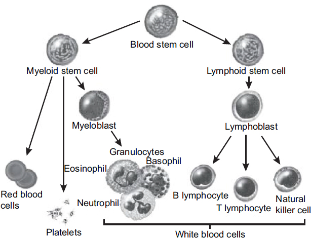
SOURCE: © Winslow, 2007. U.S. government has certain rights.
immunity, immune regulation, and the facilitation of B-cell function. Progenitor or pre-B cells mature in the bone marrow into antigen-specific B cells. On encountering their cognate antigens, B cells differentiate into antibody-secreting plasma cells involved in humoral immunity.
LHCs originate in specific pluripotent or lineage-restricted cells at different stages in hematopoiesis and immune-cell development. The normal cells are transformed into a malignant cell population through a multistep process that involves genetic and epigenetic alterations. Traditionally, LHCs have been divided into leukemias, lymphomas, myelomas, and so on, according to their cell type and site of origin (see Figure 7-1). Additional morphologic, cytochemical, and immunophenotypic data are used to characterize LHCs further and to further divide them into distinct subtypes.
Leukemias occur when a myeloid stem cell residing in the bone marrow becomes transformed, resulting in a failure of differentiation and a resistance to normal feedback on cellular proliferation. As the leukemic cells (blasts) fill the bone marrow, they actively secrete cytokines that prevent normal cellular proliferation, leading to reduced circulating normal blood cells. In addition, changes in adhesion molecules allow the release of these immature cells into the peripheral blood. Leukemias are generally classified as myeloid or lymphoid, depending on the lineage of the malignant cell population. If the original mutated cell of
a cancer of the blood arises in a lymphoid progenitor, then the malignancy is termed lymphocytic leukemia; lymphocytic leukemias have been further partitioned into acute lymphoblastic leukemia (ALL) (also known as acute lymphoid leukemia or acute lymphocytic leukemia), which is derived from precursor B or T lymphoid stem cells, and chronic lymphocytic leukemia (CLL), which is a “mature B-cell neoplasm” arising from a transformed B lymphocyte. Myeloid leukemias arise from a myeloid hematopoietic stem cell and are classified into acute (AML) and chronic (CML) forms.
Lymphoma is a general term for malignancies that arise from lymphocytes (B, T, or natural killer cells). Lymphomas generally present as solid tumors at lymphoid proliferative sites, such as lymph nodes and the spleen. As stem cells mature into B or T cells, they pass through several developmental stages, each with unique functions. The developmental stage of the malignant cell defines the subtype of lymphoma. About 85% of lymphomas are of B-cell origin, and 15% are of T-cell or natural killer–cell origin (Jaffe et al., 2001; Liao et al., 2012). There are two major types of B-cell lymphomas: HL, previously referred to as Hodgkin disease, and NHL. B cells give rise to a wide array of neoplasms, which are characterized by the stage at which B-cell development was arrested, as well as by the surface protein expression and the genetic characteristics of the malignant cells. Follicular, large-cell, and immunoblastic lymphomas result when a malignancy develops after a B cell has been exposed to antigens. CLL is a tumor of antigen-experienced (memory) B cells (Chiorazzi et al., 2005); small lymphocytic lymphoma involves the same cells as CLL, but presents primarily in lymph nodes rather than in the bone marrow and blood and is therefore a variant of the same disease (Jaffe et al., 2008).
Multiple myeloma is a lymphohematopoietic malignancy derived from antibody-secreting plasma cells, which also have a B-cell lineage, that accumulate primarily in the bone marrow but may also infiltrate extramedullary sites. The related condition amyloid light chain (AL) amyloidosis also arises from B cell–derived plasma cells and reflects an abnormal deposition of antibody-derived light chains. It occurs as a complication in 5–15% of patients with multiple myeloma, and may also occur without evidence of frank multiple myeloma. Monoclonal gammopathy of undetermined significance (MGUS) is a clonal proliferation of plasma cells condition that may progress to multiple myeloma.
The ICD system partitions these malignancies into leukemias and lymphomas based primarily on whether the cancer cells circulate in the blood (disseminated) or appear in the lymphatic tissues, respectively, before subdividing by cell type. The emerging WHO classification of lymphohematopoietic malignancies (Campo et al., 2011; Jaffe, 2009) stratifies malignancies of the blood and lymph nodes into disease categories by their cell lineages—lymphoid or myeloid—as shown in Figure 7-1. It represents a substantial advance in understanding the biologic paths by which these malignancies develop. Since the new WHO classification has not yet been definitively approved, the committee decided that it
would not be productive to reformulate this entire section to correspond to the new WHO categories. In practice, LHCs have routinely been reported in a variety of groupings, so it is a continuing challenge to parse out results, noting when results for broader groupings are presented in the supplementary tables for several more specific diagnoses, while recognizing that the specific results may be confounded by being “misclassified” with other entities. Most epidemiologic studies already in the evidentiary database that specifed diseases precisely used codes from ICD-9 or earlier versions, but some recent studies have applied ICD-10. Furthermore, the existing records that will serve as the basis of many current and even future studies will use earlier and evolving classifications, so a confounding of classification is likely to remain, even in new literature. The nomenclature has become more uniform in recent studies, but the possibility of ambiguity remains if earlier researchers did not use a unique code in accordance with some established system.
On occasion, the observed number of cases is so small that researchers cannot perform useful analyses for each type of LHC and will instead provide summary statistics for the entire group of them. In updating mortality in the Hamburg cohort in 1952–2007, Manuwald et al. (2012) found non-significant increases in mortality from LHC in both men and women, which combined to give a significant association between TCDD and all LHC deaths in the whole cohort (SMR = 1.61, 95% CI 1.03–2.40). In a Dutch cohort of workers in two phenoxy-herbicide plants, Boers et al. (2012) assessed plasma TCDD concentrations at the time of the assumed last exposure and reported a modest but not statistically significant increase in the HR for LHC in the total cohort, but there was no increase in plant A, where workers were occupationally exposed to TCDD.
Because VAO committees aim to address disease entities as specifically as possible with the available data, the overall results on the broader grouping of LHCs are of little consequence for the conclusions of association that have been drawn for the more specific entities. The committee for Update 2010 noted, however, that the common biologic origin of LHCs that have been judged to have a substantial amount of evidence supporting association with the COIs (HL, NHL, CLL, hairy-cell leukemia [HCL], multiple myeloma, and AL amyloidosis) means that the WHO approach is supportive of and consistent with these decisions on the part of VAO committees. The Update 2014 committee familiarized itself with the classification systems that have been used for lymphoid malignancies, including hearing a presentation from the International Lymphoma Epidemiology Consortium (InterLymph) describing a proposed classification of these cancers into subtypes that are particularly appropriate for epidemiologic research, including methods to harmonize data, standardized definitions of disease entities and rigorous quality control of these subtype assessments, and attempts to understand the implications of etiologic heterogeneity (Morton et al., 2014a,b). At the same time, as has been recognized by others (Saberi Hosnijeh et al., 2012c), given the type and quality of the historical data that constitute the majority of the material
available to the committee for review and judgement, little of that impressive effort can be applied to the committee’s assessment of association.
VA asked previous VAO committees to address CLL, AML, and HCL individually. A scrutiny of the entire body of epidemiologic results on leukemias for findings on particular types (as had been the most common manner of grouping) revealed several studies that showed increased risks specifically of CLL but that did not provide support for an association of AML with exposure to the COIs. The committee for Update 2002 advised VA that CLL is recognized as a form of NHL, which is already recognized as a service-related condition, whereas the committee for Update 2006 did not recognize an association between the COIs and AML. Later, the committee responsible for Update 2008 advised VA that, like CLL, HCL should be grouped as a mature B-cell neoplasm. For the current update, VA has tasked the committee to specifically address myeloproliferative neoplasms (MPNs). In light of the history and in accord with the current WHO classification, the committee for this update has incorporated data specifically on CLL and HCL into the section on NHL. After a brief synopsis of biologic plausibility of the LHCs overall, the more common cancers of the lymphatic system are described in the sections below on HL, NHL, and multiple myeloma (with a section on the related condition, AL amyloidosis), and then evidence on leukemias in general is discussed, with a focus on information regarding leukemias of myeloid origin.
Biologic Plausibility
Studies in animal models have indicated that the AHR pathway plays an integral role in B-cell maturation and that TCDD and dioxin-like chemical exposure may alter the function of these cells and lead to critical changes in the immune response. The suppression of the immune response by TCDD and similar chemicals in rodents and primates has been known for more than 30 years, but the effect on human cells is less clear. Some reports indicate that TCDD and dioxin-like chemicals elicit similar effects in humans. The activation of non-transformed human B cells results in an increase in the expression of AHR, and additional data indicate that this pathway has a role in normal B-cell function (Allan and Sherr, 2010; Sherr and Monti, 2013). Furthermore, treating these cells with benzo[a]pyrene suppresses B-cell differentiation. H. Lu et al. (2010) demonstrated that although human B cells appeared less responsive to TCDD in terms of increasing the expression of AHR pathway genes, the ability of TCDD to decrease immunoglobulin (Ig) M production is similar in both mouse and human B cells. Research that modeled the mode by which TCDD suppresses the terminal differentiation of B cells offers distinct pathways whose action can be altered by exposure (Q. Zhang et al., 2013). Data on human hematopoietic stem cells and from the use of knockout Ahr mouse models show that Ahr is critical in hematopoietic stem cell maturation and differentiation (Ahrenhoerster et al.,
2014; Fracchiolla et al., 2011; K. P. Singh et al., 2011, 2014; B. W. Smith et al., 2013). TCDD not only alters hematopoietic stem cell maturation, but also alters proliferation and migration in vivo and in vitro (Casado et al., 2011). Finally, emblematic of the potential pleotropic effects of TCDD, Hughes et al. (2014) demonstrated that AHR plays a critical role in promoting lymphocyte differentiation into mature natural killer cells. Reviews have highlighted the complex and varied nature of the interaction of TCDD with the immune system (Gasiewicz et al., 2014; Lindsey and Papoutsakis, 2012).
Saberi Hosnijeh et al. (2012b, 2013a) assessed both the immune profile and the levels of soluble immune signaling proteins in TCDD-exposed workers. Consistent with data published on U.S. ACC veterans, in 47 highly TCDD-exposed and 38 low TCDD-exposed workers they found no effect of TCDD on major leukocyte subsets or on white blood cell counts. They did note a non-significant decrease in most lymphocyte subsets, which was most prominent for B cells. In these same workers, a study of soluble CD27 and soluble CD30 in sera found no clear dose–response relationship of TCDD with the level of these signaling proteins. However, there was a significant negative association of the serum IL-1RA level with the TCDD serum level among workers without chronic disease. Taken together, these data indicate that exposure to TCDD (and the alteration of normal AHR function) may have multiple effects on immune cell differentiation and function.
No new mechanistic or biologic plausibility studies regarding lymphohematopoietic cells have been identified by the committee since Update 2014.
Hodgkin Lymphoma
HL (ICD-9 201; ICD-10 C81), also known as Hodgkin disease, is distinguished from NHL primarily based on its neoplastic cells, mononucleated Hodgkin cells, and multinucleated Reed–Sternberg cells, derived from germinal-center B cells (Küppers et al., 2002). NCI estimated that 8,500 people would receive a new diagnosis of HL in the United States in 2018 and that 1,050 men and women would die from it; it ranks 25th in most common cancer diagnoses (NCI, n.d.y).
The incidence of HL increases with age; the average age of diagnosis is 39 years. Although the incidence rate is slightly higher in men than in women of the same race, it is about the same in whites and blacks (NCI, n.d.y). In the age groups that include most Vietnam veterans, the age-adjusted modeled incidence rate of HL for men 50–64 years old of all races combined was 3.2 per 100,000 in 2014 and increased to 4.7 for 65–74-year-olds and 5.7 for men over 75 years.19
___________________
19 As calculated on the site https://seer.cancer.gov/faststats/selections.php?#Output by using the SEER 13 dataset and choosing age-adjusted rates, HL, all races, age ≥ 50 years, and male sex.
The possibility that HL has an infectious etiology has been a topic of discussion since its earliest description. A higher incidence in people who have a history of infectious mononucleosis has been observed in some studies, and a link with Epstein–Barr virus has been proposed (Balfour et al., 2015; Murray and Bell, 2015). In addition to the occupational associations discussed below, higher rates of the disease have been observed in people who have suppressed or compromised immune systems.
Conclusions from VAO and Previous Updates
Based on the 32 studies it reviewed, the committee responsible for VAO determined that there were sufficient epidemiologic data to support an association between exposure to the COIs and HL. Additional studies available to the committees responsible for subsequent updates have not changed that conclusion.
Two well-conducted Swedish studies with good exposure characterization reviewed in VAO provide the most comprehensive information on the association between exposure to phenoxy herbicides (2,4-D and 2,4,5-T), picloram, or chlorophenols and HL. Hardell et al. (1981) considered NHL and HL together, and Hardell and Bengtsson (1983) considered HL separately; they found statistically significant associations with exposure to phenoxy acids (after excluding people who were exposed to chlorophenols) and with exposure to chlorophenols. In a study of 54 HL cases, Persson et al. (1989) found a large but imprecise and not statistically significant risk associated with exposure to phenoxy acids. Several of the other case-control and occupational-cohort studies in U.S. and international populations reviewed in VAO showed an increased risk of HL, but only a few of the results were statistically significant. As with NHL, even the largest studies of production workers who were exposed to TCDD did not indicate an increased risk. The few studies of HL in Vietnam veterans tended to show increased risks, but only one (Holmes et al., 1986) was statistically significant.
Among the studies of veterans reviewed throughout the VAO series, no statistically significant increased risk of HL was found. Publications from the AFHS showed no statistically significant increases in HL or lymphopoietic cancers (AFHS, 2000; Akhtar et al., 2004). Other populations of Vietnam-era veterans likewise did not find an association (Anderson et al., 1986a,b; Holmes et al., 1986; Lawrence et al., 1985; Visintainer et al., 1995). A proportionate mortality ratio analysis that compared the experience of 33,833 U.S. Army and Marine Corps Vietnam veterans who died during 1965–1988 with that of 36,797 deceased non-Vietnam veterans found a statistically significant increase of deaths from HL in Marine Corps veterans—although not Army veterans—who had served in Vietnam (Watanabe and Kang, 1996). Cypel and Kang (2008) compared mortality from lymphopoietic cancers in female Vietnam veterans with that of female era
veterans and the U.S. population; deaths from lymphopoietic cancers were not higher in those who served in Vietnam.
Studies of Australian, New Zealand, and Korean veterans who served in Vietnam have also been reviewed. The incidence of HL was found to be statistically significantly higher than the general population when Australian veterans from all branches were combined (ADVA, 2005a), but when veterans were stratified by service branch, the only statistically significant association was between HL and service in the Army. However, no statistically significantly increased mortality from HL was found for all service branches combined, nor for any single branch (ADVA, 2005b). A comparison of deployed and non-deployed Vietnam-era Australian conscripted Army National Service veterans found no association between deployment and the incidence of or mortality from HL (ADVA, 2005c). Among a cohort of male Vietnam veterans from New Zealand, McBride et al. (2013) reported one death from and three incident cases of HL resulting in imprecise and unstable effect estimates. Among the Korean veterans who had served in Vietnam, Yi and Ohrr (2014) reported no statistically significant difference in the risk of HL in the internal comparison of the high- and low-exposure groups based on the EOI scores.
Several occupational cohorts of workers from several countries who were exposed to phenoxy herbicides and other related chemicals have been followed long term. Studies of the IARC phenoxy-herbicide cohort showed no excess of HL incidence or mortality compared with national rates (Kogevinas et al., 1993). Additional follow-up showed a non-statistically significant increase in HL in workers who were exposed to TCDD or higher chlorinated hydrocarbons but no association was found between phenoxy herbicides or chlorophenols and HL (Kogevinas et al., 1997). In a multinational IARC cohort of 60,468 pulp-and-paper-industry workers, McLean et al. (2006) found that death from HL was significantly higher in those who had ever been exposed to nonvolatile organochlorine compounds (which would include TCDD) but not in those who had never been exposed compared with the national standardized populations.
In a retrospective cohort study of Dutch production and contract workers who were exposed to phenoxy herbicides, chlorophenols, and contaminants during 1950–1976, Hooiveld et al. (1998) reported a non-significant increase in HL. Rix et al. (1998) compared mortality in a cohort of Danish paper mill workers with mortality in the general Danish population and found a statistically significant increase in men but not women. Swaen et al. (2004) extended the follow-up of mortality by 13 years in a cohort of Dutch herbicide appliers and observed no additional deaths, causing the earlier mortality risk estimate of HL in the cohort to no longer be statistically significant (Swaen et al., 1992). Studies of German manufacturing workers found no association between exposure to TCDD and HL (Becher et al., 1996). McBride et al. (2009a) examined mortality in TCP manufacturing workers in the Dow AgroSciences plant in New Zealand, but a single observed HL death yielded inconclusive results. A French,
hospital-based, case-control study of lymphoid neoplasms (Orsi et al., 2009) did not find a statistically significant increase in the risk of HL after occupational exposure to herbicides in general or after occupational exposure to phenoxy herbicides in particular.
Among U.S. production workers, the low numbers of deaths from HL and imprecise estimates limited the findings. In an update and expansion of cohorts involved in the NIOSH study, Steenland et al. (1999) observed three deaths attributed to HL, which was not statistically different from the comparison population. No deaths from HL were identified in Dow PCP workers in Midland, Michigan (Collins et al., 2009c; Ramlow et al., 1996), and only two deaths from HL occurred among the TCP workers, resulting in imprecise risk estimates (Collins et al., 2009b). Additional updates of these occupational cohorts noted very small numbers of additional cases of HL, which did not produce substantive changes in prior findings. Burns et al. (2011) reported an additional case among Dow 2,4-D production workers. Ruder and Yiin (2011) likewise reported one additional HL death in the NIOSH cohort of PCP workers.
Studies of the Seveso cohort have found few cases and no increased incidence or mortality of HL among men or women in zones A, B, or R. These findings remained consistent at the 10-year follow-up (Bertazzi et al., 1993), 15-year follow-up (Bertazzi et al., 1997), 20-year update (Bertazzi et al., 2001; Pesatori et al., 2009), and 25-year update (Consonni et al., 2008).
Several studies of HL among agricultural workers have also been reviewed by the update committees. Whereas Persson et al. (1993) reported a significant increase in the odds of HL prevalence in Swedish farmers who were exposed to phenoxy acid herbicides, no excess of HL deaths was found in a death certificate review of U.S. farmers from 23 states (Blair et al., 1993). In a nonspecific analysis of exposure to “herbicides” in a Michigan farming community, Waterhouse et al. (1996) demonstrated a statistically significant increase in the combined incidence of lymphopoietic neoplasms in a prospective study. Two reports from the AHS found no excess risk of HL in pesticide applicators, commercial applicators, or their spouses, but herbicide exposures were not specific (Alavanja et al., 2005; Blair et al., 2005a). Updates of cancer incidence (Koutros et al., 2010a) and mortality (Waggoner et al., 2011) among participants in the AHS also did not find increases of HL in private applicators or their spouses.
In the Cross-Canada Study of Pesticides and Health, P. Pahwa et al. (2006) found no association of any exposure to phenoxy herbicides, 2,4-D, Mecoprop, or MCPA and HL. Nested case-control studies of data from the Cross-Canada study found no statistically significant associations of exposure to COIs with HL (Karunanayake et al., 2012; P. Pahwa et al., 2003). A follow-up analysis of the Cross-Canada study (Navaranjan et al., 2013) that examined the incidence of STS, NHL, multiple myeloma, and HL in men ages 19 years and older found no increased risk of HL in those exposed to one, two, or three or more phenoxy
herbicides after adjusting for age and the province of residence. However, exposure specificity remains a major issue for this study.
Update of the Epidemiologic Literature
No new published literature of Vietnam veterans, environmental studies, or case-control studies that addressed exposure to the COIs and HL was identified by the committee for the current update. Reviews of the relevant studies are presented in the earlier reports. Table 24, which can be found at www.nap.edu/catalog/25137, summarizes the results of studies related to HL.
Occupational Studies Among the Dow Midland, Michigan, worker cohort that was compared with the standardized U.S. population, Collins et al. (2016) found only two deaths from HL were reported during the follow-up period and both were among TCP workers, making the reported risk estimates unreliable.
In an extension of the follow-up of UK phenoxy herbicide manufacturers and sprayers to examine the carcinogenicity of phenoxy herbicides, Coggon et al. (2015) reported only three deaths from HL, and all were among the workers potentially exposed to phenoxy acids above background levels and for more than 1 year (SMR = 1.71, 95% CI 0.35–4.99).
Other Identified Studies One other study that reported outcomes of mortality from HL was identified, but it lacked sufficient exposure specificity to be included as contributing to the evidence base of the potential effects of the COIs (Ruder et al., 2014).
Biologic Plausibility
HL arises from the malignant transformation of a germinal-center B cell and is characterized by malignant cells that have a distinctive structure and phenotype; these multinucleate cells are known as Reed–Sternberg cells (Jaffe et al., 2008). No animal studies have shown an increase in HL after exposure to the COIs. Reed–Sternberg cells have not been demonstrated in mice or rats, so there is no good animal model of HL. Thus, there are no specific animal data to support the biologic plausibility of an association between the COIs and HL.
Synthesis
The relative rarity of HL complicates the evaluation of epidemiologic studies because their statistical power is generally low. Earlier studies (Eriksson et al., 1992; Hardell et al., 1981; Holmes et al., 1986; LaVecchia et al., 1989; Persson et al., 1993; Rix et al., 1998; Waterhouse et al., 1996; Wiklund et al., 1988a) were generally well conducted and included excellent characterizations of exposure,
and they formed the basis of previous VAO committees’ conclusions. Subsequent findings have not contradicted those conclusions, especially given that most studies have had low statistical power, as was seen in the current extended follow-ups of occupational cohorts that reported two (Collins et al., 2016) and three (Coggon et al., 2015) deaths from HL. Although it has not been demonstrated in any animal models, a positive association between the COIs and the development of HL is biologically plausible because of the common lymphoreticular origin of HL and NHL (which has been demonstrated in animal models) and their common risk factors.
Conclusion
Based on the evidence reviewed here and in previous VAO reports, the committee concludes that there is sufficient evidence of an association between exposure to at least one of the COIs and HL.
Non-Hodgkin Lymphoma
NHL (ICD-9 200.0–200.8, 201, 202.0–202.2, 202.8–202.9; ICD-10 C82–85, C96.3) is a general name for malignancies of the lymphatic system other than HL or plasma cell dyscrasias. NHL consists of a large group of lymphomas that can be partitioned into acute and aggressive (fast-growing) or chronic and indolent (slow-growing) types of either B-cell or T-cell origin. B-cell NHL includes Burkitt lymphoma, diffuse large B-cell lymphoma, follicular lymphoma, large-cell lymphoma, precursor B-lymphoblastic lymphoma, and mantle-cell lymphoma. T-cell NHL includes mycosis fungoides and anaplastic large-cell lymphoma. Precursor T-lymphoblastic lymphoma is not considered a type of NHL and is considered instead part of T-lymphoblastic lymphoma/leukemia, a precursor lymphoid neoplasm included with the broad group of “acute lymphoid leukemias,” which can be of either T-cell or B-cell origin.
In response to requests from VA to address CLL, HCL, and AML specifically and individually, CLL and HCL have been recognized as subtypes of NHL (demonstrating B-cell origin and immunohistochemical properties consistent with B-cell lymphoma). The proposed WHO classification of NHL notes that CLL (ICD-9 204.1; ICD-10 C91.1) and its lymphomatous form, small lymphocytic lymphoma, are both derived from mature B cells (Chiorazzi et al., 2005; IARC, 2001). The committee for Update 2012 determined that it is more appropriate to consider those lymphatic malignancies with other forms of NHL. Therefore, the discussion of CLL and HCL has been moved into the NHL grouping.
NCI estimated that 74,680 people would receive a new diagnosis of NHL in the United States in 2018 and that 19,910 men and women would die from it; it ranks as the seventh most common cancer diagnosis (NCI, n.d.z). The incidence of NHL increases with age; the average age of diagnosis is 67 years. The incidence rate is about 50% higher in white and black men than in women of the
same race and is highest for whites. In the age groups that include most Vietnam veterans, the age-adjusted modeled incidence rate of NHL for men 50–64 years old of all races combined was 36.3 per 100,000 in 2014 and increased to 91.1 for 65–74-year-olds and 147.8 for men over 75 years.20
In addition, NCI estimated that 20,940 men and women would receive a diagnosis of CLL in the United States in 2018 and that 4,510 people would die from it. Nearly all cases occur after the age of 50 years, and the median age of diagnosis is 70 years. The incidence rate is about two times higher in men than in women of the same race and is highest for whites (NCI, n.d.aa).
The causes of NHL are poorly understood. People who have suppressed or compromised immune systems are known to be at higher risk, and some studies show an increased incidence in people who have HIV, human T-cell leukemia virus type I, Epstein–Barr virus, surface antigen positive hepatitis B, or gastric Helicobacter pylori infections. The human retrovirus HTLV-1 causes adult T-cell lymphoma, but early reports that HTLV-2 might play a role in the etiology of HCL have not been substantiated. A broad spectrum of behavioral, occupational, and environmental risk factors have been proposed as contributors to the occurrence of NHL, but given the diversity of malignancies included under this name and the evolving classification of the subtypes (to date, minimal exploration of etiologic heterogeneity has been done), it is not too surprising that—aside from infectious agents, immune problems, and particular chemotherapies—specific risk factors have not been definitively established (Morton et al., 2008, 2014a,b; Wang and Nieters, 2010).
Conclusions from VAO and Previous Updates
The committee responsible for VAO concluded that there was sufficient evidence to support an association between exposure to at least one of the COIs and NHL. Additional information available to the committees responsible for later updates has not changed that conclusion. Update 2002 was the first to discuss CLL separately from other leukemias. The epidemiologic studies indicated that farming, especially with exposure to 2,4-D and 2,4,5-T, is associated with significant mortality from CLL. Many more studies support the hypothesis that herbicide exposure can contribute to NHL risk. Most cases of CLL and NHL reflect the malignant transformation of germinal-center B cells, so these diseases could have a common etiology.
As with HL, the epidemiologic data reviewed by previous VAO committees suggest that it is the phenoxy herbicides (including 2,4-D) rather than TCDD that may be associated with NHL. The original VAO committee concluded that a
___________________
20 As calculated on the site https://seer.cancer.gov/faststats/selections.php?#Output using the SEER 13 dataset and by choosing age-adjusted rates, non-Hodgkin lymphoma, all races, age groups 50–64, 65–74, and ≥ 75 years, and male sex.
positive association existed between exposure to herbicides and the development of NHL, and studies reviewed by later committees have continued to support that finding. A large, well-conducted case-control study in Sweden by Hardell (1981) examined NHL and HL together and found, on the basis of 105 cases, a statistically significantly increased risk associated with exposure to phenoxy acids or chlorophenols. Those results were replicated in further investigations of the validity of the exposure assessment and potential biases (Hardell, 1981). Hardell et al. (1994) also examined the relationship between occupational exposure to phenoxyacetic acids and chlorophenols and various characteristics related to NHL—including histopathologic measures, stage, and anatomic location—on the basis of the NHL cases in a previous study (Hardell et al., 1981). Similar data by Persson et al. (1989) showed, based on a logistic regression analysis of 106 cases, an increased risk of NHL in those exposed to phenoxy acids. Other case-control studies of NHL conducted in Sweden found a statistically significantly elevated risk of NHL (Eriksson et al., 2008; Hardell and Eriksson, 1999; Hardell et al., 2001, 2002; Olsson and Brandt, 1988).
Several case-control studies of NHL incidence, risk, or mortality in other exposed populations have also been reviewed by VAO committees, including studies from New Zealand (Pearce et al., 1985, 1986b, 1987), Italy (Amadori et al., 1995; Nanni et al., 1996), and Canada (Ng et al., 2010; Spinelli et al., 2007). Various U.S. populations have also been studied for associations of herbicides with NHL (Cantor, 1982; Cantor et al., 1992; Colt et al., 2009; Hartge et al., 2005; Tatham et al., 1997; Zahm et al., 1993); NHL and multiple myeloma (Burmeister et al., 1983); and STS and NHL (Woods and Polissar, 1989; Woods et al., 1987).
Studies of production workers have shown some association between TCDD exposure and NHL. A larger study of 21,863 workers in the IARC phenoxyherbicide cohort followed from 1939 to 1992 found a non-significant increase in NHL risk (Kogevinas et al., 1997). Other studies of Danish and Dutch phenoxyherbicide workers who were part of the IARC cohort have shown a non-significant increased risk of NHL (Boers et al., 2010; Bueno de Mesquita et al., 1993; Hooiveld et al., 1998; Lynge, 1993). A cohort of 2,479 workers in four plants in Germany with exposure to phenoxy herbicide and contaminants (dioxins and furans) had a significantly increased risk of NHL on the basis of five cases (Becher et al., 1996). Non-statistically significant increases in risk have also been found in the NIOSH mortality study (Steenland et al., 1999). Risks were not significantly increased among the Dow Chemical Company Midland, Michigan, or Plymouth, New Zealand, chemical production workers, phenoxy-herbicide sprayers, or 2,4-D production workers (Bloemen et al., 1993; Bodner et al., 2003; C. J. Burns et al., 2001; Collins et al., 2009b,c; McBride et al., 2009a,b; Ramlow et al., 1996; 't Mannetje et al., 2005). A multinational IARC cohort study of paper- and-pulp workers found a statistically significant increase in workers who were exposed to chlorophenols (McLean et al., 2006).
Studies of farmers and agricultural workers have been generally positive for an association between herbicides or TCDD and NHL; however, only a few were statistically significant. A meta-analysis of several studies of the association between employment as a farmer in the central United States and NHL showed a statistically significant risk (Keller-Bryne et al., 1997). All of the studies of U.S. agricultural workers that were reviewed showed increased risk ratios, and two NCI studies of farmers in Kansas and Nebraska (Hoar et al., 1986; Zahm et al., 1990) showed patterns of an increased risk linked to use of 2,4-D. A third NCI study of NHL was conducted in four Surveillance, Epidemiology and End Results program centers (Detroit, Iowa, Los Angeles, and Seattle) from 1998 to 2000 (Pronk et al., 2013). The researchers used residential history from 15 years prior to diagnoses to link residence to EPA databases of dioxin-emitting facilities, studying 969 cases and 749 controls. Proximity to any dioxin-emitting facility was not associated with NHL. A study of a subcohort of Hispanic workers in a larger cohort of 139,000 California members of the United Farm Workers of America (Mills et al., 2005) and a population-based case-control study in Italy of NHL and CLL cases (combined) identified during 1991–1993 (Miligi et al., 2006) both showed statistically significant associations with 2,4-D. An additional occupational exposure study using the EPI-LYMPH multicenter study (conducted in the Czech Republic, France, German, Ireland, Italy, and Spain from 1998 to 2004) assessed pesticide use in applying a crop-exposure matrix with lymphoma diagnoses in a case-control design (Cocco et al., 2012). No statistically significant association was observed between any kind of pesticide or herbicide use and lymphomas or its subtypes, including between exposure to phenoxy acids and CLL.
The Cross-Canada Study of Pesticides and Health, a large, well-conducted, population-based, case-control study, reported on pesticide use and NHL incidence in men identified from cancer registries of six Canadian provinces from 1991 to 1994. Statistically significant associations were found between exposure to phenoxy herbicides, 2,4-D, or Mecocrop (MCPA) and NHL. A reanalysis of the data from that study confirmed the findings on phenoxy herbicides but found that the association with 2,4-D, although still increased, was no longer statistically significant (McDuffie et al., 2001). A study of the participants who had a first diagnosis of STS, NHL, multiple myeloma, or HL during the original study period were followed through mailed and telephone interviews to examine the joint effects of asthma, allergies, or asthma and allergies and hay fever combined with pesticide exposure in the genesis of NHL. Incident NHL cases (n = 513) diagnosed between 1991 and 1994 were compared with the experience of 1,506 controls. Subjects with asthma, allergies, or hay fever had elevated risks that were not statistically significant associated with the use of phenoxy herbicides, MCPA, or 2,4-D. The results overall were not supportive of any major effect modification by these immune conditions.
A population-based case-control study conducted in 2000–2001 of men and women 20–74 years old living in New South Wales, Australia, found an
increased risk of NHL associated with “substantial” exposure to phenoxy herbicides (Fritschi et al., 2005). Spinelli et al. (2007) reported on a population-based case-control study in Vancouver and Victoria, British Columbia, which found strong monotonic increases in serum concentrations of two dioxin-like PCBs (PCB 118 and PCB 156). Chiu et al. (2004) and W. J. Lee et al. (2004a) conducted a pooled analysis of two case-control studies that were carried out in three midwestern U.S. states—Iowa and Minnesota (Cantor et al., 1992) and Nebraska (Zahm et al., 1990)—and found that NHL risks were increased in farmers by the use of herbicides, including 2,4-D and 2,4,5-T. In a study of NHL incidence in people who lived near 13 French municipal waste incinerators, Viel et al. (2008b) found a small but statistically significant increase in the risk of NHL and evidence of a dose–response relationship with increased exposure to dioxin. A case-control study of NHL rates in people who lived near a municipal solid-waste incinerator in Bensaçon, France, found that the incidence of NHL was significantly increased in the area determined to have the highest dioxin contamination, but no increases were found in the low and intermediate categories (Floret et al., 2003). A French hospital-based case-control study of lymphoid neoplasms (Orsi et al., 2009) did not find the occurrence of NHL to be associated with occupational or domestic use of pesticides or phenoxy herbicides in particular. With 25 years of follow-up of the Seveso population and a relatively small number of observed cases, there is no evidence of an increased incidence of NHL or mortality in any of the exposed zones (Bertazzi et al., 1989b, 1993, 1997, 2001; Consonni et al., 2008; Pesatori et al., 1992, 2009).
The findings of several PCB-focused studies (Bertrand et al., 2010; Engel et al., 2007; Laden et al., 2010) are consistent with the associations with NHL repeatedly observed in connection with the COIs in the VAO series. However, the extent of intercorrelation of these persistent organic pollutants greatly curtails the degree to which any effect can be specifically attributed to dioxin-like activity.
Evidence of an association between the COIs and NHL in Vietnam veterans, the primary population of interest in the VAO updates, has been lacking. The CDC Selected Cancers Study (CDC, 1990a,b) showed a significantly increased risk of NHL in all Vietnam veterans; however, in an analysis that considered branch of service, Army and Air Force personnel were found not to be at increased risk. Marine Corps veterans had a higher mortality in the CDC Selected Cancers Study and significantly increased risks in several other studies (Breslin et al., 1988; Burt et al., 1987; Watanabe and Kang, 1996; Watanabe et al., 1991), but the implications of these findings are unclear. No increased risk has been found in Ranch Hand veterans (AFHS, 2000; Akhtar et al., 2004; Michalek et al., 1990; Wolfe et al., 1990) or in members of the ACC (Boehmer et al., 2004).
In studies of Vietnam veterans from New Zealand and Korea, no statistically significant increased risk of NHL was found in either group. The New Zealand study also reported on lymphoid leukemia (which would include any cases of CLL) and found a statistically significant increase (McBride et al., 2013). Yi et
al. (2014b) reported a modestly increased risk for NHL mortality between the high- versus low-exposure groups of the Korean veterans and a small increased risk with the individual log-transformed EOI scores, but neither was statistically significant. Lymphoid leukemia showed a nonstatistically significant decreased risk based on nine low-exposure and five high-exposure deaths.
Update of the Epidemiologic Literature
Several new studies of exposure to the COIs and NHL were identified including two studies of Vietnam veterans and CLL. Reviews of the relevant studies are presented in the earlier reports. Table 25, which can be found at www.nap.edu/catalog/25137, summarizes the results of studies related to NHL.
Vietnam-Veteran Studies A retrospective study of veterans diagnosed with CLL who attended the Minneapolis VA at least one time between 2001 and 2010 was conducted to determine whether exposure to Agent Orange alters features of CLL disease presentation or its prognostic features, including stage at diagnosis, lymphocyte doubling time, or cytogenetics (Baumann Kreuziger et al., 2014). Of the 195 veterans identified as having a diagnosis of CLL, 33 (17%) were determined to have been exposed to Agent Orange. (Veterans submit a claim of exposure that is reviewed by VA using service records to confirm whether the veteran was stationed in an area that was sprayed with herbicides during the service period.) Except for age at diagnosis, demographic factors and laboratory counts were similar between the veterans determined to have been exposed and those reported as unexposed. Compared with veterans who were reported as unexposed to Agent Orange, the exposed patients showed a statistically significantly lower age of presentation (61 versus 72 years; p = 0.001), and the time to first treatment was also shorter in the exposed patients (9.6 versus 30 months, p = 0.02), but the number of therapies and the rate of transformation were not different between exposed and unexposed patients. However, providers were not blinded to exposure status, and, because patients were younger, these factors may have influenced the therapy paradigm used. Overall, survival was not statistically different between exposed and unexposed patients, and this remained the case after adjusting for age (HR = 1.8, 95% CI 0.7–4.5, p = 0.24).
A second retrospective study among U.S. Vietnam veterans was identified that examined CLL prognosis, treatment, and survival and their association with exposure to Agent Orange (determined by benefits and compensation officers who use service records to confirm if the locations and timeframes of deployment correspond to sprayed areas). Mescher et al. (2018) identified 2,052 Vietnam veterans (20.4% determined to have been exposed to Agent Orange) who were diagnosed with CLL between 2009 and 2013 from the National VA Tumor Registry. Information on demographics, laboratory and disease-related parameters at the time of the initial diagnosis, and the type and number of treatments received was taken from
medical records. Overall survival was defined as time from CLL diagnosis to death from any cause or else to date of last follow-up. The Cox proportional hazards model included age, Rai stage, and baseline laboratory parameters. Prognostic factors did not differ based on exposure. Compared with the unexposed, veterans exposed to Agent Orange were diagnosed at younger age (63.2 versus 70.5 years, p < 0.0001) and had longer overall survival (p < 0.001). CLL prognostic factors and cytogenetics did not differ between the groups, although more of the exposed group had cytogenetics available. Time to initial treatment was shorter in exposed patients. Median overall survival was longer in exposed patients—quite possibly because they were treated more aggressively because they were young.
Occupational Studies Among the Dow Midland, Michigan, worker cohort, Collins et al. (2016) found that compared with the standardized U.S. population, no differences in mortality for NHL were found for the TCP workers (n = 10; SMR = 1.08, 95% CI 0.52–1.99) or the PCP workers (n = 8; SMR = 1.92, 95% CI 0.83–3.79).
In an extension of the follow-up of UK phenoxy herbicide manufacturers and sprayers examining the carcinogenicity of phenoxy herbicides and their association primarily with HL, STS, and CLL, Coggon et al. (2015) calculated standardized mortality ratios using person-years and the general populations of England and Wales as the comparison group. Nested case-control analyses compared men with incident or fatal NHL/CLL (n = 74) and matched controls (up to 10 per case). A total of 29 NHL/CLL deaths were reported. The risk of death from NHL/CLL was not statistically significantly elevated among all workers (SMR = 0.96, 95% CI 0.64–1.38) or among workers exposed to herbicide levels above background (n = 26; SMR = 1.15, 95% CI 0.75–1.68). However, death from NHL was statistically significantly higher for men who were exposed to phenoxy acids above background for 1 year or longer (n = 19; SMR = 1.85, 95% CI 1.12–2.89). No other statistically significant associations or trends were reported for mortality from other types of cancers. Although a statistically significant excess of deaths from NHL was reported, the authors believe that greater weight should be given to the nested case-control analysis, which included both non-fatal and fatal cases and men with CLL as well as solid lymphomas. That analysis found no statistically significant increased risk with exposure to phenoxy herbicides; the highest risk estimate was for workers with high exposure (OR = 1.22, 95% CI 0.61–2.46). The committee believes that the caveat here is that the researchers seem to use CLL as their surrogate for NHL, which may or may not be correct. A cleaner analysis would have been to look specifically at other lymphomas (diffuse large B cell, follicular lymphomas, etc.). However, as the data linking exposure to phenoxy herbicides and NHL have been inconsistent, the committee agrees with the authors that if there really is an increased risk of NHL/CLL, it is likely very small.
Case-Control Studies Czarnota et al. (2015) conducted an analysis of 27 correlated chemicals (5 PCBs, 7 PAHs, and 15 pesticides) measured in house dust samples and NHL risk using data collected from 1998 to 2000 as part of the population-based case-control study of NHL in four SEER study sites (Detroit, Iowa, Los Angeles, and Seattle). Eligible cases were 20–74 years of age, diagnosed with a first primary NHL, and uninfected with HIV. The study recruited 1,321 people with NHL who were frequency-matched to 1,057 controls by sex, age (within 5-year groups), race, and study site. Computer-assisted personal interviews were conducted in the home of each participant and included questions on demographics, house characteristics, pesticide use in the home and garden, and residential and occupational histories. Dust samples were collected and analyzed from vacuum cleaners for participants who had used their vacuum cleaner within the past year and owned at least half of their carpets or rugs for 5 years or more. The chemicals relevant to the work of the VAO committee that were analyzed were PCB 105, 2,4-D, and dicamba. Pairwise comparisons were performed to obtain a weighted quantile sum for an index of 27 environmental chemicals. The 27 chemicals were not weighted equally in the index, overall or by site. The weighted quantile sum index was statistically significantly associated with NHL (OR = 1.30, 95% CI 1.08–1.56). Concentrations of 2,4-D and dicamba were found to be elevated in the Iowa site. The only individual chemical that was associated with NHL was non-dioxin-like PCB 180. However, associations with NHL were negative and statistically significant for 2,4-D (OR = 0.36, 95% CI 0.19–0.68) and dicamba (OR = 0.48, 95% CI 0.26–0.90) in the Iowa site and 2,4-D (OR = 0.53, 95% CI 0.29–0.97) and dicamba (OR = 0.41, 95% CI 0.22–0.76) in the Seattle site.
Kelly et al. (2017) used data from the EnviroGenoMarkers study, which is nested within two prospective cohorts in Italy and Sweden, to determine the association between NHL and prediagnositic blood plasma concentrations of 10 environmental pollutants, including two dioxin-like PCB congeners (118 and 156). During 16 years of follow-up, 270 incident cases of B-cell NHL (including 76 cases of multiple myeloma) were diagnosed. Cases were matched to 270 healthy controls by center, age, gender, and date of blood collection. Cases were categorized into ordered quartiles of exposure for each persistent organic pollutant based on the distribution of exposure in the control population. No difference in median exposure levels was observed between cases and controls for any of the chemical exposures (p > 0.05) or, specifically, for the dioxin-like PCBs: PCB 118 (p = 0.991) and PCB 156 (p = 0.186). The quartile analysis similarly did not show an association between the dioxin-like PCBs and NHL (only the comparison of cases and controls in the third quartile was statistically significant, but there was no significant association in the test for trend). These were not heavily exposed individuals, so the levels may be uniformly low. Overall, this study does not provide evidence that higher body burden of the two dioxin-like PCBs (or
the other persistent organic pollutants measured) increases the risk of subsequent NHL diagnosis.
Other Identified Studies Six other studies of NHL in occupational cohorts were identified. Two examined participants of the AHS: in the first study none of the COIs were included in the analysis of exposures (Alavanja et al., 2014), and the second paper (Goodman et al., 2017) was an updated meta-analysis of 2,4-D and NHL that included 10 studies of the AHS but did not contribute original data. A third study examined cause-specific mortality, including NHL, among U.S. workers exposed to mixed PCBs (Ruder et al., 2014), the fourth study analyzed health outcomes among Brazilian agricultural workers exposed to unspecified pesticides (Boccolini et al., 2017), and the fifth study was a small case-control study designed to investigate the effects of exposure to pesticides (determined by occupation as a farm worker) on NADPH (nicotinamide adenine dinucleotide phosphate, a cofactor used in biochemical reactions), including its level and relationship with the Th1/Th2 ratio, in patients diagnosed with NHL in Algeria (Zahzeh et al., 2015). A sixth study (Aras et al. 2014) was a case-control study conducted at a cancer center in France to determine whether patient diagnosed with NHL had reported exposure to pesticides (types of insecticides and herbicides, if known; frequency; and duration), and other known or suspected risk factors. However, all these studies lacked sufficient exposure specificity to be included as contributing to the evidence base of the potential effect of the COIs.
Biologic Plausibility
The diagnosis of NHL encompasses a wide variety of lymphoma subtypes. In humans, about 85% are of B-cell origin and 15% of T-cell origin. In commonly used laboratory mice, the lifetime incidence of spontaneous B-cell lymphomas is about 30% in females and about 10% in males. Although researchers seldom note the subtypes of B-cell lymphomas observed, lymphoblastic, lymphocytic, follicular, and plasma-cell lymphomas are seen in mice and are similar to the types of NHL seen in humans. Laboratory rats, on the other hand, are less prone to develop lymphomas, although Fisher 344 rats do have an increased incidence of spontaneous mononuclear-cell leukemia of non-specific origin. The lifetime incidence of leukemia is about 50% in male rats and about 20% in female rats. Neither mice nor rats develop T-cell lymphomas spontaneously at a predictable incidence, but T cell–derived tumors can be induced by exposure to some carcinogens.
Several long-term feeding studies of various strains of mice and rats have been conducted over the past 30 years to determine the effects of TCDD on cancer incidence. Few of them have shown effects of TCDD on lymphoma or leukemia incidence. NTP (1982a) reported no increase in the overall incidence of lymphoma in female B6C3F1 mice exposed to TCDD at 0.04, 0.2, or
2.0 μg/kg per week for 104 weeks, but it did find that histiocytic lymphomas (now considered to be equivalent to large B-cell lymphomas) were more common in the high-dose group. No effects on lymphoma incidence were seen in Osborne–Mendel rats treated with TCDD at 0.01, 0.05, or 0.5 μg/kg per week. Sprague Dawley rats treated with TCDD at 0.003, 0.010, 0.022, 0.046, or 0.100 μg/kg per day showed no change in the incidence of malignant lymphomas. Nor has long-term exposure to phenoxy herbicides or cacodylic acid resulted in an increased incidence of lymphomas in laboratory animals. Thus, few laboratory animal data support the biologic plausibility of the promotion of NHL by TCDD or the other COIs, but it should be noted that the standard rodent models are not particularly sensitive for the detection of chemicals that cause lymphohematopoietic cancers.
In contrast, more recent studies at the cellular level indicate that activation of AHR by TCDD inhibits apoptosis, a mechanism of cell death that controls the growth of cancer cells. Vogel et al. (2007) studied human cancer cells in tissue culture and showed that the addition of TCDD inhibited apoptosis in histiocytic-lymphoma cells, Burkitt-lymphoma cells, and NHL cell lines. The reduction in apoptosis was associated with an increase in the expression of Cox-2, C/EBP β, and Bcl-xL mRNA in the cells. Those genes code for proteins that protect cells from apoptosis. The effects of TCDD on apoptosis were blocked when an AHR antagonist or a Cox-2 inhibitor was added to the culture; this demonstrated the underlying AHR-dependent mechanism of the effects. More important, when C57Bl/10J mice were given multiple doses of TCDD over a period of 140 days, premalignant lymphoproliferation of B cells was induced before the appearance of any spontaneous lymphomas in the control mice. When the B cells were examined, they were found to manifest changes in gene expression similar to those induced by TCDD in the human cell lines, which provided support for this mechanism of lymphoma promotion by TCDD. Work by Phadnis-Moghe et al. (2015) found that TCDD exposure in human B-cell culture impairs B-cell lymphoma-6 activity, which may contribute to the development of NHL.
It is well established that AHR activation by TCDD in human breast and endocervical cell lines induces sustained high concentrations of the IL-6 cytokine, which has tumor-promoting effects in numerous tissues (Hollingshead et al., 2008). IL-6 plays a role in B-cell maturation and induces a transcriptional inflammatory response. It is known to be increased in B-cell neoplasms, including multiple myeloma and various lymphomas, and especially diffuse large B-cell lymphomas (Hussein et al., 2002; Kato et al., 1998; Kovacs, 2006).
An alternative link that could help explain the association between TCDD and NHL has been explored in human studies. Chromosomal rearrangements, with the consequent dysregulation of the expression of various genes, are prevalent in B-cell lymphomas, and the t(14;18) reciprocal translocation, which juxtaposes the BCL2 with the locus of the immunoglobin heavy chain, is found in tumor cells in most cases of follicular lymphoma. Roulland et al. (2004)
investigated the prevalence of the t(14;18) translocation, which is characteristic of most cases of follicular lymphoma, in 53 never-smoking and pesticide-using men in a cohort of French farmers whose pesticide exposures and confounding information had previously been well characterized; blood samples had been gathered from 21 of them during periods of high pesticide use, and samples from the other 32 had been collected during a period of low pesticide use. The authors found a higher prevalence of cells carrying the translocation in the farmers whose blood had been drawn during a period of high pesticide use than in those whose blood had been drawn during a low-use period. Baccarelli et al. (2006) reported an increase in t(14;18) chromosomal translocation in lymphocytes from humans who were exposed to TCDD in the Seveso accident. In most cases of follicular lymphoma, tumor cells carry the t(14;18) chromosomal translocation, and there is evidence that an increased frequency of lymphocytes from the peripheral blood carrying this tumor marker may be a necessary but not sufficient step toward the development of follicular lymphoma (Roulland et al., 2006).
More recently, Saberi Hosnijeh et al. (2011, 2012a,b, 2013a,b) have published a series of papers examining factors associated with immune regulation and possibly related to B-cell neoplasms and serum TCDD levels in Dutch production workers from a subcohort of the IARC study sample. The mortality status of the entire subcohort was updated, and blood samples were gathered in 2007–2008 from a small number of survivors—45 who had TCDD exposure in factory A, 39 whose jobs in factory A did not expose them to TCDD, and 69 in factory B that produced phenoxy herbicides not subject to TCDD contamination (Boers et al., 2010). Boers et al. (2012) modeled the resulting contemporary TCDD serum levels to back-extrapolated TCDD concentrations at the end of employment for each worker. When examining immunoglobulin (IgG, IgA, IgM, IgD, and IgE) and complement (C3 and C4) concentrations measures of humoral immunity, Saberi Hosnijeh et al. (2011) found a consistent pattern only for C4, which was negatively associated with both measured current and estimated maximum TCDD serum concentrations. Limiting the analyses to workers from factory A and examining serum concentrations of 16 cytokines, 10 chemokines, and 6 growth factors, Saberi Hosnijeh et al. (2012a) found that most analytes were negatively associated with the current and estimated past maximum TCDD levels. Saberi Hosnijeh et al. (2012b) found that for both cell counts and lymphocytes, the results were similar between high- and low-exposed workers from factory A, except for a non-dose-dependent increase in the CD4/CD8 ratio among the high-exposed workers. Most lymphocyte subsets, in particular the B-cell compartment, showed decreases with higher levels of both current and estimated maximum levels of TCDD. Saberi Hosnijeh et al. (2013a) addressed plasma levels of CD27, CD30, and IL-1RA, which are proteins that regulate immune function and are thought to be involved in lymphopoeitic neoplasms, and they found a tendency toward decreased levels with increasing TCDD concentrations, which would be consistent with immune suppression. Similarly, Saberi Hosnijeh et al. (2013b) investigated
the possibility of a relationship between TCDD levels and serum metabolites, but they found no notable patterns. Overall, this set of findings in a group of workers with an elevated incidence of NHL at its most recent mortality update provides some insight into the biological processes, particularly immunological ones, that TCDD might stimulate on the path to this malignancy.
Synthesis
The first VAO committee concluded that there was sufficient evidence of an association between exposure to at least one of the COIs and NHL. The evidence was drawn from occupational and other studies in which subjects were exposed to a variety of herbicides and herbicide components. Although the new studies of NHL incidence and mortality that were reviewed in this update (Coggon et al., 2015; Collins et al., 2016; Czarnota et al., 2015) mostly found increased, but not statistically significant, risks of NHL after exposure to at least one of the COIs, based on the results of studies previously reviewed, the committee maintains the conclusion of sufficient evidence of an association.
Individual findings on CLL are fairly few compared with the considerable number of studies supporting an association between exposure to the COIs and NHL. Two new studies of CLL in U.S. Vietnam veterans were reviewed. Baumann Kreuziger et al. (2014) found that veterans who reported having been exposed to Agent Orange had a statistically significantly lower age of presentation and time to first treatment, but the number of therapies and the rate of transformation were not different between exposed and unexposed patients. In the second study, Mescher et al. (2018) used data from the National VA Tumor Registry to identify cases of CLL. Similar to the findings of Baumann Kreuziger et al., veterans presumed to have been exposed to Agent Orange were diagnosed at a younger age and had shorter time to initial treatment, but in this analysis those exposed had significantly longer overall survival. Results of some high-quality studies show that exposure to 2,4-D and 2,4,5-T appears to be associated with CLL; these studies include the incidence study of Australian veterans (ADVA, 2005a), the case-control study by Hertzman et al. (1997) of British Columbia sawmill workers who were exposed to chlorophenates, the Danish-gardener study (Hansen et al., 1992), and the population-based case-control study in two U.S. states by Brown et al. (1990) that showed increased risks to be associated with any herbicide use and, specifically, with the use of 2,4,5-T for at least 20 years before the interview. Other studies that showed positive associations have lacked exposure-specificity in the the populations studied.
Conclusion
Based on the evidence reviewed here and in previous VAO reports, the committee concludes that there is sufficient evidence of an association between exposure to at least one of the COIs and NHL.
Plasma Cell Dyscrasias
Plasma cell dyscrasias are a heterogeneous group of disorders characterized by the presence of monoclonal immunoglobulins in the serum, which reflects a monoclonal proliferation of lymphoplasmacytic cells in the bone marrow (Wahed and Dasgupta, 2015). Plasma cell neoplasms are lymphoid neoplasms of terminally differentiated B cells, all of which exhibit the expansion of a single clone of Ig-secreting plasma cells. Included in this group are MGUS, multiple myeloma, Waldenstrom’s macroglobulinemia, immunoglobulin deposition diseases (such as AL amyloidosis and primary amyloidosis), plasmacytoma, and plasma cell leukemia.
Monocolonal Gammopathy of Undetermined Significance
MGUS is a precursor condition of multiple myeloma. However, only an estimated 1% of MGUS cases progress to multiple myeloma each year (Kyle et al., 2018; Mateos and Landgren, 2016). It is a clinically silent condition, meaning that there are no apparent signs or symptoms or other clinical manifestations, although MGUS has been associated with osteoporosis, hip fractures, and peripheral neuropathy (Bida et al., 2009). MGUS is defined by the presence of a monoclonal antibody, antibody heavy chain, or antibody light chain in the blood or urine of a person lacking the symptoms or signs of a more serious plasma cell dyscrasia. MGUS is categorized into subtypes based on the identity and levels of the myeloma proteins detected as well as the prognosis for progressive disease. IgG and IgA MGUS are precursor conditions of multiple myeloma, whereas IgM MGUS is a precursor of Waldenstrom’s macroglobulinemia and other lymphoproliferative disorders (van de Donk et al., 2014). The condition is typically discovered as an incidental finding when a protein electrophoresis test is performed for reasons unrelated to plasma cell dyscrasias.
A prospective longitudinal study of 1,384 (predominantly white) patients who were residing in southeastern Minnesota and who were diagnosed with MGUS at the Mayo Clinic from 1960 through 1994 were followed for progression to multiple myeloma or another plasma-cell or lymphoid disorder. The patients were stratified into IgM and non-IgM MGUS to determine their risk factors for progression. The presence of one of two factors (an abnormal serum free light-chain ratio and a high serum M protein level [≥ 1.5 g per deciliter]) was associated with a higher risk of progression in both groups; IgM MGUS patients
had an even higher risk than the non-IgM MGUS patients. After an adjustment for competing causes of death, the risk of progression was 10% at 10 years, 18% at 20 years, 28% at 30 years, 36% at 35 years, and 36% at 40 years (Kyle et al., 2018). Using the same population in another analysis, the prevalence of MGUS in people 50 years and older was found to be 3.2% and 5.5% in people over 70 years (Kyle et al., 2007).
A systematic review and meta-analysis of MGUS conducted in 2010 found that the prevalence of MGUS ranged from 0.05% to 6.1% in 11 different studies. The prevalence of MGUS increases with age and is affected by race (higher among blacks than whites and Japanese), sex (higher in men than women), family history, immunosuppression, and pesticide exposure (Wadhera and Rajkumar, 2010).
The risk factors for the progression of clinical MGUS to multiple myeloma that have been reported are the size and type of serum M protein and the presence of an abnormal serum free light chain ratio (Rajkumar et al., 2005), immune status (with higher prevalence among immunosuppressed and immunocompromised patients), hereditary or familial factors (Landgren et al., 2006; Vachon et al., 2009), and some occupational and environmental factors such as exposure to certain pesticides and fertilizers in the AHS (Landgren et al., 2009), but those associations have been inconsistent. Heavy tobacco smoking has also been associated with the prevalence of MGUS (Pasqualetti et al., 1997).
Update of the Epidemiologic Literature MGUS was not considered as a separate outcome in any of the prior VAO updates primarily because most relevant studies focused on multiple myeloma and did not include MGUS. The present committee determined that because it is a precursor condition of multiple myeloma, it would be considered as a separate outcome with respect to exposure to the COIs. It was felt that an increased incidence of MGUS, while itself clinically silent, could be a harbinger of an increase in the incidence of multiple myeloma; however, there are not yet any data to support this inference. One published study that specifically examined the prevalence of MGUS in Vietnam veterans who were responsible for spraying herbicides was identified and reviewed; no other studies of MGUS and the COIs were identified. Table 27, which can be found at www.nap.edu/catalog/25137, summarizes the results of studies related to plasma cell dyscrasias and includes the studies of MGUS.
In a new analysis using data and biospecimens collected from the AFHS, Landgren et al. (2015) examined the association between serum TCDD levels and the presence of MGUS. Data and biospecimens were collected prospectively from individuals to whom structured questionnaires and physical exams were given at set times over 20 years, with the final exam conducted in 2002. The study included 479 Ranch Hand veterans (who conducted aerial spray missions of the herbicides from 1962 to 1971) and 479 controls (comparison veterans who were also in the Air Force and had similar job duties and were deployed to
Southeast Asia during the same period) who participated in the 2002 follow-up examination and had given a serum specimen and were at least 50 years old at the 2002 follow-up. Individuals with a history of multiple myeloma, Waldenstrom macroglobulinemia, solitary plasmacytoma, or amyloidosis were excluded. All serum specimens were tested without knowledge of the exposure status. TCDD concentrations were either measured in the 1987 exam or reconstructed from the measurement in the nearest subsequent follow-up (1992, 1997, 2002 follow-ups). Logistic regression was used for analyses, and models were adjusted for age, race, BMI at the 2002 examination, and changes in BMI between 2002 and the time of blood draw for the TCDD measurement; additional variables of military occupation, smoking history, drinking history, and history of radiation therapy or chemotherapy for cancer treatments were also considered. The Ranch Hand veterans and comparison veterans had similar demographic and lifestyle characteristics and medical histories. The crude prevalence of overall MGUS was 7.1% in Ranch Hand veterans and 3.1% in comparison veterans, which is equivalent to a 2.4-fold increased risk for MGUS in Ranch Hand veterans versus the comparison veterans, after adjustment (OR = 2.37, 95% CI 1.27–4.44, p = 0.007). The risk of MGUS was significantly increased in veterans younger than 70 years (OR = 3.4, 95% CI 1.46–8.13, p = 0.004), whereas no significant increase in the risk was seen in those 70 years or older (OR = 1.4, 95% CI 0.55–3.63, p = 0.63), although both estimates were imprecise. When compared with the veterans in the lowest TCDD level (≤ 3.65 ppt), the crude ORs for having MGUS were statistically significant for veterans with TCDD levels of 5.81–10.92 ppt and more than 10.92 ppt, but an adjustment eliminated the significant effect of TCDD at the highest level. In both Ranch Hand veterans and comparison veterans, the prevalence of MGUS increased with age. This study is the first to correlate objective measurement of levels of TCDD exposure with MGUS. This study was well designed and is strengthened by its use of objective measurements of serum TCDD levels in both exposure groups, long-term follow-up with prospective data and biospecimen collection, and the use of standard laboratory analyses. Although the serum samples taken to measure TCDD levels were collected at least 15 years after military exposure and it was not possible to measure exposure to 2,4-D or 2,4,5-T, TCDD levels may be used as a surrogate of exposure for the Ranch Hands and comparison veterans. However, the findings strongly support an association between TCDD exposure and MGUS, and therefore, multiple myeloma.
Multiple Myeloma
Multiple myeloma (ICD-9 203.0; ICD-10 C90.0) is characterized by a proliferation of bone marrow cells that results in an excess of neoplastic plasma cells and in the production of excess immunoglobulin protein. Multiple myeloma is sometimes grouped with other immunoproliferative neoplasms (ICD-9 203.8;
ICD-10 C90.2). The American Cancer Society estimated that 16,400 men and 14,370 women would receive diagnoses of multiple myeloma in the United States in 2018 and that 6,830 men and 5,940 women would die from it (ACS, 2018e). The incidence of multiple myeloma is highly age-dependent and is relatively low in people under 40 years old. The incidence is slightly higher in men than in women; for all races the incidence rate for 2009–2013 was 8.0 per 100,000, and for women it was 5.2 per 100,000. The difference becomes more pronounced with age. In the age groups that include most Vietnam veterans, the age-adjusted modeled incidence rate of myeloma for men 50–64 years old of all races combined was 13.1 per 100,000 in 2014, increasing to 39.0 for 65- to 74-year-olds and 61.1 for men over 75 years.21
An increased incidence of multiple myeloma has been observed in several occupational groups, including farmers and other agricultural workers and those with workplace exposure to paint strippers, petroleum, and certain metals, minerals, and chemical substances (Sergentanis et al., 2015). People who have high exposure to ionizing radiation and those who have other plasma-cell diseases, such as MGUS or solitary plasmacytoma, are also at greater risk (Sergentanis et al., 2015).
Conclusions from VAO and Previous Updates The committee responsible for VAO concluded that there was limited or suggestive evidence of an association between exposure to the COIs and multiple myeloma. Few studies of multiple myeloma have been conducted among Vietnam veterans, and most of them have reported no cases of or decreased risks of multiple myeloma (Akhtar et al., 2004; Boyle et al., 1987; Breslin et al., 1988; Cypel and Kang, 2008; Goun and Kuller, 1986; Wolfe et al., 1990). Follow-up analyses of the New Zealand cohort of veterans who served in Vietnam found no statistically significant increase in the risk of deaths from or incidence of multiple myeloma when compared to the standardized general New Zealand population (McBride et al., 2013). Likewise, in the Korean cohort of veterans who served in Vietnam, there was no statistically significant increase in multiple myeloma in the internal comparison of the high- and low-exposure groups based on EOI scores (Yi and Ohrr, 2014). Similarly for multiple myeloma mortality, Yi et al. (2014b) reported a decreased risk for the high- versus low-exposure groups and with the individual log-transformed EOI scores.
However, several occupational cohort studies with well-characterized exposures to one or more of the COIs found a statistically significant increased incidence or mortality of myeloma among agricultural workers and farm workers (Blair et al., 1993; Boffetta et al., 1989; Brown et al., 1993; Burmeister et al., 1983; Eriksson and Karlsson, 1992; La Vecchia et al., 1989; Morris et al., 1986).
___________________
21 As calculated on the site https://seer.cancer.gov/faststats/selections.php?#Output using the SEER 13 dataset and by choosing age-adjusted rates, myeloma, all races, age groups 50–64, 65–74, and ≥ 75 years, and male sex.
Additional studies reviewed since VAO have reported increased risks of incidence or mortality, but in general the findings have not been statistically significant. Most recently, a follow-up analysis of the Cross-Canada Study of Pesticides and Health was reviewed by the Update 2014 committee. The association between lifetime use of multiple pesticides and multiple myeloma risk was examined among men 19 years and older who worked in agriculture and had a first diagnosis of STS, NHL, multiple myeloma, or HL during the original study period of 1991–1994 (Kachuri et al., 2013). Pesticides were grouped by type, chemical class, and their carcinogenic potential. The investigators recruited 342 cases and 1,357 controls. Odds ratios were calculated and adjusted for age, residence, medical history, and smoking. A statistically significant increased risk of multiple myeloma was observed with exposure to Mecoprop but not with exposure to 2,4-D. An evaluation of days per year of mixing or applying phenoxy herbicides was not statistically significant for any category (≤ 2 days per year, 2–5 days, and > 5 days).
Update of the Epidemiologic Literature No new studies of multiple myeloma in Vietnam veterans or environmental or case-control studies of exposure to the COIs and multiple myeloma have been published since Update 2014. Reviews of the relevant studies are presented in the earlier reports. Tables 26 and 27, which can be found at www.nap.edu/catalog/25137, summarize the results of studies related to CLL and plasma cell dyscrasias and include the studies of multiple myeloma.
Occupational Studies In an extension of the follow-up of UK phenoxy herbicide manufacturers and sprayers to examine the carcinogenicity of phenoxy herbicides and their association primarily with HL, STS, and CLL, Coggon et al. (2015) also reported mortality from other types of cancer, including multiple myeloma. A total of 18 deaths from multiple myeloma were reported, but although risk estimates were slightly elevated, none were statistically significant for any of the groups of workers: all workers (n = 18; SMR = 1.04, 95% CI 0.62–1.64), workers exposed to herbicide levels above background (n = 15; SMR = 1.18, 95% CI 0.66–1.94), or workers exposed for more than 1 year at levels above background (n = 6; SMR = 1.01, 95% CI 0.37–1.86).
Other Identified Studies One other study that reported mortality outcomes from multiple myeloma was identified, but it lacked sufficient exposure specificity to be included as contributing to the evidence base of the potential effect of the COIs (Ruder et al., 2014).
Biologic Plausibility No animal studies have reported an association between exposure to the COIs and multiple myeloma. AHR activation by TCDD in human
breast and endocervical cell lines induces sustained high concentrations of IL-6, which has tumor-promoting effects in numerous tissues (Hollingshead et al., 2008). IL-6 plays a role in B-cell maturation and induces a transcriptional inflammatory response. It is known to be increased in B-cell neoplasms, including multiple myeloma and various lymphomas (Hussein et al., 2002; Kovacs, 2006).
In comparing the frequency of specific variants of several metabolic genes between multiple myeloma cases and controls, Gold et al. (2009) found some indication of differences, particularly in CYP1B1 and AHR alleles, that might reflect an increased susceptibility to multiple myeloma after exposure to particular chemicals. A biochemical link to the COIs, however, is far from being established.
Synthesis Previous VAO reports found limited or suggestive evidence of an association between exposure to at least one of the COIs and multiple myeloma. The evidence of an association between the COIs and lymphomas (NHL, HL, and CLL/HCL) has been classified as sufficient. Most of these cancers also arise from B cells, so the committee hypothesized that it would be etiologically plausible for the association with multiple myeloma to belong with the lymphomas in the sufficient category. Although many studies of exposure to pesticides in general and multiple myeloma found strong or at least positive associations, a review of studies that addressed an association between exposure to the specific COIs and multiple myeloma found that the results were considerably weaker than those for the other B-cell neoplasms; in particular, the results did not justify advancing multiple myeloma out of the limited or suggestive category. No animal studies have reported an association between exposure to the COIs and multiple myeloma. However, given the close relationship between myeloma and other lymphoproliferative neoplasms, biologic and mechanistic studies implying a connection between the COIs and NHL likely link to similar pathways in plasma cell dyscrasias. Two well-designed studies of well-characterized cohorts were reviewed in the current update. In an analysis using data and preserved serum samples from the AFHS, Landgren et al. (2015) found a 2.4-fold increased risk of MGUS in Ranch Hand veterans compared with a matched group of veterans who did not participate in aerial herbicide spraying missions. In both Ranch Hand veterans and comparison veterans, the prevalence of MGUS increased with age. This study is the first to provide objective evidence of an association between higher levels of TCDD exposure and MGUS, and therefore, possibly multiple myeloma. In the extended follow-up of UK men who worked manufacturing or spraying phenoxy acids in the United Kingdom, Coggon et al. (2015) reported slightly increased, but not statistically significant, risks of multiple myeloma, and the increased risk was lowest in the most highly exposed group. No toxicologic studies on exposure to the COIs and MGUS or multiple myeloma have been identified.
Amyloid Light Chain Amyloidosis
AL amyloidosis is a rare condition that is a complication of multiple myeloma. The committee responsible for Update 2006 moved the discussion of AL amyloidosis from the chapter on miscellaneous non-neoplastic health conditions to the cancer chapter to put it closer to related neoplastic conditions, such as multiple myeloma and some types of B-cell lymphomas. The conditions share several biologic features, notably the clonal hyperproliferation of B cell–derived plasma cells and the production of abnormal amounts of immunoglobulins.
The primary feature of amyloidosis (ICD-9 277.3; ICD-10 E85) is the accumulation and deposition in various tissues of insoluble proteins, or amyloids. Amyloid protein accumulates in the extracellular spaces of various tissues. The pattern of organ involvement depends on the nature of the protein; some amyloid proteins are more fibrillogenic than others. Amyloidosis is classified according to the biochemical properties of the fibril-forming protein. Excessive amyloid protein can have modest clinical consequences, or it can produce severe, rapidly progressive multiple-organ-system dysfunction. The Amyloidosis Foundation estimates that approximately 4,500 new cases are diagnosed each year (Amyloidosis Foundation, 2018). It usually affects people from ages 50 to 80 years and occurs more often in males than in females.
AL amyloidosis is the most common form of systemic amyloidosis; the A stands for amyloid, and the L indicates that the amyloid protein is derived from immunoglobin light chains. AL amyloidosis results from the overproduction of immunoglobulin light-chain protein from a monoclonal population of plasma cells; some of these light chains can form a beta-pleated sheet, engendering protein deposition in tissue. Clinical findings can include excessive AL protein or immunoglobulin fragments in the urine or serum, renal failure with nephrotic syndrome, liver failure with hepatomegaly, heart failure with cardiomegaly, marcroglossia, carpal tunnel syndrome, and peripheral neuropathy. Bone marrow biopsies commonly show an increased density of plasma cells, which suggests a premalignant state. Historically, bone marrow biopsies emphasized routine histochemical analysis, but modern immunocytochemistry and flow cytometry now commonly identify monoclonal populations of plasma cells with molecular techniques. AL amyloidosis can progress rapidly and is often far advanced by the time it is diagnosed (Buxbaum, 2004).
Conclusions from VAO and Previous Updates The committees responsible for Update 2000, Update 2002, and Update 2004 concluded that there was inadequate or insufficient evidence to determine whether there is an association between exposure to the COIs and AL amyloidosis. Although there are few epidemiologic data specifically on AL amyloidosis, the committee responsible for Update 2006 changed the categorization to limited or suggestive evidence
of an association on the basis of commonalities in its cellular lineage with multiple myeloma and B-cell lymphomas. Later committees have not changed that categorization.
Epidemiologic results for amyloidosis were reported for the first time in Vietnam veterans in the publication from the Korean Veterans Health Study (Yi et al., 2014a) on the prevalence of diseases as confirmed by insurance records. From the internal comparison of veterans in the category with high EOI scores (nine cases) to those in the low-potential-exposure group (six cases) with adjustments for age, rank, smoking, drinking, physical activity, domestic herbicide use, education, income, and BMI, a statistically significantly elevated, but imprecise, risk of amyloidosis (OR = 3.02, 95% CI 1.02–8.93) was found. When regression with the same adjustments was performed on the logarithms of the individual EOI scores for the entire set of veterans, a statistically significant increased risk was again found but it was lower in magnitude (OR = 1.32, 95% CI 1.02–1.71).
Update of the Epidemiologic Literature No new studies of exposure to the COIs and AL amyloidosis (human or animal) have been identified since those reviewed in Update 2014. Reviews of the relevant studies are presented in the earlier reports. Table 27—contained in the supplementary tables available at www.nap.edu/catalog/25137—summarizes the results of studies related to plasma cell dyscrasias and includes the studies of amyloidosis.
Biologic Plausibility A 1979 study reported the dose-dependent development of a “generalized lethal amyloidosis” in Swiss mice that were treated with TCDD for 1 year (Toth et al., 1979). That finding has not been validated in 2-year carcinogenicity studies of TCDD in mice or rats, but the use of different strains may explain the discrepancies. Thus, few animal data support an association between TCDD exposure and AL amyloidosis in humans, and no animal data support an association between the other COIs and AL amyloidosis.
It is known, however, that AL amyloidosis is associated with B-cell diseases, and 15–20% of cases of AL amyloidosis occur with multiple myeloma. Other diagnoses associated with AL amyloidosis include B-cell lymphomas (Cohen et al., 2004), MGUS, and agammaglobulinemia (Rajkumar et al., 2006).
Synthesis AL amyloidosis is very rare, and previous VAO committees have thought that it was unlikely that population-based epidemiology will ever provide substantial direct evidence regarding its causation. The assignment of this condition to the “limited or suggestive” category of association has been based on the biologic and pathophysiologic features linking AL amyloidosis, multiple myeloma, and some types of B-cell lymphomas—especially the clonal hyperproliferation of plasma cells and abnormal immunoglobulin production—thus indicating that AL amyloidosis is pathophysiologically related to these conditions. No new epidemiologic or mechanistic studies of exposure to the COIs and AL
amyloidosis have been identified since Update 2014, and therefore the committee maintains the conclusion of limited or suggestive evidence of an association.
Conclusion on Plasma Cell Dyscrasia
Based on new information from an analysis of the AFHS that examined MGUS and used objective measures of dioxin exposure in a well-characterized exposed group of Vietnam veterans, the committee concludes that there is sufficient evidence of an association between exposure to at least one of the COIs and MGUS. However, the committee notes that the clinical and health implications of a diagnosis of MGUS are minimal and that only a small percentage of people with MGUS progress to multiple myeloma. Although an increase in MGUS may be inferred to predict an increased incidence of multiple myeloma, there is no scientific evidence at this time to confirm that the elevated rates of MGUS seen in the exposed population will translate to higher rates of multiple myeloma. Therefore, based on the evidence reviewed here and in previous VAO reports, the committee concludes that there remains limited or suggestive evidence of an association between exposure to at least one of the COIs and multiple myeloma and AL amyloidosis. The close association of these diagnoses to a previous diagnosis of MGUS strengthens this association; however, further study would be needed to establish definitively that the increased prevalence of MGUS will translate to higher levels of these clinically significant plasma cell neoplasms.
Leukemias
Leukemias (ICD-9 202.4, 203.1, 204.0–204.9, 205.0–205.9, 206.0–206.9, 207.0–207.2, 207.8, 208.0–208.9; ICD-10 C90.1–C95.9) have traditionally been divided into four primary types: acute and chronic lymphocytic leukemias and acute and chronic myeloid leukemias. There are numerous subtypes of acute myeloid leukemia (ICD-9 205; ICD-10 C92), which is also called acute myelogenous leukemia, granulocytic leukemia, or acute non-lymphocytic leukemia.
NCI estimated that in the United States in 2018, 60,300 people would receive a new diagnosis of and 24,370 men and women would die from some form of leukemia. Collectively, leukemias were expected to account for 3.7% of all new diagnoses of malignancies and 4.1% of deaths from malignancy in 2017 (NCI, n.d.bb). Grouping all different forms of leukemias into a single group is not informative because the different forms have different patterns of incidence and different risk factors.
Myeloid Leukemias
In adults, the majority of acute leukemias are AML (ICD-9 205.0, 207.0, 207.2; ICD-10 C92.0, 92.4–92.5, 94.0, 94.2). NCI estimated that 19,520 people
would receive a new diagnosis of AML in the United States in 2018 and that 10,670 men and women would die from it. Overall, AML is slightly more common in men than in women of the same race and incidence is highest in whites and lowest in American Indians/Alaska Natives (NCI, n.d.cc). Risk factors associated with AML include high doses of ionizing radiation, occupational exposure to benzene, and exposure to some medications used in cancer chemotherapy (such as melphalan) (Deschler and Lubbert, 2006; IARC, 2016). Several bone marrow failure syndromes (severe congenital neutropenia, Fanconi anemia, SchwachmanDiamond syndrome, and dyskeratosis congenital) and Down syndrome are associated with an increased risk of AML, and tobacco use is thought to account for about 20% of AML cases.
Vietnam veterans have expressed concern about whether myelodysplastic syndromes, which can transform to AML, are associated with Agent Orange exposure. However, no results on those conditions in conjunction with the COIs have been found in VAO literature searches. Epidemiologic research on those hematologic disorders has been undertaken fairly recently; for instance, the LATIN case-control study (Maluf et al., 2009) has undertaken an investigation of aplastic anemia in South America, but the reported exposures have been only as specific as “herbicides” and “agricultural pesticides.”
The incidence of CML increases steadily with age in people older than 30 years. NCI estimated that 8,430 people would receive a new diagnosis of CML in the United States in 2018 and that 1,090 men and women would die from it. Its lifetime incidence is roughly equal in whites and blacks and is slightly higher in men than in women of the same race and ethnicity (NCI, n.d.dd). CML accounts for about one-third of cases of leukemias in people in the age groups that include most Vietnam veterans (60–80 years). It is associated with an acquired chromosomal translocation known as the Philadelphia chromosome, for which exposure to high doses of ionizing radiation is a known risk factor.
Lymphoid Leukemias
ALL is a disease of young children (peak incidence at the age of 2–5 years) and of people over 70 years old. NCI estimated that 5,960 people would receive a new diagnosis of ALL in the United States in 2018 and that 1,470 men and women would die from it (NCI, n.d.ee). The lifetime incidence of ALL is slightly higher in whites than in blacks and about the same for men and women. Exposure to high doses of ionizing radiation is a known risk factor for ALL, but there is little consistent evidence on other factors.
CLL is more appropriately classified as a form of B-cell NHL given its immunohistochemistry, B-cell origin, and progression to an acute, aggressive form of NHL. Therefore, the committee considers it in the section above on NHL, as classified in the WHO system.
Conclusions from VAO and Previous Updates
The committee responsible for VAO concluded that there was inadequate or insufficient information to determine whether there is an association between exposure to the COIs and all types of leukemias. Additional information available to the committees responsible for Update 1996 through Update 2010 did not change that conclusion. The committee responsible for Update 2002, however, considered CLL separately and judged that there was sufficient evidence of an association with the herbicides used in Vietnam and CLL alone, and Update 2008 noted that HCL is closely related to CLL.
The committee responsible for Update 2006 considered AML individually but did not find evidence to suggest that its occurrence is associated with exposure to the COIs, and there is still not sufficient evidence to support such an association, so AML has been retained with other non-CLL leukemias in the category of inadequate or insufficient evidence.
The Update 2014 committee assessed two cohorts of veterans who served in Vietnam. In the New Zealand cohort, McBride et al. (2013) reported four leukemia deaths, which was not statistically different from the general standardized population. They also reported the incidence of 21 leukemias overall, which resulted in a statistically significantly elevated standaridized incidence ratio. Results were stratified by incident non-lymphoid and lymphoid leukemias, but only the lymphoid leukemia standardized incidence ratio was statistically significant. From the Korean Health Study, Yi et al. (2014b) reported 107 leukemia deaths, but neither the low-exposure nor the or high-exposure hazard ratios were statistically significant. That included 5 ALL, 46 AML, and 15 CML deaths. The small number of low-exposure deaths makes the reported estimates quite unstable. When examined using EOI scores, the adjusted hazard ratios for myeloid leukemia in the low-exposure and high-exposure groups were not statistically significant (Yi and Ohrr, 2014).
Update of the Epidemiologic Literature
No studies of leukemia of any type in Vietnam veterans have been published since Update 2014. Likewise, no environmental or case-control studies of the COIs and leukemia were identified. Reviews of the relevant studies are presented in the earlier reports. Table 28, which can be found at www.nap.edu/catalog/25137, summarizes the results of studies related to leukemia.
Occupational Studies Among the Dow Midland, Michigan, worker cohort, Collins et al. (2016) reported categories of leukemia and leukemia (C91–C95), total lymphoid leukemia (C91–C95), total myeloid leukemia (C92), acute non-lymphatic leukemia (92.0, 93.0, 94.0), and all other leukemia (C93–C95). Compared with the standardized U.S. population, an increased risk of mortality for
all leukemias was found for the TCP workers (n = 17; SMR = 1.78, 95% CI 1.04–2.85) but not for the PCP workers (n = 3; SMR = 0.67, 95% CI 0.14–1.96). Likewise, an increased risk of death was seen among TCP workers for total myeloid leukemia (C92) (n = 10; SMR = 2.42, 95% CI 1.16–4.46) and acute non-lymphatic leukemia (C92.0, 93.0, 94.0) (n = 9; SMR = 2.88, 95% CI 1.32–5.47), but no difference was found for PCP workers, for which there were only two reported deaths from each of those leukemias. No differences in mortality for total lymphoid leukemia was found for either the TCP workers (n = 5; SMR = 1.99, 95% CI 0.64–4.64) or the PCP workers, for which there was only one death. Only two deaths among TCP workers and none among the PCP workers were found for all other leukemias.
Coggon et al. (2015) extended the follow-up period of a large IARC-sponsored study and examined the carcinogenicity of phenoxy herbicides and their association primarily with HL, STS, and CLL, but deaths from other types of cancer were also reported. A total of 40 leukemia deaths were reported. Although risk-of-death estimates from leukemia were slightly elevated for all three groups of workers, none reached statistical significance: all workers (SMR = 1.27, 95% CI 0.91–1.73), workers exposed to herbicide levels above background (n = 30; SMR = 1.29, 95% CI 0.87–1.84), and workers exposed to phenoxy acids above background for 1 year or longer (n = 11; SMR = 1.04, 95% CI 0.52–1.86). Mortality from myeloid leukemia specifically (n = 22) was also not statistically significant for any of the groups of workers: all workers (SMR = 1.21, 95% CI 0.76–1.84), workers exposed to herbicide levels above background (n = 15; SMR = 1.11, 95% CI 0.62–1.83), or workers exposed for more than 1 year at levels above background (n = 6; SMR = 0.98, 95% CI 0.36–2.13).
Other Identified Studies Several other studies (occupational, environmental, and case-control designs) were identified that examined leukemia outcomes, but all lacked sufficient exposure specificity (e.g., mixtures to several chemicals with no concentrations measured, general herbicide exposure, or exposure determination based on broad categories of occupation with no further details collected) to be included further as contributing to the evidence base of the potential effect of the COIs (Akahane et al., 2017; Levine et al., 2016; Maryam et al., 2015; Poynter et al., 2017; Punjindasup et al., 2015; Ruder et al., 2014).
Biologic Plausibility
Leukemia is a relatively rare spontaneous neoplasm in mice, but it is less rare in some strains of rats. A small study reported that 5 of 10 male rats fed TCDD at 1 ng/kg per week for 78 weeks showed an increased incidence of various cancers, one of which was lymphocytic leukemia (Van Miller et al., 1977). Later studies of TCDD’s carcinogenicity have not shown an increased incidence of lymphocytic leukemia in mice or rats.
Two studies that used cells in tissue culture suggested that TCDD exposure does not promote leukemia. The proliferation of cultured human bone marrow stem cells (the source of leukemic cells) was not influenced by the addition of TCDD to the culture medium (van Grevenynghe et al., 2005). Likewise, Mulero-Navarro et al. (2006) reported that the AHR promoter is silenced in ALL—an effect that could lead to a reduced expression of the receptor, which binds TCDD and mediates its toxicity. No reports of animal studies have noted an increased incidence of leukemia after exposure to the phenoxy herbicides or other COIs. AHR plays a role in hematopoetic stem cell expansion as well as in erythroid and megakaryocytic differentiation (B. W. Smith et al., 2013). In this context, information in a letter to the editor of the American Journal of Hematology from Nguyen-Khac et al. (2014) is interesting. The researchers described a chromosomal translocation found in a human acute leukemia that recombines the TEL gene with the ARNT (Arylhydrocarbon Receptor Nuclear Translocator) gene, producing a fusion gene product. This recent functional work strongly suggests that the translocation impairs the normal functions of ARNT, potentially contributing to leukemogenesis.
Synthesis
Several studies of the incidence of or mortality from leukemia have been reviewed in the VAO series. Among U.S. Vietnam veterans, findings have been null, and risk estimates have been less than 1.0; among international cohorts of Vietnam veterans (Australia, New Zealand, and Korea), risk estimates have been mixed in direction, but most continue not to be statistically significant. No new studies of leukemia in Vietnam veterans were identified for the current update. Two mortality updates of occupational cohorts were reviewed that examined all leukemia and subtypes. Collins et al. (2016) extended the follow-up period of workers who were exposed to dioxins during the manufacturing process of PCP and TCP in Midland, Michigan, and found a statistically significant elevated risk of mortality from all leukemias for the TCP workers, but not for PCP workers compared with the standardized U.S. population. Likewise, a statistically significant increased risk of death compared with the standardized U.S. population was seen among TCP workers for total myeloid leukemia and acute non-lymphatic leukemia, but no difference was found for PCP workers for which there were only two reported deaths from each of those leukemias. A small number of deaths from total lymphoid leukemias and all other leukaemia were reported for both TCP and PCP workers, and neither estimate was statistically significant. In the follow-up study of UK workers who manufactured or sprayed phenoxy herbicides, SMR estimates from all leukemia and myeloid leukemia specifically were slightly elevated for all three groups of workers, but none reached statistical significance (Coggon et al., 2015). Given the new mixed findings from the well-characterized occupational cohorts and the lack of reported findings of increased incidence
of leukemia after exposure to the phenoxy herbicides or other COIs in animal models, the committee maintains the conclusion of inadequate or insufficient evidence of an association between exposure to the COIs and leukemias in general.
Conclusion
Based on the evidence reviewed here and in previous VAO reports, the committee concludes that there is inadequate or insufficient evidence to determine whether there is an association between exposure to the COIs and leukemias in general. An exception is the specific leukemia subtypes of chronic B-cell hematoproliferative diseases, including CLL and HCL, which are more appropriately grouped with lymphomas.
Other Myeloid Diseases
Myelodysplastic syndromes (MDSs) are a collection of clonal diseases (ICD-9 238.7; ICD-10 D46) that involve dysplastic maturation and cytopenias. The malignant potential of MDS is variable, but with time, many patients will progress to AML. MDS was not required to be reported to cancer registries until 2001. U.S. incidence was estimated at 4.9 per 100,000 people per year for 2007–2011, but this is thought to be an underestimate due to underreporting and underdiagnosis (Cogle, 2015). NCI has estimated the incidence rate of MDS to be even higher in older age groups: 13.5 per 100,000 in those aged 65–69 years to 63.6 per 100,000 in those aged 85 years and older. The median age of diagnosis is 70 years (Montalban-Bravo et al., 2016). In addition to increasing incidence with older age, MDS is most prevalent in individuals who are white and male. It has been conservatively estimated that 10,000 new cases of MDS occur in the United States annually and that more than 60,000 individuals in the United States are currently living with MDS (Ma, 2012).
Various factors determine the prognosis for MDS, and several scoring systems are used. Most involve the number of cytopenias, dependence on transfusion, cytogenetic abnormalities, and the number of blasts in the marrow. For low-risk disease, the median survival is about 7 years; for high risk, it is less than 1 year. MDS does not always progress to AML, and the incidence of progression varies with the risk category. Of cases with high-risk MDS, around 25–35% progress to AML. More people die from complications of infection or bleeding related to cytopenias than through transformation to AML.
Myeloproliferative neoplasms (MPNs) (ICD-9 205.1, 238.4, 289.89, 289.9; ICD-10 D47.1) are clonal diseases of the myeloid lineage associated with hyperproliferation and elevated blood counts, usually associated with constitutive tyrosine kinase activation. These syndromes include polycythemia vera, essential thrombocytosis, myelofibrosis, and CML. The non-CML MPNs have a closely-related pathogenesis. Nearly all of patients with polycythemia vera have a point
mutation in the JAK2 gene (JAK2V617F), which is also present in about half of patients with essential thrombocytosis and myelofibrosis. The majority of JAK2-negative essential thrombocytosis and myelofibrosis are associated with mutations in the calreticulin gene.
The non-CML myeloproliferative neoplasms are characterized by abnormal peripheral blood counts with increased reticulin fibrosis in the bone marrow; the degree to which the abnormalities relate to red cells (polycythemia vera), platelets (essential thrombocytosis), and fibrosis (myelofibrosis) determines the phenotype. All of these closely related illnesses are associated with increased clotting and bleeding risk, and all have the potential to progress to marrow failure (secondary to myelofibrosis) and AML.
Aplastic anemia (ICD-9 284; ICD-10 D60–D61) is a bone marrow failure syndrome associated with the primary loss of stem cells or the immunologic suppression of stem-cell proliferation and maturation leading to pancytopenia. Exposures to radiation, a number of drugs, and some industrial chemicals (such as benzene) are recognized as risk factors for this condition, but it may also arise from an autoimmune disease.
Conclusions from VAO and Previous Updates
MDS was not reviewed as a separate category until Update 2014, which reviewed only one paper that included MDS as an outcome. Yi and Ohrr (2014) assessed cancer incidence among Korean veterans who had served in Vietnam between 1964 and 1973 and reported a non-significant increased risk of MDS in the internal comparison of the high- and low-exposure groups, based on the EOI scores.
Update of the Epidemiologic Literature
Although VA charged the committee with specifically considering and reviewing the evidence of exposure to the COIs and MPNs, no published studies were identified (in humans or in animals or cell lines). There have been no published data on the relationship between the COIs and MPNs; therefore no evaluation of the literature could be performed.
Biologic Plausibility
K. P. Singh et al. (2014) explored the relationship of the absence of the AHR locus and changes in hematopoetic stem cells associated with aging. They followed Ahr-null mice, showing that they have diminished survival, splenomegaly, leukocytosis, and anemia. The hematopoetic stem cells showed diminished self-renewal capacity, with somatic changes compatible with a profile of accelerated aging and hematopoetic stem cell exhaustion.
Synthesis
There are minimal data to assess the role that specific COIs may play in the occurrence of the various nonmalignant bone marrow–derived diseases. Based on the one null study of Korean veterans who served in Vietnam that was reviewed in Update 2014 and the paucity of new studies, the committee maintains the conclusion of inadequate or insufficient evidence of exposure to any of the COIs and other myeloid diseases including MPNs.
Conclusion
Based on the available information, the committee concludes that there is inadequate or insufficient evidence to determine whether there is an association between exposure to the COIs and other myeloid diseases including myeloproliferative neoplasms.


