
3
Quantum for Biology
The second day of the workshop focused on quantum for biology including the use of quantum tools and technologies for the sensing and imaging of biological processes. Although Marlan Scully, a member of the faculty in physics and astronomy at Texas A&M University, Princeton University, and Baylor University, spoke on the first day of the workshop, his keynote on applications of quantum laser spectroscopy are described here based on the topics covered. The second day opened with a joint keynote on the possibilities of quantum-enabled electron microscopy, given by Elizabeth Villa, assistant professor of biological sciences at the University of California, San Diego, and Karl K. Berggren, professor of electrical engineering, leader of the Quantum Nanostructures and Nanofabrication Group, and director of the Nanostructures Laboratory at the Research Laboratory for Electronics at the Massachusetts Institute of Technology. The keynote address was followed by three panel discussions covering the topics of quantum principles for enhanced measurement and imaging in microscopy, broadband spectroscopies of collective dynamics in biology, and ultrafast spectroscopy and biological reporters. During the panels, each discussant was given 7 minutes of opening remarks followed by a moderated audience question-and-answer session.
NEW QUANTUM THEORY APPLICATIONS FOR BIOLOGY
Marlan Scully
During this keynote address on the first day of the workshop, Scully delivered a presentation on quantum laser spectroscopy, COVID-19 applications, and superradiance.
Laser Spectroscopy
Scully engineered a laser spectroscopic technique, Femtosecond Adaptive Spectroscopic Technique for Coherent Anti-Stokes Raman Scattering (FAST CARS) which was used for rapid identification of anthrax spores (Scully et al., 2002). This technique was built using the Raman effect, the spontaneous effect of light scattering by a molecular system, a quantum mechanical process (see Figure 3-1).
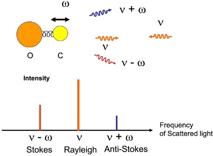
SOURCE: Image by Marlan Scully.1
The Raman effect is embodied by the coherence between a molecule’s ground state and the first excited state. However, different molecules emit the same scattered frequency, making it hard to tell them apart (Pestov et al., 2007). Manipulating lasers via FAST CARS enhanced the signal and suppressed noise, and when FAST CARS was applied to surface Raman, it created astonishing improvements in sensitivity (Lis and Cecchet, 2014; Voronine et al., 2012).
COVID-19 Applications
Scully and his collaborators developed an enhanced technique, Femtosecond Adaptive Spectroscopic Technique with Enhanced Resolution for Coherent Anti-Stokes Raman Scattering (FASTER CARS) and applied it to identify SARSCoV-2, the virus that causes COVID-19 (Deckert et al., 2020). FASTER CARS can scan a single RNA or DNA strand and measure the Raman signal to accurately determine the sequence of its nucleotide bases (He et al., 2020). Scully’s team is currently investigating improvements to this technology to better understand the mechanics and increase its accuracy.
___________________
1 Thank you to Dr. Marlan Scully for allowing us to use his image.
Superradiance
Another topic under study is whether superradiance has any function in the brain, one of many ideas put forth by Fröhlich that several other researchers are pursuing (Fröhlich, 1968; Nardecchia et al., 2018; Reimers et al., 2009; Zhang et al., 2019). Scully outlined the differences between superradiance and laser coherence. Laser coherence is a dynamical stimulated emission, whereas superradiance is spontaneous, cooperative, and possibly coherent. The possibility of quantum coherence and superradiant states in brain microtubules inspired Scully to investigate timed Dicke states to create superradiance over long distances (Celardo et al., 2019; Jibu et al., 1994; Mavromatos, 2011; Scully and Svidzinsky, 2009). Scully said superradiance is still a very open topic, with exciting research advancements on the horizon.
Discussion
Taekjip Ha, Johns Hopkins University, asked about decay times. Scully responded that the superradiant state decays very quickly, before environmental phonon-induced decoherence can cause decay. He and Ha agreed that the short decoherence time may help couple quantum and biological processes.
When asked about the broader applications of this work, Scully replied that his team is one of several investigating improvements to antibody detection and methods to study SARS-CoV-2 surface proteins (Peng et al., 2020).
QUANTUM-ENABLED ELECTRON MICROSCOPY FOR BIOLOGICAL STUDY
Electron Microscopy and Cryogenic Electron Microscopy Development, Technologies, Applications, and Challenges
Elizabeth Villa
Cryogenic electron microscopy (Cryo-EM) could become the highest resolution technique for cell biology. For the first part of this joint keynote presentation, Villa discussed the development of the electron microscopy (EM) and cryo-EM technologies, current cryo-EM capabilities, and biomedical applications and challenges.
EM and Cryo-EM Development
Early electron microscopes were pioneered almost 100 years ago (see Figure 3-2) and led to the development of several tools enabling increasingly sophisticated three-dimensional (3D) reconstructions of two-dimensional (2D) biological images (Driest and Muller, 1935; Dubochet et al., 1988; Henderson and Unwin, 1975; Klug and Finch, 1965; Taylor and Glaeser, 1973; van Heel and Frank, 1981). Today, researchers can use single-particle cryo-EM to solve the structures of macromolecules with atomic resolution. In this technique, hundreds of thousands of structurally identical macromolecules are isolated, flash frozen in different spatial orientations on a monodispersed layer and photographed to create 3D reconstructions.
Solving structures with this level of atomic resolution can improve understanding of biochemical processes and aid drug design. However, the samples are very low contrast, making it hard to distinguish the images. Using higher doses of radiation to create higher contrast damages the materials, which are already very sensitive to radiation. Continuing to improve the quality of EM images requires finding a careful balance between higher contrast and radiation damage (Grant and Grigorieff, 2015).
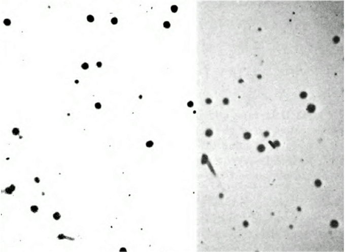
SOURCE: Courtesy of the U.S. National Library of Medicine.
After the “Resolution Revolution”
In the early 2010s, the cryo-EM “resolution revolution” brought faster direct detector devices and the ability to create high-resolution “movies” to review structures (McMullan et al., 2014). The signal-to-noise ratio (SNR) also increased, allowing the application of sophisticated algorithms to analyze data (Zhang et al., 2017). Cryo-EM leaped to the forefront of structural biology, without any advances in optics, and still has many facets where there is room for improvement. Adding quantum effects to cryo-electron tomography, an imaging technique used to make 3D reconstructions of entities such as cells, can allow for the analysis of thicker samples that have more crowded environments and low SNR (Villa et al., 2013).
EM Applications and Challenges
Villa’s laboratory uses cryo-EM to study the Leucine-Rich Repeat 2 (LRRK2) protein, the most common mutations in genetically-driven Parkinson’s disease, and thus a major drug target. Until recently, researchers have been unable to obtain its structure (Gaskill, 2019). By leveraging this protein’s microtubule-binding properties, Villa’s team successfully obtained the LRRK2 structure. The process involved computationally extracting sections of LRRK2-decorated microtubules, increasing resolution by subtomogram averaging, then using integrative modeling to determine the protein’s architecture and study its interactions (Deniston et al., 2020; Kett et al., 2012; Watanabe et al., 2020).
Cryo-EM technologies have many other exciting applications, but there are challenges; studying macromolecules, including LRRK2, and their cellular pathways would be easier with higher-resolution tomography, minimal radiation, and improved SNR. Addressing those challenges will enable researchers to examine the entire molecular landscape in cells, transforming structural cell biology, Villa said.
EM Improvements, Applications, Challenges, and Opportunities
Karl K. Berggren
For the second part of this joint keynote, Berggren discussed improvements to EM, EM applications, and challenges and opportunities in this field.
Improvements to EM
Early EM designers were concerned about damage, quantum mechanics, and resolution limitations. However, resolution in biological systems is primarily limited not by optics, but rather by the maximum allowable dose of radiation
(Egerton, 2014). To make a microscope that causes less damage to biological samples, researchers turned to quantum mechanics, quantum measurements, and deeply counterintuitive concepts such as the quantum zeno effect, interaction-free measurement (IFM), and squeezed states of light (Bell, 1964; de Broglie, 1924; Degasperis et al., 1974; Einstein et al., 1935; Heisenberg, 1927; Yuen, 1975). In particular, IFM, which detects a sample without interacting with it, can be exploited with Mach–Zender interferometry and electrons (Agarwal et al., 2017; Elitzur and Vaidman, 1993; Turner et al., 2020).
EM Applications
Researchers have taken strides toward developing a microscope that effects a quantum zeno process by creating coupled waveguides and using mirrors to effectively create identical copies of a sample (Kwiat et al., 1995; Putnam and Yanik, 2009). This led Berggren and collaborators to pursue using free-space electron optics (including electron mirrors) for quantum EM (Kwiat et al., 1995; Putnam and Yanik, 2009). Finally, research suggests that an EM that does not require electron-splitting or a qubit-based system could be used as a coherent probe of quantum systems (Juffmann et al., 2017; Okamoto, 2012).
Challenges and Opportunities
Berggren’s electron mirror could be developed for in-depth biological or environmental applications. Entanglement and quantum coherence also offer opportunities to improve performance and broaden scientific approaches. However, there are challenges. Electron optics such as mirrors, switches, and appropriate beam splitters are not widely available, and theoretical work around black-and-white versus grayscale approaches needs further development.
Discussion
Prineha Narang, Harvard University, moderated a discussion between attendees, Villa, and Berggren that addressed quantum-enabled EM and tracking coherence and cell interactions.
Quantum-Enabled EM
Narang asked how to build quantum-enabled EM for imaging cells. Berggren replied that Juffman and colleagues’ (2017) multipass approach combined with multiparticle EM is the most promising technique to improve SNR and build real quantum tools in the next 5 to 10 years, although it may not work with fixed or thick samples. Berggren said that EMs with single-pass and multipass IFM could
see small-percentage improvements in SNR, but an idea analogous to squeezing is needed to go further. Prem Kumar, Northwestern University, noted that increasing dosing and resolution can be problematic, and Berggren agreed that those were issues that needed more analysis, although he said that he is nevertheless convinced that nonclassical mechanisms are involved.
In response to a question, Berggren noted that challenges to using quantum systems to image every single molecule in a cell include a lack of instrument automation (especially for slicing thin films), unknown data pathways, and the arbitrary orientation of molecules. Villa agreed, adding that if quantum systems can enable a balance of SNR and dosing, they could create pattern-matching technology improvements over the current exhaustive 2D matching process. Villa noted that a transformation in data quality is also needed to bring this goal closer to reality, but as the field’s landscape currently stands, it is expensive and likely out of reach.
Philip Kurian, Howard University, asked if it mattered whether protein data bank files were derived from cryo-EM instead of x-ray crystallography. Villa replied that the process was less important than the resulting resolution and interpretation of the structure, which rely on original data and can be of varying resolution depending on the sample. One important advantage to cryo-EM is that this method can more easily examine large and complex samples, such as membrane proteins.
Tracking Coherence
Asked what would help improve coherence with EM, Berggren responded that lens scale and adaptive measuring tools are more important than the EM itself for tracking coherence. In the initial stages, the resolution limitations will be challenging, but adding computing power and prior information will create SNR improvements and enable subangstrom resolution that could improve coherence.
Cell Interactions
When asked if the hierarchy of cell interactions was settled, Villa replied that exactly how electrons interact with biological matter is an open question with no consensus yet, for example, in the case of beam-induced motion. There is an opportunity to enhance the qualitative study of how imaging is affected by the interactions of the electron beam with the vitrified sample.
QUANTUM PRINCIPLES FOR ENHANCED MEASUREMENT AND IMAGING IN MICROSCOPY
Prem Kumar, Northwestern University, moderated the workshop’s fourth session and gave a short introduction to the panel topic. The other panelists were Theodore Goodson III, Richard Barry Bernstein College Professor of Chemistry and Molecular Science and Engineering at the University of Michigan; Ted Laurence, deputy group leader for laser materials interaction science at the Lawrence Livermore National Laboratory; and Kevin Eliceiri, investigator at the Morgridge Institute for Research and associate professor of biomedical engineering at the University of Wisconsin–Madison and leader of the Laboratory for Optical and Computational Instrumentation at the University of Wisconsin–Madison Carbone Cancer Center.
Quantum Imaging
Prem Kumar
The concepts of squeezed light and quantum imaging microscopy have existed for decades (Kolobov, 1999; Kolobov and Kumar, 1993; Kumar and Kolobov, 1994). Kumar said that key issues in the field include raster casting versus multimode image rendering, ghost imaging and its variants, quantum illumination, and the Laser Interferometer Gravitational Wave Observatory, which is unusual because it can go beyond unique quantum effects (Barzanjeh et al., 2015; Ferri et al., 2010; Lloyd, 2008; Padgett and Boyd, 2017; Shapiro, 2020; Shapiro and Boyd, 2012; Shih, 2008; Tsang, 2013).
Some concepts are progressively more quantum than classical (see Figure 3-3). For example, superposition can occur as a classical behavior, but a superposition of single particles, where particle duality comes into place, is on the classical–quantum boundary. Concepts such as quantum computing are truly quantum when they require superposition, measurement, and entanglement. Kumar said that progress in imaging and microscopy will require innovation at the classical–quantum boundary despite fundamental quantum limits and varying environments.
Finally, Kumar urged attention to fair comparisons between classical and quantum approaches by considering improvements at a system level: a quantum digit offering 1,000× improvement should not be chosen over a classical digit offering 1,000,000× improvement. He also noted that there are unique scenarios where the quantum nature of the digit takes a higher priority, and he urged the consideration of all difference factors when making decisions about the utility of different methods.
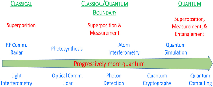
NOTE: RF Comm. = radio frequency communication.
SOURCE: Image by Prem Kumar.
Quantum Light Spectroscopy
Theodore Goodson III
Goodson’s laboratory uses quantum light spectroscopy with photon entanglement to create new tools for investigating biological systems. This emerging field is an alternative to classical excitation, with both challenges and promise for biological imaging, especially if coupled with theoretical advances in quantum light.
Quantum light spectroscopy with entangled photon pairs has many uses. As Goodson noted, these include the high degree of correlation that can be leveraged to achieve enhanced sensitivity; entanglement transfer that could enable new material states; entangled photon pairs that could enable new dimensions to study, such as time, area, pathways, or dynamics; new molecular properties that could be activated and probed; and noise characteristics that can be improved. In addition, the lower number of photons needed to see quantum effects resolves past issues such as tissue transparency.
Goodson’s team has studied two-photon absorption and linear dependence in organic molecules, entangled-photon fluorescence, and biological systems such as flavoprotein structures. They have used those findings to construct a microscope that offers nonlinear imaging capabilities with excitation intensity 106 lower than that necessary for classical light (Eshun et al., 2018; Harpham et al., 2009; Lee and Goodson, 2006; Upton et al., 2013; Varnavski and Goodson, 2020; Varnavski et al., 2017; Villabona-Monsalve et al., 2018).
Quantum Principles for Enhanced Measurement and Imaging
Ted Laurence
Developing correlations from quantum measurements, such as single photon counting, fluorescence transitions, and lasers, can take longer to produce the same results as classical measurements. They do not require calibration, however, and can enable new research areas, such as photon antibunching and quantum-entangled photons. For example, fluorescence correlation spectroscopy was combined with photon antibunching to determine the number of apolipoprotein A-1 proteins in one nano lipoprotein in a hydrated state (Ly et al., 2011). Laurence suggested that antibunching could be impacted by cooperative effects such as superradiance.
Laurence’s group is also pursuing ghost imaging, a technique where light being measured has never directly interacted with the object of interest, using correlations between entangled photon pairs and polarizations to produce an image. Ghost imaging produces the same images as classical means but could provide different observables with quantum measurements, higher resolution, and higher sensitivity. The two detectors require a correlation, however, which presents a challenge; in addition, Laurence said that higher-rate sources, better detectors, and better detection strategies are needed.
Imaging Cell Autofluorescence for Clinical Treatment Guidance
Kevin Eliceiri
Quantum optics in cell imaging has potential for improving the understanding of the role that cells and their autofluorescent signatures play in cancer progression. Adapting multiphoton-based technologies to exploit intrinsic metabolic signatures from human tumor biopsies, Eliceiri’s team has found that the levels of nicotinamide adenine dinucleotide hydrogen (NADH), an autofluorescent coenzyme that can be both free and protein-bound, can detect the Warburg effect, one of cancer’s principal metabolic actions (Stringari et al., 2017; Yu and Heikal, 2009).
These multiphoton-based imaging techniques can achieve very specific excitation at the focal spot, deeper imaging, and better viability than one-photon methods. However, the required low excitation intensity can limit imaging speeds and imaging depth and there is a risk of photodamage. In addition, better techniques are needed to use FLIM to differentiate nicotinamide adenine dinucleotide phosphate hydrogen from NADH, Eliceiri noted.
Engaging with the quantum community could solve some of these problems and bring enhanced tools to biological imaging. Autofluorescence-based quantum imaging could improve optical sectioning, depth, and viability. Several studies have demonstrated proof of concept of entangled photon microscopy but
not yet in a biological context (Lemos et al., 2014; Magaña-Loaiza et al., 2019; Scarcelli and Yun, 2007; Varnavski and Goodson, 2020).
Discussion
Kumar moderated a discussion that covered quantum light receivers, quantum light resolution, and sensitivity and entangled light.
Quantum Light Receivers
Bern Kohler, The Ohio State University, asked about the best receiver for quantum light spectroscopy. Goodson answered that for two-photon spectroscopy, he has tried aggregates, polymers, dimers, and more. He has found that what is most important is the ability to image large molecular cross-sections and understand energy levels, excitation, and intermediate states. Laurence added that for one-photon excitation, nothing special is needed.
Kumar asked the panelists what would enable quantum light to advance practical quantum sensing and imaging. Goodson replied that theory and experimentation need to create a better shared understanding of the structural and functional relationship between molecules.
Quantum Light Resolution
Asked about the theoretical resolution of quantum light techniques, Goodson replied that it could be used for spatial resolution, but would not necessarily create dramatic increases. A bigger impact in biological systems would be a reduction in the number of photons, although the field is still in the early stages.
Sensitivity and Entangled Light
Kumar, asking about sensitivity, reiterated that quantum does not always have a speed advantage over classical techniques. Goodson agreed, adding that low efficiency is one challenge of using entangled photons, and low-light excitation is still out of reach. Kumar suggested using the low number of photon pairs in innovative ways, and Goodson supported this idea, noting that nonclassical light is still an emerging area.
Berggren asked how relevant the transparency window is for entangled light, and Goodson replied that while the issue is still being investigated, it appears to be a trade-off between the molecule absorption and the number of photons. Photodamage can be extensive, and reducing it requires shifting the wavelength and examining different spectral regions outside the linear absorption to find molecules that have promising entangled two-photon cross-sections.
BROADBAND SPECTROSCOPIES OF COLLECTIVE DYNAMICS IN BIOLOGY
Narang moderated the workshop’s fifth session, which focused on how the nonclassical properties of light can be used in metrology, microscopy, and biological system dynamics to improve quantum-enhanced sensing and imaging.
The panelists were Philip Hemmer, professor of electrical and computer engineering at Texas A&M University; Prem Kumar, Northwestern University; Kim Lewis, professor of physics and associate dean of research at Howard University; and Michelle O’Malley, professor in the Department of Chemical Engineering at the University of California, Santa Barbara.
Nanoparticles for Biosensing
Philip Hemmer
Nanoparticles can be used to amplify a target molecule’s signal to enable identification and tracking. They offer advantages over fluorescent proteins, but also have some disadvantages. For example, although nanoparticles last longer than fluorescent proteins and are less prone to bleaching or blinking, they cannot be programmed into or expressed by genes the way proteins can.
For magnetic sensing, nitrogen-vacancy-center (NV-center) diamonds have proved effective because they have up to 30 percent suppressed fluorescence, depending on magnetic spin state. This large fluorescent contrast is what distinguishes them from the competition, but its precise mechanism is still not known (Nizovtsev et al., 2003). Nonetheless NV-center in diamonds has already been applied to magnetic resonance imaging (MRI), a superresolution technique, by replacing the complex MRI apparatus with a single molecule that can sense external spins and image molecules (Grotz et al., 2011). Researchers are still working on a way to image complex molecules, and Hemmer suggested that novel color centers in diamond are a promising avenue.
Hemmer’s team also avoids biofluorescence interference by using upconversion nanoparticles. These particles sense temperature and certain biochemicals with high sensitivity (Hao et al., 2011). Here multimodal sensing is critical for accuracy because it identifies false signals. In the future, nanoparticles may also work in cell membranes, for electric field sensing. Hemmer aims to create novel multimodal sensors by growing diamond shells around upconversion particles and vice versa. Hemmer pointed out that these and other sensors have potential for quantum-enhanced imaging applications (Kolesov et al., 2012), but to use them to look for innate quantum effects in biological systems will require new protocols.
Generation of Photonic Entanglement in Green Fluorescent Proteins
Prem Kumar
Genetic engineering techniques may be able to optimize physical characteristics that are quantumly, but not classically, heuristic. Kumar’s group studies quantum biology techniques and processes, such as photosynthesis and light harvesting, that have nonbiological applications (Engel et al., 2007; Li et al., 2005; Maeda et al., 2008; Sarovar et al., 2010; Sharping et al., 2006).
Because green fluorescence proteins (GFPs) can be used to generate entanglement, they are a promising method for creating a heuristic approach to study new sources of entangled photon pairs for nonbiological applications (Shaner et al., 2007; Yang et al., 1996). Although their molecular structure and behavior are still not fully understood, polarization filtering does improve the coincidence-to-accidental ratio (a measurement reflecting the number of pulse excitations of four-wave mixing in which a coincidence count was detected in the signal and idler arms versus what could have been attributed to accidental coincidence) (Shi et al., 2014, 2016). Knowing that photon pairs can be generated from a protein, Kumar’s team, turning energy time entanglement into polarization entanglement by delayed choices, was able to create and measure the quantum interference of the generated entangled photon pairs (Shi et al., 2017, 2018). Future collaborations will explore why GFPs performed better than ordinary dyes and how they can be fused to other biological systems.
Broadband Spectroscopies of Collective Dynamics
Kim Lewis
Scanning tunneling microscopy, conductive atomic force microscopy, electromigration, and inelastic electron tunneling spectroscopy (IETS) are techniques to study electron transport and nanoscale junctions in order to build molecular electronics (i.e., electronic circuits that behave like conventional silicon devices). With these techniques, researchers have identified conductance mechanisms, extracted the electron attenuation coefficient, and investigated electron-phonon coupling and molecular vibration modes (Esposito, 2017; Saha et al., 2011).
IETS, the most relevant of these techniques to biology, tracks molecule changes in nanojunctions, enabling identification of vibrational modes linked to electron transport or electron-phonon coupling, to prove the presence of a molecule in a junction (Troisi and Ratner, 2006). Challenges abound: a well-defined molecular junction has yet to be fabricated, good results require very low temperatures in a vacuum, there needs to be a way to ensure that measurements come from the device in use, and molecules need to be properly attached to electrodes. Once those challenges are met, theoretical calculations are needed to
determine chemical structure or bonds. Finally, once scientists can understand and control a molecule, Lewis said, new instrumentation will be needed to create short- and long-term biological sensing and imaging applications.
Unlocking the Biotech Potential of “Weird” Microbes
Michelle O’Malley
Better, noninvasive imaging approaches are needed to understand complicated systems, such as how microbial communities in the digestive tracts of herbivores accomplish lignocellulose hydrolysis, a very complex and slow decomposition process. GFPs do not work well because they require oxygen and this system is anaerobic.
Microbes employ a divide-and-conquer strategy to accomplish big tasks such as carbon recycling and plant and plastic degradation. Better imaging would enable researchers to understand the 3D structures and behavior involved and then replicate it to create value out of commingled waste. Researchers have investigated the process, but many questions remain regarding how enzymes and other proteins break down plant matter, their behavior and unpredictability, polymer resistance, and the role of the environment (Rubin, 2008).
Lignocellulose microbes thrive in the digestive tract of large herbivores, where a very diverse structure of anaerobic microorganisms live in a cross-kingdom relationship (Haitjema et al., 2014; O’Malley et al., 2011). New sequencing tools have contributed some insights, but O’Malley said that more imaging tools are needed to capture gut microbes’ interactions and enable researchers to engineer microbial communities that create added value.
Discussion
Narang moderated a discussion that covered new techniques, isotopic effects and chlorophyll, the brain–gut microbe connection, and vibrational modes and high-temperature limits.
New Techniques
Narang asked the panelists what techniques could be developed to improve the understanding of complex, multicomponent systems. Kumar answered that minimizing loss of all kinds would open a quantum advantage for studying any system. In addition, some molecule behavior is captured in the reconstructive density matrix, which could provide a photonic readout of short-timescale electronic processes. Hemmer added that nanoparticles can follow and study molecules, but they cannot determine quantum properties.
Ralph Jimenez, University of Colorado Boulder, asked if photosynthetic systems required photon-limited sensitive samples. Kumar replied that sensitive
detectors that can measure single photons, pixelate, and accomplish spatial resolution were more important.
Isotopic Effects and Chlorophyll
A participant asked Hemmer if he had studied isotopic effects in NV-center diamonds for biological imaging. He replied that at low temperatures, diamonds without carbon-13s work best.
Brain–Gut Microbiome Connection
Kurian asked O’Malley about the brain–gut microbiome connection, and she theorized that research into how memory proteins and serotonin receptors relay responses between the brain and gut during the divide-and-conquer process would be valuable. One promising idea is to use biosensors made from G protein–coupled receptors to mediate and understand communication.
Vibrational Modes and High-Temperature Limits
In response to a question, Lewis said that it is possible to excite specific vibrational modes, but it depends on the material and environment in use. In addition, IETS has a high-temperature limit of 77 K. At that level, it gets very noisy, and researchers have been testing various methods to reduce the noise. This temperature optimization would be critical for more extensive use of IETS in biological settings.
ULTRAFAST SPECTROSCOPY AND BIOLOGICAL REPORTERS
Kurian moderated the workshop’s sixth session, focused on ultrafast spectroscopy and biological reporters. He noted that intrinsic chromophores could be used in quantum reporting to examine local and distributed quantum effects across the brain and gut, which could have several implications for nutrition and disease. Applying ultrafast spectroscopy to biological systems is moving research to the femtoscale, elucidating the complex, subtle effects of electromagnetic field and relaxation on biology.
The panelists were Majed Chergui, head of the Laboratory of Ultrafast Spectroscopy and founding director of the Lausanne Centre for Ultrafast Science at École Polytechnique Fédérale de Lausanne; Dongping Zhong, Robert Smith Professor in the Department of Physics and professor in the Department of Chemistry and Biochemistry at The Ohio State University; Michelle Y. Sander, associate professor in the Department of Electrical and Computer Engineering,
Boston University; and Bern Kohler, professor in the Department of Chemistry and Biochemistry at The Ohio State University.
Probing Interchromophoric Interactions in Biosystems
Majed Chergui
The deep UV range of light occurs below 300 nanometers, and is important because DNA, RNA, and amino acids absorb at that wavelength or below. To use naturally occurring chromophores to prove biosystem dynamics, researchers have developed two deep-UV spectroscopic techniques with femtosecond to picosecond resolution: one-dimensional (1D) and 2D transient absorption spectroscopy and time-resolved circular dichroism, in addition to broadband fluorescence spectroscopy (Auböck et al., 2012a,b; Cannizzo et al., 2007; Oppermann et al., 2019).
With these tools, Chergui’s group was able to learn more about intraprotein transient electric fields, intraprotein energy–electron transfer and energy transfer, spin and structural dynamics in heme proteins, and excited-state chirality (Bacellar et al., 2020; Consani et al., 2013; Kinschel et al., 2020; Leonard et al., 2009; Monni et al., 2015; Schenkel et al., 2006). Chergui’s team plans to extend these techniques to monitor spectroscopic changes that could be used as markers of drug–target and protein–protein interactions.
Biological Photoenzymes and Photoreceptors
Dongping Zhong
Nature repairs UV-induced DNA damage with photolyases. Using femtosecond spectroscopy combined with absorption and fluorescence, Zhong’s team was able to reverse DNA damage and produce high-resolution detailed movies of the electron tunneling and hopping that took place during the repair process (Li et al., 2010; Zhang et al., 2016). Zhong has also studied photoreceptors with femtosecond spectroscopy to investigate details of light harvesting and energy transfer for dynamic proteins (Li et al., 2020).
In response to a question, he noted that his experiments used adenine, but it is not the adenine itself that is critical, but the unusual fact that its flavin is folded instead of stretched, which has many functions and plays an important role in biology.
Photothermal Material Interactions for Modulation and Imaging Using Infrared Light
Michelle Y. Sander
Infrared nerve manipulation and vibrational infrared photothermal amplitude and phase signal (VIPPS) imaging are two optical techniques based on
spatiotemporal temperature gradients for either label-free neuronal modulation or chemical identification and microscopy. The waveform of action potentials (APs) is reduced or completely blocked by pulsed infrared light (Zhu et al., 2019), impacting neuronal communication and downstream physiological outputs. Blocked APs result in complete and reversible termination of the muscle response while suppressed AP waveforms can recover, leaving the synaptic transmission unchanged (Zhu et al., 2020).
VIPPS imaging can be used to explore structural and chemical composition based on infrared vibrational bands. Relying on thermal lensing effects, detection can occur at visible and near-infrared wavelengths, thus resulting in subdiffraction-limited resolution (Samolis and Sander, 2019; Totachawattana et al., 2015, 2017). In addition, this technique is able to capture, distinguish, and characterize weakly absorbing features and can offer insights into thermal diffusion dynamics (Samolis et al., 2020). When applied to cell imaging, both the nanoscale cell and nuclear membrane can be localized and secondary protein conformation can be monitored (Timmel and Hore, 1996).
These multidimensional, label-free modulation and imaging techniques could be integrated across different platforms to offer novel capabilities to simultaneously analyze biological phenomena across different spatial and temporal resolutions and obtain quantum molecular, cellular, and ensemble information, Sander said.
Photodamage and DNA
Bern Kohler
The constant photodamage of DNA by UV light happens over many timescales and is a problem of great biological importance (Schreier et al., 2007). To learn about the photophysics of monomeric nucleobases, Kohler’s team used femtosecond transient absorption methods and discovered that their excited states have very short lifetimes. These short lifetimes are associated with a lower risk of photodamage and are several hundred femtoseconds in duration (Crespo-Hernández et al., 2004; Middleton et al., 2009; Pecourt et al., 2000, 2001). The special photoproperties of the DNA nucleobases may be the legacy of repeated cycles of absorption, synthesis, and destruction, which led to intrinsically photostable building blocks critical to the start of life on Earth (Beckstead et al., 2016).
DNA is a multichromophoric assembly, and because of this, its structure takes a role in dissipating electronic energy (Ostroumov et al., 2013). Using infrared spectroscopy to detect DNA radicals has provided rich information about charge transfer states (Zhang et al., 2015, 2016a,b). These states move rapidly from more delocalized excited states known as excitons, but the precise events are unclear. There is interest in whether there is quantum mechanical or coherent
transport of DNA excitons that affect photodamage. There are different kinds of coherence, and it is important to consider coherences that arise from natural sunlight. Coherence within eigenstates, a quantum mechanical value, can create delocalized excited states using solar illumination, but sunlight cannot create coherences between eigenstates (Mukamel, 2013). This distinction is important for understanding how electronic energy deposited by sunlight can move about in DNA. Kohler noted that quantum biology effects must be driven by mechanisms that are evolutionarily advantageous.
Discussion
Kurian moderated a discussion covering applications for quantum reporting, longer timescales, UV absorption, channel-forming proteins, and melanin.
Applications for Quantum Reporting
Kurian asked what applications for quantum reporting with intrinsic biological chromophore networks were most promising. Chergui pointed to in vitro studies of interactions between biological systems. Sander added that at the neuronal level, it is important that in vitro experiments optimize the environment and eliminate perturbations to elicit accurate measurements and signal interpretations to be closer to in vivo quantum measurements and scalable to neuronal communication.
Longer Timescales
Noting that it will be important to understand how ultrafast technology can report on longer-scale biological time lengths, Kurian asked the panelists how scientists can bridge the gap between femtoscale measurements and nano or microscale biology. Chergui answered that there are many examples of longer-timescale biological cascades that are induced by an initial action of light-driven processes such as photosynthesis. Kohler added that one challenge of ultrafast spectroscopy is that outcomes, while rare and short-lived, are biologically significant; therefore, the timescales must be bridged.
Zhong stated that for photoreceptors, inducing long-range changes by ultrafast approaches can help, but first it is necessary to understand the initial dynamics and then trace conformation changes. Sander agreed, noting that longer timescales are found in larger biological hierarchies and systems. To image and manipulate these events, it is necessary to understand how individual mechanisms interact.
UV Absorption
Alistair Nunn, University of Westminster, asked about the effect of plant compounds that absorb UV light on electric fields. Chergui noted that in his research, he sensed chromophore response in the UV. Zhong replied that he had the same result.
Channel-Forming Proteins
When asked if she had studied channel-forming proteins, Sander replied that several unknowns in the biophysical mechanisms need to be clarified first. Currently, classical microscopy is used to understand the chain of events that trigger an output, but drilling deeper into the individual protein or channel response level is promising. In addition, her team has considered studying solitonic vibrations, but has not yet been able to monitor them.
Melanin
Kurian asked how Kohler would design an entangled photon experiment with melanin. He replied that melanin is especially challenging, because it is one of the last biopolymers whose complex microscopic structure and individual chromophores are unknown. His team is using spectroscopy to deepen the understanding of melanin, which he said is a very rich, heterogeneous system that overlaps with carbonaceous nanomaterials (Ju et al., 2019).


