
4
Biology for Quantum
The third day had a theme of biology for quantum, which focused on the current state of biological imaging in order to understand where quantum advances might be most useful. Michelle O’Malley, professor of chemical engineering at the University of California, Santa Barbara, gave a keynote on imaging and sensing needs for anaerobic microbes. Following the keynote, there were two panel discussions that covered the topics of current capabilities and limitations in plant imaging, and measurement and sensing needs for microbial communities. During the panels, each discussant was given 7 minutes of opening remarks, followed by a moderated audience question-and-answer session.
EXPLOITING ANAEROBES FOR BIOMASS BREAKDOWN AND SUSTAINABLE CHEMISTRY
Michelle O’Malley
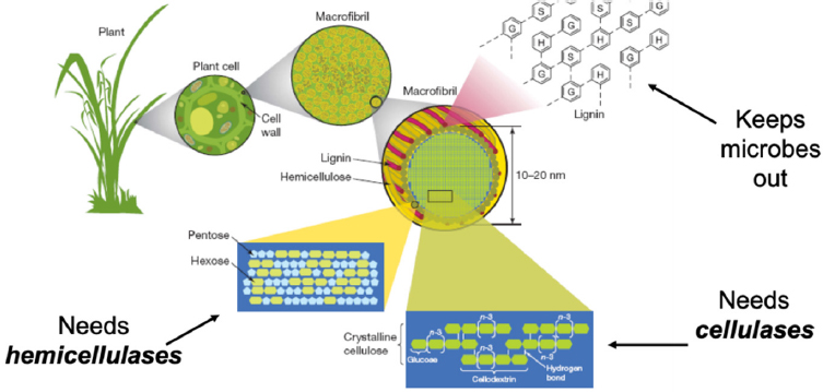
O’Malley started the day with a keynote presentation on research into processes for sustainable chemistry. If researchers could understand and overcome the ability of lignans, energy-rich plant metabolites, to avoid the decomposing processes of microbes and enzymes, they could engineer enzymes that would enable plant waste to become value-added material. Studying these organisms can also fill important gaps in understanding biology and help to engineer sustainable energy sources.
The natural breakdown process of plants is slow, messy, and difficult to study (Rubin, 2008). One promising environment for research is the digestive tract of large herbivores. In this environment, teams of anaerobic microorganisms work in a cross-kingdom partnership to break through lignans to extract nutrition from forage (Haitjema et al., 2014; O’Malley et al., 2011). O’Malley’s group has isolated, investigated, and analyzed anaerobes from the digestive tracts of herbivores, and is now building imaging tools as a step toward bioengineering microorganisms that can degrade plant waste faster (see Figure 4-1).
Isolate and Investigate
First, O’Malley’s team needed to isolate and investigate the actions of biomass-degrading microorganisms. Neocallima stigomycota, a phylum of anaerobic fungi, are primitive and, thus far, found only in large herbivores’ stomachs (Doi, 2008; Grigoriev et al., 2011; Orpin, 1975; Trinci et al., 1994).
SOURCE: Reprinted by permission from RightsLink: Springer Nature, Genomics of cellulosic biofuels by Rubin et al., © 2008.
Studying the growth and behavior of these fungi was very difficult, because there was no genomic information, proteomic characterization, or lab-ready strain available. In addition, these fungi are difficult to grow, cryopreserve, and genetically transform. Overcoming these challenges, O’Malley’s team isolated anaerobic fungal spores directly from their animal sources, cultured them, and sequenced their genomes. Within these fungi’s genomes, they found the largest and most comprehensive array of biomass-degrading enzymes among sequenced microorganisms, a genomic profile that likely enables these fungi to assist in the breakdown of lignans (Haitjema et al., 2017; Henske et al., 2017; Mondo et al., 2017; Solomon et al., 2016). A growing community of researchers is studying these enzymes’ biotechnological potential, but there is still much to learn.
Analyze
Next, her team focused on understanding the function of these biomass-degrading enzymes via transcriptomic- and genomic-based (“omics”) analyses. They discovered unique enzymes that interweave with the substrate they degrade and leave behind unidentifiable materials, but the structure, composition, and enzyme-tethering mechanism within cellulosome scaffolding is still unknown (Gilmore et al., 2015; Haitjema et al., 2014). Combining several “omics” approaches, the team discovered approximately 400 genes involved in the process, most of which are carbohydrate-active enzymes originating in bacteria through horizontal gene transfer (Haitjema et al., 2017).
Build
This work still has many unanswered questions, making the building of tools very challenging. However, O’Malley said that her laboratory is working with the U.S. Department of Energy to build imaging tools capable of nondestructively capturing anaerobic enzyme actions in vivo to demystify the lignan degradation process. They plan to synthesize quantum dots attached to nanobodies, direct these nanobodies to cellulosomes in vivo, image their kinetics via multimodal methods, and adapt existing imaging techniques to create 3D structures (Podolsky et al., 2019). Those structures will then be used as a platform to measure kinetics and enzyme rearrangement. The team is also transforming anaerobic fungi to conjugate quantum dots to cellulosomes for enzyme tracking in real time. More broadly, this process could accelerate in vivo imaging characterization (Lillington et al., 2021).
O’Malley concluded that developing new, real-time, nondestructive bioimaging techniques will advance the understanding of microbe communities, and requires interdisciplinary collaborations and an openness to learning about understudied, real-world systems. The payoff is that these advances can fill critical gaps in biological research and have wide-ranging health, energy, and sustainability applications.
Discussion
Asked if the spatial organization of these microbes mattered, O’Malley answered that she had not studied the question directly, but suspected that microorganism location and spread were very important as they spread out and appear to form films when cultured. In reply to another question, she noted that she had not studied the potential quantum electron transport function of ferritin.
CURRENT CAPABILITIES AND LIMITATIONS IN PLANT IMAGING
Jason West, professor of ecology and conservation biology at Texas A&M University, moderated the workshop’s seventh session, covering advances and limitations for imaging plant biology.
The panelists were Ross Sozzani, professor of plant and microbial biology and director of the plant improvement platform for the North Carolina Plant Sciences Initiative at North Carolina State University; Keiko Torii, Johnson & Johnson Centennial Chair in Plant Cell Biology at The University of Texas at Austin, a Howard Hughes Medical Institute Investigator, and member of the Institute of Transformative Biomolecules at Nagoya University in Japan; and Christopher Topp, assistant investigator at the Donald Danforth Plant Research Center.
The Goldilocks Principle: Just the Right Amount of Growth
Ross Sozzani
The root of the model plant Arabidopsis has many features that reduce complexity, making it a tractable system to study (Fisher and Sozzani, 2016). It also presents biologists with the opportunity to collaborate with mathematical modeling and imaging experts, whose work is crucial to learning more about biological systems, improving imaging techniques, and advancing the field.
One key aspect of Arabidopsis root stem cell regulation is its transcription factors. These move between cells and form protein complexes with different stoichiometries to create the “just right” conditions for stem cell division (Benfey et al., 1993; Cruz-Ramírez et al., 2012; Di Laurenzio et al., 1996; Sozzani et al., 2010). Quantifying the movement of transcription factors makes it possible to create space, time, and stoichiometric maps, and fluorescence correlation spectroscopy sheds light on transcription factors’ diffusion coefficient, oligomeric state, and protein interactions (Clark and Sozzani, 2017; Clark et al., 2016).
Quantitative measurements of transcription factors’ specific movements also show differential representation of both the movements and the factors’ interacting partners, justifying close examination of cell types’ specific mechanisms (Clark et al., 2020). In addition, the measurements make it possible to create accurate multiscale hybrid models at the molecular and cellular level that track and predict cell divisions, and then validate those predictions (Van den Broeck et al., 2021). Imaging these movements required building microfluidic devices and software to track cell division (Buckner et al., 2018, 2020; de Luis Balaguer et al., 2016; Madison et al., 2020).
Visualizing the Cellular Decision-Making Process During Plant Epidermal Development
Keiko Torii
Stomata, a plant’s epidermal pores, can teach researchers important lessons about how cells differentiate. When a precursor cell divides into stem cells and neighboring cells, either could become stomata, but at some point this flexibility ends and a cell’s fate is determined. During this time, the plant cells cannot move or rearrange themselves, and the process cannot be regained, even if researchers disturb it.
Torii’s team learned that the key regulators of this process are the polarity component and peptide signaling that enforce proper transcription factor actions. To better understand peptide functions, Torii and collaborators developed a tool that visualizes peptides to monitor cell activity and determine how receptors move, a collective set of actions that determines the cell’s fate (Zeng et al.,
2020). Some of this process can be visualized in real time or with time-lapse imaging, but will require bridging the imaging technology gap for visualization over different scales.
Technical challenges to this work include strong autofluorescence from chlorophyll, organelle behavior, and interference from some of the techniques employed, such as optogenetics or optophysiology, which can affect a plant’s ability to sense and respond to low-level light. In addition, most imaging tools are optimized for animals, not plants. More collaborations with physicists, chemists, computer scientists, and engineers are needed to use quantum imaging to more fully understand plant cell dynamics.
Deep Phenotyping of Root and Rhizosphere Biology
Christopher Topp
Understanding the molecular and growth processes of plant roots, and how they affect whole-plant functioning, holds enormous potential for enhanced crop productivity and sustainable agriculture. Modern agriculture’s drive for high yields with unsustainable inputs ignores the plant’s root system, which provides all of the water and nutrients for the shoots and grain (Donald, 1968). As a primary source of carbon, roots also drive physical, chemical, and biological changes in the soil.
Studying roots is very difficult, especially given that methods to access and observe root systems in the field have not substantively changed in the last century. Furthermore, root–soil–microbe interactions are complex and dynamic, one primary root can lead to hundreds of thousands of roots, and each root has local temporal processes and interactions that add to the macro-level process. A better understanding of time dimensions will enable the creation of models for interactions, growth patterns, and environmental conditioning.
Through x-ray microscopy and large-format, high-resolution x-ray tomography, Topp is able to identify root systems’ structural details, functional information, and interactions (Duncan et al., 2020). Using positron emission tomography, it is possible to measure a whole plant’s carbon and nitrogen allocation dynamics in real time, as well as other dynamic physiological processes, although the resolution is not fine enough to enable differentiation.
Current limitations, such as studying plants in natural soil, could be overcome with emerging quantum technologies such as quantum dots or other nanoparticles. Topp’s lab is working to capture the 3D structure of an entire freely grown root system via multimodal sensing to create digital models that can be used to study plant–environment dynamics. The ability to bridge timescales and design functional, high-resolution imaging systems would enable in situ, thick-sample imaging and sensing and further advance knowledge of whole-plant, root and shoot, systems.
Discussion
West moderated a discussion that covered tools, facilitating collaborations, and remote plant sensing.
New Versus Existing Tools
West asked the panel which was needed more—new tools or adaptations to existing tools? Sozzani replied that both were necessary. Some animal tools work well for plants, but she suggested that scientists should collaborate with engineers to develop plant-specific sensing tools as needed, especially because they could have a large societal and economic impact.
Torii agreed, adding that she has learned about good tools from both animal scientists and stem cell biologists. There are challenges, such as timescale and detecting underground behavior, but large, free-growing plants cannot yet be studied in the wild. Tools that bridge timescales, combine and analyze data, and are optimized for plants will make the study of whole-plant organisms possible, she suggested. Topp also agreed that plant science can and should adapt advanced medical tools, with the understanding that there will be limitations.
Facilitating Collaborations
West asked how best to facilitate collaborations to advance research in this space. Torii replied that the best collaborations happen when the project has mutual advantages that are able to show off everyone’s talents, each team finds the work interesting, and partners develop a camaraderie to pursue new knowledge.
Sozzani suggested that funding agencies, which are starting to understand that the most impactful collaborations integrate basic and applied research, could recommend including an industry partner to prioritize solution-driven science. Topp added that funders should recognize that this work requires continuous long-term commitment and mutual learning. In addition, he said collaborations should be incentivized and recognized, because successes can elevate each field.
Remote Plant Sensing
Margaret Ahmad, Sorbonne University, asked if remote plant sensing was possible. Topp replied that reporters attached to autonomous robots and low-flying drone sensors, built in collaboration with engineers, have worked well. Not every prototype has been successful, and investment here is risky, but more tools, and experts who can help build them, are needed.
Sozzani agreed, adding that in addition to time and expense, sensing tools create questions about data, data management, and cyber infrastructure. On the
plus side, data enable predictions, and if plant science becomes more interdisciplinary, it can take advantage of those abilities. Torii also agreed, noting that hyperspectral camera drones are being tested for monitoring plant health, but more analysis and machine learning techniques are needed to correlate images and measurements and provide meaningful information.
MEASUREMENT AND SENSING NEEDS FOR MICROBIAL COMMUNITIES
Jennifer Pett-Ridge, leader of the Environmental Isotope Systems Group at the Lawrence Livermore National Laboratory, moderated the workshop’s eighth session, which focused on critical knowledge gaps in the understanding of microbial communities and their interactions in the environment, and the tools needed to address these gaps.
The panelists were Victoria Orphan, James Irvine Professor of Environmental Science and Geobiology, Allen V. C. Davis and Lenabelle Davis Leadership Chair at the Center for Environmental Microbial Interactions, and director of the Center for Environmental Microbial Interactions at the California Institute of Technology; Alice Dohnalkova, environmental microbiologist in the Environmental Molecular Science Laboratory at the Pacific Northwest National Laboratory; and Elizabeth Shank, professor of microbiology and physical systems at the University of Massachusetts Medical School.
Syntrophic Interspecies Microbial Interactions and Extracellular Electron Transfer in the Environment
Victoria Orphan
Orphan studies how microorganism interactions—specifically the direct passage of electrons between organisms known as extracellular electron transfer—affect the cycling of carbon and nutrients through the biosphere. This cycling process is known to be important in microbial metal respiration and was recently discovered to be important to interspecies syntrophy, the process by which different species pass metabolites and nutrients to each other. For example, in methane-oxidizing archaea, the full range of this process is responsible for sequestering up to 80 percent of methane flux from ocean sediments.
These microorganisms are challenging to study because they are uncultured and live in deep-sea sediment in diverse communities, coexisting with hundreds of other species. Orphan and colleagues use multiple techniques to identify, isolate, and glean knowledge about interaction constraints of extracellular electron transfer within methane-consuming syntrophic associations (He et al., 2021). In the future, her team plans to use multimodal imaging to study these
microorganisms at different scales in their environmental context, one of the Grand Challenges of environmental microbial ecology.
In situ microbial communities are inherently complex, and to study them will require field-portable, nondestructive, precise tools to identify microbes, track their dynamics, and image and sample them in opaque environments over time, space, and varied chemical, mineral, and metabolic processes. Although technologies such as quantum dots and gas vesicles are promising, Orphan noted that these are still in the very early stages of development, and there exists a need for more advanced sensing capabilities to tackle these challenges (Farhadi et al., 2019; Whiteside et al., 2019).
Revealing Mechanisms for Stabilizing Organic Carbon by Microbial–Mineral Interactions: Innovative Chemical Imaging
Alice Dohnalkova
Dohnalkova uses electron microscopy, its coupled analyses, multiscale imaging correlated with multiple methodologies, and chemical imaging to study microbial processes in biogeochemical environments such as soil. Although they appear on the macroscale, most biogeochemical processes are driven by micro- or nanoscale processes that must be studied to be understood.
To learn how microbes stabilize carbon in soil, Dohnalkova’s team sequenced and imaged microbial–mineral associations naturally recruited from forest soil. Imaging and analyzing organic macromolecules is challenging with electron beam methods, making correlation a labor-intensive process. Using scanning transmission x-ray microscopy with x-ray absorption near edge structure, a quantum-based method, the team was able to trace the signatures for calcium and carbon in the microbe and the extracellular polymeric substance, demonstrating the role of calcium bridges in carbon stabilization in soils (Lawrence et al., 2003).
With aloof beam electron energy loss spectroscopy, originally developed for materials science, her team was also able to image away from samples (thus avoiding sample degradation) with very high energy resolution, adding more unprecedented insights from microbe–environment interactions. In addition, Dohnalkova noted that machine learning and artificial intelligence techniques, increasingly applied to microscopy, are promising ways to manage the large amounts of data that automated robotics processes generate (Broderick et al., 2018).
Microbial Interactions and Quantum Bioimaging
Elizabeth Shank
Quantum bioimaging may enhance the study of microbial interactions, carbon degradation, and interkingdom interactions in complex, heterogeneous, opaque
environments such as soil. These processes happen on a microscale but have large impacts on an ecosystem’s biogeochemistry, plant health, and soil health.
Natural soil is a challenging study environment for bioimaging. However, Shank’s team can use bioimaging tools on transparent soil model systems (see Figure 4-2) to realistically and nondestructively investigate the physiology and transcriptional states of microbial interactions, and how they affect the larger, heterogeneous environment over time (Downie et al., 2012; Sherrard et al., 2018). Transparent soil also enables the use of fluorescence microscopy, Raman spectroscopy, and mass spectrometry to get a spatial and temporal image of the entire 3D microcosm, including microbes, cells, and the activity of metabolites and molecules (Sharma et al., 2020).
Advancements are needed in identifying and tracking microbes within complex, native communities; imaging multiple organisms simultaneously; combining multiple types of datasets; and imaging more deeply in live samples, over longer timescales, and with wild microbes. Two-photon quantum approaches may be able to image deeper into samples, study larger interaction areas, and identify transcriptional states of different microbes and their heterogeneous gene expression. Quantum dots are promising because they have been used to monitor multiple fluorophores over longer lifetimes, but they can be destructive. These may also be useful in studying carbon degradation because they can track nitrogen movement through plants and fungi, Shank noted (Whiteside et al., 2009).
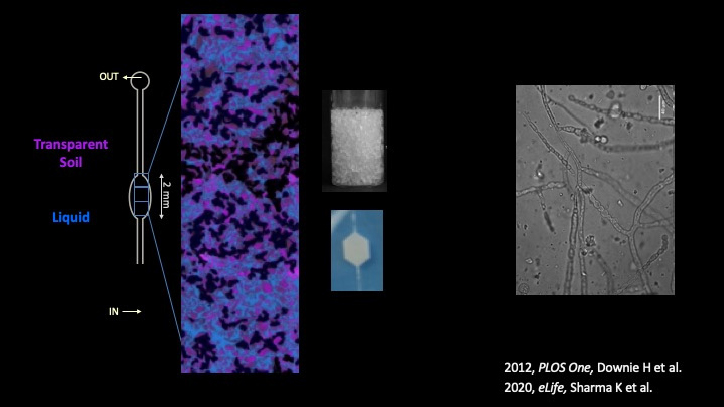
SOURCE: Credit for this image goes to Kriti Sharma and Elizabeth Shank (left); Downie et al., 2012 CC by 4.0 (top middle); Sharma et al., 2020 CC by 4.0 (bottom middle and right).
Discussion
Pett-Ridge moderated a discussion touching on current hurdles, quantum trade-offs, bioluminescence versus fluorescence, short-term interactions, and infrared photodetectors.
Current Hurdles
Pett-Ridge asked panelists to name current hurdles to advancing their work. Orphan pointed to the inability to replicate the wild environment in the laboratory, which necessitates new quantum tools and sensors to image deep structures in real ecosystems. Dohnalkova agreed that the laboratory environment is different, adding that the dehydration process, required for imaging, unnaturally collapses plant systems.
Shank also agreed that laboratory work is extrapolation, although she noted the value of building model systems. She also noted the importance of achieving unlabeled identification of wild microbes, noninvasive and long-term natural-environment study methods, and more sensitive detection methods as critically needed advances.
Quantum Trade-offs
Pett-Ridge asked the panelists if quantum approaches are truly needed, and what trade-offs would be acceptable. Dohnalkova replied that while researchers are pushing the limits, and there is a strong drive for bigger and better tools, many fields may be able to improvise with existing tools.
Shank stated that trade-offs already exist for every technique, so in most places they would be acceptable. As to whether quantum approaches are needed, she expressed ambivalence. Quantum dots could be more widely used, and striving to design new tools can add to an existing suite of techniques for answering different questions, she said.
Orphan agreed that a suite of imaging tools that includes quantum techniques would advance the field, but the biggest hurdle would be to correlate images across different platforms, each with its own limitations and benefits. Adding computational power in the post-processing stage would also advance the field.
Pett-Ridge asked if tools should be more widely accessible. Shank replied that many tools are not used enough to require wider distribution, and there is a value in having experts who understand the tools’ full capabilities. She and Orphan agreed that increased awareness of these tools’ existence could close communication and technology gaps among scientists.
Bioluminescence Versus Fluorescence
Shank noted that she has used bioluminescence as a reporter, and although it is better than fluorescence at detecting weak signals, it is only one-color and thus cannot be used to look at multiple moieties simultaneously, like fluorescence. In addition, microscopes are not set up for bioluminescence, and so imaging and sensing only work for bulk cellular measurement, not individual cells.
Orphan noted that in her work, she gets around fluorescence issues by using deep UV Raman to image mineral substrates outside the usual range for fluorescence. She added that some solutions developed for aerospace applications could be adapted for environmental systems. Pett-Ridge agreed that the current fluorescence techniques are tedious and require customization.
Short-Term Interactions
Pett-Ridge asked how it may be possible to see fluxes and compounds that are short-lived. Orphan replied that some researchers are using genetically tractable organisms as sensors to create readouts. Fast interactions may drive carbon and nutrient cycling, and their study could yield more insight than studying chemical residues. Shank noted that items of low abundance are very difficult to measure or retrieve without disrupting system balance.
Infrared Photodetectors
An attendee asked Shank if infrared photodetectors could help penetrate soil opacity. She replied that it may be possible, and Dohnalkova added that the challenges are with imaging and detecting speed and resolution. Pushing the technology into the femtosecond range, or faster, which is when these processes occur, would be a major development, she added.


