


Below is the uncorrected machine-read text of this chapter, intended to provide our own search engines and external engines with highly rich, chapter-representative searchable text of each book. Because it is UNCORRECTED material, please consider the following text as a useful but insufficient proxy for the authoritative book pages.
214 Repeatability and Reproducibility of FTIR Spectra Evaluating the repeatability and reproducibility of the results produced by the portable Fourier transform infrared (FTIR) equipment was necessary before making any recommenda- tions for the further use of this equipment in the field study in Phase 3. Therefore, these parameters were evaluated in Phase 2 as discussed in the following text. The results of the first round of testing showed that repeat- ability of FTIR results for pure materials and components and simple compounds (i.e., those materials that can be easily âfingerprintedâ) was not an issue. Therefore, the second round of testing in the repeatability and reproducibility study concerned composite materials, such as structural coating systems, pavement markings, portland cement concrete with admixtures, and polymer-modified binders. All of the afore- mentioned products were prepared in the laboratory accord- ing to the proportions summarized in Table P.1. More details on the results for particular materials from Table P.1 are pro- vided in this appendix. Reproducibility Study on Structural Coatings and Pavement Markings All epoxy-based paint samples were first mixed using the component proportions prescribed by the manufacturer. Next, a ready mixture was placed on a metal pad with dimensions of 4 in. by 4 in. and allowed to dry for 24 h. The samples were tested by three operators independently using a Bruker attenuated total reflectance (ATR) FTIR spectrometer. Each operator tested five probes from one sample of each of the three test materials (Carbozinc 859, Scotchkote, and Epo- plex LS50). The FTIR spectra were obtained for each probe and ana- lyzed in terms of repeatability and reproducibility. The repeatability was measured by the coefficient of variation (COV) in the absorbance of the primary components (major peaks) between the probes produced by one operator. The reproducibility was evaluated by COV in mean absorbance between the independent operators who tested the same material. Tables P.2 and P.3 show an example of within and between variations for Carbozinc 859 structural paint. It was noted that the variation in the pressure applied to a sample placed on the ATR crystal before scanning signifi- cantly affected the absorbance values obtained for spectra of Epoplex LS50. This was not an issue for Carbozinc 859 and Scotchkote, which could be explained by the fact that Epoplex produced a thicker and less flexible film than Scotchkote. The stiffer film prevented full contact between the sample and ATR crystal, which interfered with the spectra. This observa- tion should be reflected in developing a test procedure for pavement markings. Repeatability and Reproducibility Study on Portland Cement Concrete with Admixtures This study concerned the repeatability of FTIR scanning of wet portland cement concrete (PCC) samples prepared with air entrainer (Air 200) and retarder (Retarder 75), separately. Three replicate tests were done for each mixture and the vari- ation in absorbance of major peaks (primary components) was evaluated. Tables P.4 and P.5 show an example of within (repeatability) and between (reproducibility) variations of PCC ready mix with the addition of air entrainer. Repeatability Study on Polymer-Modified Asphalt Binders The polymer-modified asphalt binders (PMABs) were pro- duced by adding Kraton linear styreneâbutadieneâstyrene (SBS) in concentrations of 1%, 3%, and 6% to the Nu-Star PG 64-22 neat binder (PG 64-22 East). PMABs were pro- duced by adding a designated amount of SBS to a binder A p p e n d I x p Reliability of Spectroscopic Measurements
215 Table P.1. List of Materials Tested by Attenuated Total Reflectance FTIR in Reproducibility and Repeatability Study Material Category Brand/Material Name Composition Material State Test Objective Structural coatings Carbozinc 859 2Part A : 1Part B:10 Filler 1Part A : 1Part B Powder Reproducibility study Scotchkote 1Part A : 1Part B Solid Reproducibility study Pavement markings 3M White 100% Ready Liquid Reproducibility study Epoplex LS50 Yellow 2Part A : 1Part B Solid Reproducibility study Portland cement concrete with admixtures Lafarge Type 2 cement/ local aggregates/ Air 200/Retarder 75 8.0% Water 17% Cement 31.9% Stone 35.2% Sand 0.2% Air 200 Wet mix Repeatability study Lafarge Type 2 cement/ local aggregates/ Retarder 75 8.0% Water 17% Cement 31.9% Stone 35.2% Sand 0.3% Air 200 Wet mix Repeatability study Reproducibility study Polymer-modified asphalt binders PG 64-22 modified by Kraton SBS PG 64-22 East + 1% SBS PG 64-22 East + 3% SBS PG 64-22 East + 6% SBS PG 64-22 West + 1% SBS PG 64-22 West + 3% SBS PG 64-22 West + 6% SBS Viscous solid Repeatability study Table P.2. Within Variation of Major Band Absorbance for Paint Carbozinc 859 Major Bands (cmî²1) Operator 1 Operator 2 Operator 3 Mean Concentration SD COV (%) Mean Concentration SD COV (%) Mean Concentration SD COV (%) 825â830 4.30 0.84 20 4.79 1.21 25 4.58 0.93 20 1,115â1,120 3.76 0.65 17 3.33 0.79 24 2.06 0.28 14 1,180â1,185 4.65 0.75 16 4.54 0.23 5 4.09 0.77 19 1,235â1,240 15.32 1.29 8 16.37 0.48 3 15.48 3.24 21 1,285â1,290 5.98 0.49 8 5.03 0.45 9 5.36 0.53 10 1,455â1,460 5.27 0.56 11 4.30 0.19 4 4.73 0.14 3 1,505â1,510 12.73 1.13 9 12.20 0.40 3 12.64 0.87 7 within the first 2 min followed by high-shear mixing (with a speed of about 4,500 rpm) at a temperature of 180°C to 195°C for 2 h. Three samples from each batch were scanned by a Bruker ATR FTIR spectrometer and the variability in the absorbance of major peaks (primary components) was ana- lyzed (see Table P.6). The values of COVs in Table P.6, when compared with mean concentration values, clearly indicate an increase in variability with a decrease in the concentration of primary component (major band). In addition, one can notice that the variation in major band concentration associated with SBS (e.g., 966 cm-1 and 700 cm-1) increases from 7% to 13% at 1% SBS content to 23% to 24% at 6% SBS content. This phenomenon needs to be further investigated, because it may affect the results of the quantitative analysis of the chemical composition of polymer-modified binders. For the moment, this phenomenon can be attributed to the higher level of variability observed in absorbance bands of small concentra- tions or to the nonuniform distribution of the polymer net- work inside the binder phase.
216 Table P.3. Between Variation of Major Band Absorbance for Paint Carbozinc 859 Major Bands (cmî²1) Mean Concentration SD COV (%) 825â830 4.56 0.99 22 1,115â1,120 3.05 0.57 18 1,180â1,185 4.43 0.59 13 1,235â1,240 15.72 1.67 11 1,285â1,290 5.46 0.49 9 1,455â1,460 4.77 0.30 6 1,505â1,510 12.52 0.80 6 Table P.4. Within Variation of Major Band Absorbance for PCC with Air 200 Batch 1 Batch 2 Batch 3 Major Bands (cmî²1) Mean Concentration SD COV (%) Mean Concentration SD COV (%) Mean Concentration SD COV (%) 505â515 25.69 0.25 1 25.94 2.83 11 21.81 8.78 40 595â605 4.30 0.74 17 6.14 0.77 13 3.46 2.76 80 665â675 2.12 0.37 18 2.70 1.06 39 2.18 0.62 29 875â885 1.82 0.31 17 2.07 0.34 16 1.40 0.38 27 925â935 3.13 1.06 34 3.32 0.18 5 3.24 0.63 21 1,115â1,125 16.02 1.78 11 19.34 5.01 26 16.61 2.28 14 1,445â1,455 1.72 0.13 7 1.62 0.74 46 1.21 0.78 65 1,640 33.70 2.23 7 24.53 1.35 5 32.51 5.69 18 Comparison of Portable and Stationary FTIR Results The evaluation of the spectroscopic equipment in Phase 2 included a verification of FTIR results obtained with the por- table device (Bruker ALPHA ATR FTIR) by repeating the tests for all materials on the stationary equipment available in the Institute of Material Science at the University of Connecticut (Nicolet Magna 560 transmission FTIR). Two main differ- ences in the procedures should be pointed out: 1. Technological concept. On the one hand, portable FTIR uses the principle of ATR, which measures changes that occur in an internally reflected infrared beam when it comes in contact with a sample placed directly on the infrared (IR) window. On the other hand, the transmission IR measures the IR energy that passes through a sample when it is placed in the transmission window located between the source of IR radiation and the detector. 2. Sample preparation. Regardless of the state of material (liquid, solid, or powder), no sample preparation was made for the portable ATR cell, whereas for transmission FTIR testing, a sample was prepared as a thin film and put between two potassium bromide (KBr) disks. Although a different number of scans were performed on a sample by the portable and stationary spectrometers (16 scans and 24 scans, respectively), the resolution of the scans was similar (1.9 cm-1 and 1.4 cm-1 for portable and stationary FTIR, respectively). Also, the IR spectrum of the sample was analyzed in the same frequency region between 4,000 cm-1 and 400 cm-1. Therefore, it was initially assumed that a direct comparison of the spectrograms obtained with the portable and stationery FTIR equipment was possible. However, the analysis of the portable and stationary spectra revealed that they yielded different absorbance values for the same sample, yet at the same frequency. This is explained by the difference in path length. The path length of the IR window Table P.5. Between Variation of Major Band Absorbance for PCC with Air 200 Major Bands (cmî²1) Mean Concentration SD COV (%) 505â515 24.48 3.95 17 595â605 4.63 1.42 37 665â675 2.33 0.68 29 875â885 1.76 0.34 20 925â935 3.23 0.62 20 1,115â1,125 17.32 3.02 17 1,445â1,455 1.52 0.55 39 1,640 30.25 3.09 10
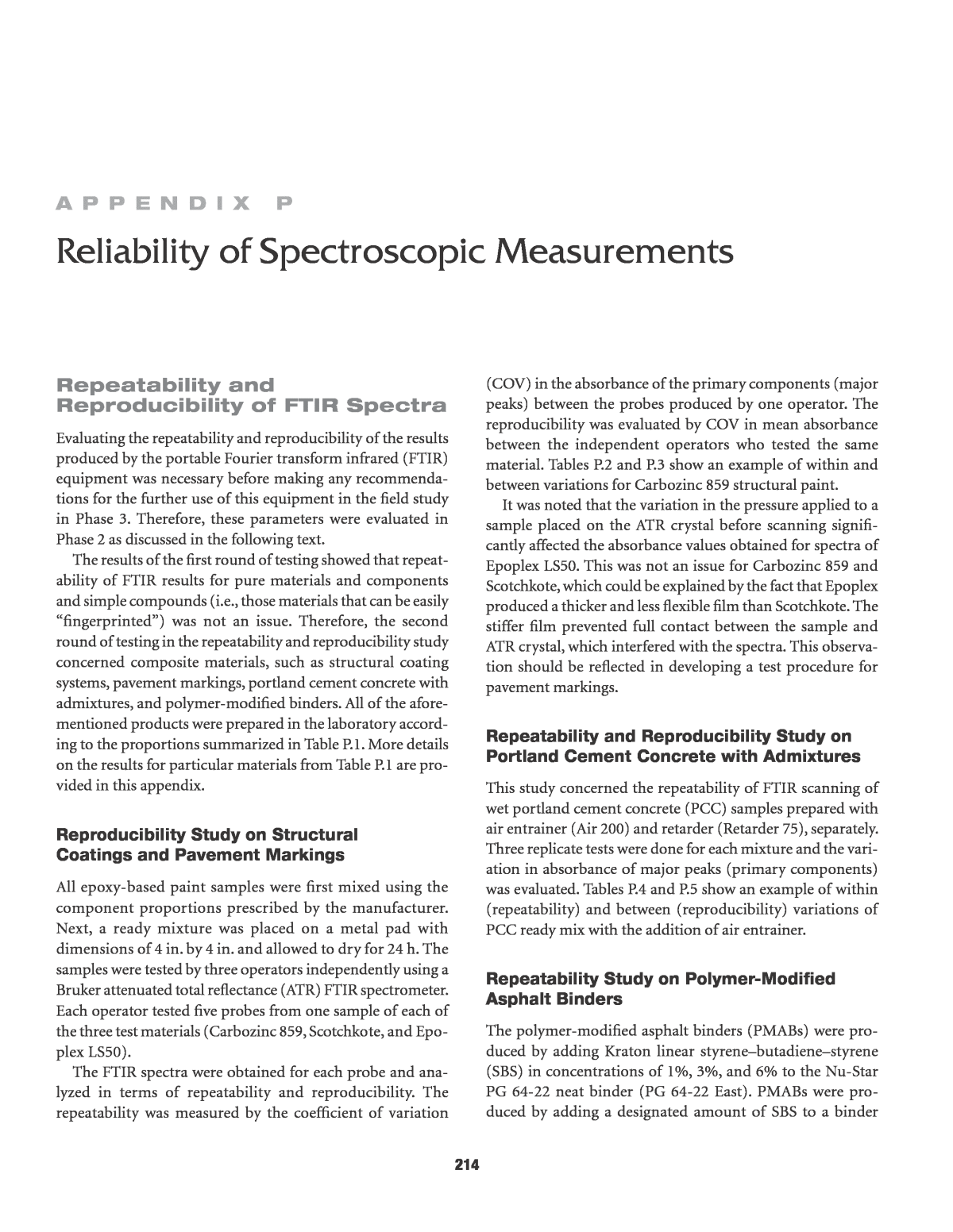
217 varied significantly because of the nonuniform thickness of samples in the KBr pellets, while in ATR refl ectance mode, the path length was set to a constant value of 1 µm. Because the path length of transmission IR samples remained unknown, it precluded a quantitative comparison of the con- centrations of different functional groups as yielded by the two spectrometers. However, it was found that the stationary FTIR (Nicolet Magna 560) identified absorbance peaks at the same wavelengths as the portable ATR FTIR (Bruker ALPHA). There- fore, it was logical to conclude that the portable spectrometer was able to identify the unique spectra corresponding to the chemical composition of all tested materials at the same level of quality as the stationary device. Examples of the analysis for liquid and solid samples are provided below to justify this conclusion. Liquid Sample Figure P.1 shows the fingerprinting region of the IR spectra for Part A of Carbozinc 859 structural coating system with major absorbance bands as identified by the ATR and sta- tionary IR spectrometers. One can observe that the major bands identified by the ATR match those identified by the Table P.6. Summary of Variation of Major Band Absorbance for PG 64-22 Binder with Different SBS Concentrations Batch 1 (1% SBS) Batch 2 (3% SBS) Batch 3 (6% SBS) Major Bands (cmî²1) Mean Concentration SD COV (%) Mean Concentration SD COV (%) Mean Concentration SD COV (%) 2,960 2.00 0.02 1 2.02 0.02 1 1.93 0.05 3 2,921 36.64 0.15 0 36.48 0.37 1 35.54 0.53 1 2,853 14.28 0.05 0 14.31 0.11 1 13.94 0.13 1 1,703 0.47 0.05 11 0.45 0.16 36 0.39 0.11 29 1,601 1.51 0.02 1 1.52 0.02 1 1.53 0.03 2 1,458 22.92 0.27 1 22.67 0.12 1 22.38 0.34 2 1,377 7.48 0.09 1 7.41 0.06 1 7.48 0.11 1 1,310 0.54 0.02 3 0.64 0.09 15 0.65 0.10 15 1,162 0.60 0.08 14 0.58 0.07 12 0.55 0.07 12 1,031 1.00 0.18 18 0.79 0.09 12 0.68 0.04 6 968 0.84 0.11 13 1.37 0.18 13 1.90 0.44 23 866 2.28 0.07 3 2.37 0.13 6 2.55 0.02 1 815 4.19 0.02 0 4.07 0.15 4 4.05 0.22 6 751 1.03 0.04 4 1.05 0.01 1 1.03 0.03 3 725 1.10 0.02 2 1.05 0.05 5 1.07 0.04 4 699 0.08 0.01 7 0.39 0.05 12 0.61 0.15 24 431 0.61 0.05 8 0.67 0.03 5 0.66 0.01 2 stationary IR at exactly the same wavelengths. In addition, the stationary IR spectra clearly show much higher absor- bance values, which indicates that a longer path is followed by the IR beam through the thickness of sample (as was mentioned in a preceding paragraph, the ATR path length is only 1 µm). Solid Sample The major absorbance bands as determined by the ATR and stationary IR for styreneâbutadiene rubber (SBR) latex- modified PG 52-34 binder are compared in Figure P.2. The binder was chosen to illustrate the commonality and dis- crepancies between the results of the two devices when used for the identification of complex mixture, such as a polymer- modified binder. At the first glance, one can see that the major aliphatic bands at ~2,960, ~2,920, ~2,850, ~1,460, and ~1,380 cm-1 are equally identified by the two methods. The aromatic bands at ~1,600 and between ~870, ~815, ~755, and ~724 cm-1 also do not show any discrepancy between ATR and IR spectra in terms of absorbance intensity. A small but significant difference in spectra (see âRegion of differenceâ in Figure P.2) can be identified around ~1,542 and 1,508 cm-1,
218 Figure P.1. Comparison of major absorbance bands identified by ATR and IR spectrometers for Carbozinc 859 Part A. Region of difference Aliphatic Bands Aromatic Bands SBR Latex Figure P.2. Comparison of major absorbance bands identified by ATR and IR spectrometers for PG 52-34 1.5% SBR latex-modified binder. where the stationary IR identifies a small amount of nitrate (-NO2), while the portable ATR ignores these bands. One significant difference shown is the amount of carbonyl (>C=O) at ~1,739 cm-1, which was measured by the ATR to be at a much greater amount than by the stationary IR (minor peaks at ~1,773, 1,736, and 1,700 cm-1). The dis- crepancy in identifying nitrate and carbonyl are most likely connected to the difference in sample preparation and environment during the testing, rather than to the composi- tion of binder. Nevertheless, both portable and stationary spectrometers are able to identify even a small (1.5 wt %) amount of the polymer added to a binder (SBR latex band at ~970 and ~1,230 cm-1). In summary, it is concluded that the portable ATR FTIR spectrometer is capable of producing at least the same quality of IR spectra as a stationary IR spectrometer.
219 Variability in x-Ray Fluorescence Results A two-phase approach was adopted for all materials. Single measurements were first employed to establish proof of con- cept, that XRF can yield measurable results for each material. When an element was detected in prominent concentrations that would not likely have interferences from other field materials and were significantly higher than the method detection limit, XRF was considered a promising quality assurance/quality control (QA/QC) method. In the next step, 10 measurements were obtained for each material in order to determine the precision of the method. It should be noted that, although XRF spectra are generated through software, the user does not see them unless explicitly requested, nor does the user have access to the spectrum manip- ulation to generate data. XRF calibration is a complex process that is performed at the production facility to which the user has no access; one simply performs the measurement and gets a summary table with the quantitative results (element concen- trations in mg/kg for the soil mode and in wt % for the mining mode). Therefore, only quantitative results are provided in this report, as generated by the equipment software. The portable XRF device owned by the team was deemed to be successful to different extents in the evaluation of struc- tural coatings, pavement markings, curing compounds for PCC, portland cement, and aggregates. Therefore, only the testing results for the aforementioned material categories are discussed. Structural Coatings Scotchkote 3M Table P.7 shows the ingredients of this product according to its material safety data sheets (MSDS). XRF can measure the titanium content of this material as the most likely candidate to conduct QA/QC. This material is supplied by the manufacturer as two sepa- rate components: (a) Part A is a clear gel substance and (b) Part B is a green emulsion. These components were tested separately. Table P.8 shows the concentrations of metals detected in both Parts A and B. Based on the results, the gel material appeared to be an organic substrate type of material. Part B was likely represen- tative of the actual mixed coating. The Ti concentration in Part B was found to be 10.8% by weight, with an instrument- based error of 0.1%. This corresponds to a TiO2 concentra- tion of 18.9%. This far exceeds the TiO2 concentration cited in the MSDS; however, the MSDS refers to the final emulsion, not Part B alone. Based on the results, it was concluded that XRF is a useful method for conducting QA/QC of Scotchkote 3M and further testing was initiated on the mixed product. Three separate mixes were prepared according to the manu- facturerâs instructions, as shown in Table P.9. Table P.7. MSDS-Based Composition of Scotchkote 3M Ingredient C.A.S. No. Wt% Di(4-Hydroxyphenol) Isopropylidene Diglycidyl Ether-Di(4-Hydroxyphenol) Isopropylidene Copolymer 25036-25-3 55â75 Quartz silica 14808-60-7 10â20 Potassium aluminosilicate 1327-44-2 5â15 Mica-Group minerals 12001-26-2 5â15 Titanium dioxide 13463-67-7 1â10 Cyanoguanidine 461-58-5 1â10 C.I. pigment green 7 1328-53-6 1â10 4,4â²-Isopropylidenediphenol 80-05-7 0.1â1 Table P.8. XRF-Measured Composition of Scotchkote 3M Materials Tested with Soil Mode Metal Detected Part A (mg/kg) Part B (mg/kg) Cl 698 ± 40 n.d.a K 192 ± 13 596 ± 176b Ca n.d.a 280,721 ± 2,716 Ti n.d.a 108,161 ± 1,079 Fe n.d.a 18,332 ± 177 Co n.d.a 128 ± 27 Cu n.d.a 87 ± 5 Zn n.d.a 7 ± 2 Sr 12 ± 1 56 ± 1 Zr 32 ± 1 n.d.a Mo 39 ± 1 n.d.a Sb 31 ± 9 n.d.a I 50 ± 6 n.d.a Ba n.d.a 1,468 ± 74 Note: Materials were tested with soil mode. a n.d. = not detected. b Standard deviations represent the instrument error. Table P.9. Mixing Design for Scotchkote Testing Mix 5 10 20 Part A 5.0 g 10.0 g 20.0 g Part B 6.65 g 13.3 g 26.6 g
220 Several replicates of each mix were investigated for both the error associated with preparing different mixes and the vari- ability within a single mix. Samples are named accordingly (i.e., Sample Mix # - Replicate #). Additionally, one mix was pre- pared when the mining mode calibration was available to test the difference between the two modes, since the Ti content was in the range of both calibrations. The results are shown in Table P.10. Trace metals are not shown. Standard deviations for each measurement are generated by the XRF equipment and are based on counting statistics. The overall average and standard deviation of the 10 measurements are calculated at the bottom of the table. Data are given in the units provided directly by the XRF (mg/kg for soil mode and wt % for mining mode). The average Ti concentration was 6.33% with the soil mode and 6.91% with the mining mode, so that both calibra- tions yield reasonable results. These values correspond to 10.55% and 11.5% TiO2. This is in agreement with the upper TiO2 content provided by the manufacturer. The standard deviation was 1.6% of the average value and very close to the instrument standard deviation. Thus, repeatability of the experiment and the accuracy of the method are considered very high. Other elements, such as Ca and Fe, also had very consistent measurements and low standard deviations. The Fe concentration measured by both modes was again close: 1.4% by the soil mode and 1.6% by the mining mode. Thus, XRF is considered to be a highly suitable method for the QA/ QC of Scotchkote material, with only one caveat: the TiO2 content range provided by the manufacturer is extremely wide (1% to 10%), which would let dilutions of the epoxy go undetected if the entire range were considered to be within tolerable limits. It is recommended that more stringent crite- ria be developed for such materials. The team attempted to determine the Ti concentration inde- pendently by sending samples out to Alamo laboratories for an evaluation of the total metal digestion and inductive coupled plasma/optical emission spectrometry, but the reported Ti con- centrations were extremely low (129, 132, and 108 mg/kg for triplicate samples). It is clear that Ti recovery with the total digestion method is extremely poor and thus not reliable. Carbozinc 859 This material was received in three parts. One part was described as a zincfiller and was a gray fine powder. The other two parts, designated Parts A and B, were a clear yel- low liquid. The mixture of the three produced the spread- able coating. The zinc content of the material is cited to be 81% ± 2% in dry film by the product specification sheet. Thus, Zn is the target compound of interest for the XRF device. Table P.11 shows the mix design for the Carbozinc product. Only the mining mode was employed, because the high Zn concentration precluded the use of the soil mode for this material. Results for major elements (Zn and Fe) are shown in Table P.12. Table P.10. Replicate XRF Results for Scotchkote 3M Tested with Both Modes Soil Mode (mg/kg) Mining Mode (wt %) Sample ID K Ca Ti Fe Ti Fe Scotchkote 20-1# 484 ± 109 134,115 ± 1,096 62,209 ± 530 11,206 ± 90 7.04 ± 0.11 1.62 ± 0.02 Scotchkote 20-2# <LOD 137,183 ± 1,114 63,812 ± 540 11,372 ± 91 7.25 ± 0.11 1.67 ± 0.02 Scotchkote 20-3# <LOD 137,684 ± 1,134 63,627 ± 546 11,402 ± 93 7.25 ± 0.11 1.64 ± 0.02 Scotchkote 20-4# <LOD 133,303 ± 1,082 61,642 ± 522 10,999 ± 88 7.19 ± 0.11 1.65 ± 0.02 Scotchkote 20-5# 417 ± 109 138,639 ± 1,129 64,091 ± 543 11,405 ± 92 7.17 ± 0.11 1.72 ± 0.02 Scotchkote 10-1# 529 ± 109 136,325 ± 1,111 62,860 ± 534 11,242 ± 91 6.62 ± 0.10 1.58 ± 0.02 Scotchkote 10-2# 388 ± 106 134,688 ± 1,085 62,232 ± 523 11,160 ± 89 6.67 ± 0.10 1.57 ± 0.02 Scotchkote 10-3# 646 ± 110 136,432 ± 1,107 63,546 ± 537 11,226 ± 90 6.71 ± 0.11 1.59 ± 0.02 Scotchkote 5-1# 444 ± 111 141,321 ± 1,160 65,039 ± 556 11,631 ± 94 6.62 ± 0.10 1.58 ± 0.02 Scotchkote 5-2# 606 ± 110 137,357 ± 1,113 63,574 ± 537 11,252 ± 90 6.60 ± 0.11 1.62 ± 0.02 Average 502 136,705 63,263 11,291 6.91 1.62 SD 97 2,339 1,023 172 0.29 0.05 Note: LOD = limit of detection. Table P.11. Mix Design for Carbozinc 859 1 2 3 Part A 1.591 mL 3.182 mL 4.773 mL Part B 0.909 mL 1.818 mL 2.727 mL Zinc filler 6.67 g 13.34 g 20.01 g
221 The average Zn content was 74.3%, which is in reasonable agreement with the 81% quoted in the MSDS, given that the XRF was measured for the wet paint and not the dry film (dry- ing of the paint will have a concentrating effect on the Zn con- tent). The results were again very consistent within and across the mixes, with the standard deviation being lower than the equipment accuracy. Thus, XRF is deemed a suitable QA/QC method for metal-based coating systems, such as Carbozinc 859, as long as a consistent Zn content of the wet emulsion is established. The independent analyses of Zn by acid digestion and induc- tive coupled plasma performed by Alamo Analytical yielded 66.1%, 67.4%, and 61.5% for triplicate samples. These are slightly lower in comparison to the XRF data; however, it is unclear what the acid digestion recovery rates are at these high concentrations. Again, it is recommended that if XRF is to be used for the QA/QC evaluation of metal-based paints in the field, then it is necessary to first establish the QA/QC criteria for the prod- uct in laboratory conditions using the same method. At high metal concentrations, different methods have different levels of recovery. In order to have a sound basis for comparison, it is recommended that the same method be used by the central DOT laboratories to develop QA/QC criteria for the selected materials. Pavement Markings Two pavement marking brands by Epoplex were evaluated in Phase 2 (LS50 White and LS50 Yellow). The MSDS of these materials cites a TiO2 content of 18% to 25% in the white pigment, making Ti the element of interest for XRF analysis. The remaining components are polymers, glass beads, and silica, all of which do not have a significant metal content that can be identified by a portable XRF. Both paints were tested with the mining mode because of the anticipated high Ti content. Table P.13 shows the resul- tant replicate concentrations of Ti for both LS50 White and LS50 Yellow. The TiO2 content of the white pigment was 50.7%, sig- nificantly higher than the MSDS value. The yellow pigment TiO2 content was 8.7%, also not within the quoted range. Both sets of measurements were very consistent, with the overall standard deviation being close to the instrument resolution. This indicates that XRF is a suitable method for QA/QC of the two Epoplex paints. The main issue is the establishment of the appropriate QA/QC criteria, given that the MSDSs do not provide accurate information to use as criteria for QA/QC. It is therefore recommended that individual DOTs establish criteria for each type of material approved for field use before implementing XRF-based QA/QC procedures. Again, the acid digestion method con- ducted by Alamo had extremely poor recovery rates and could not be used for comparison. Aggregates Two aggregates were tested using XRF: the sand and the gravel used to produce PCC samples in other experiments. Both materials had low concentrations in most elements, so they were tested in the soil mode. Mining mode results are pro- vided only for Fe, which had a higher concentration. The rep- licate results are shown in Tables P.14 and P.15 for the sand and stone, respectively. Table P.12. Replicate XRF Results for Carbozinc 859 Sample ID Zn (wt %) Fe (wt %) Carbozinc 1-1 72.06 ± 0.12 1.67 ± 0.02 Carbozinc 1-2 73.73 ± 0.13 1.66 ± 0.02 Carbozinc 1-3 74.54 ± 0.13 1.71 ± 0.02 Carbozinc 2-1 77.39 ± 0.13 1.67 ± 0.02 Carbozinc 2-2 71.86 ± 0.12 1.65 ± 0.02 Carbozinc 3-1 77.57 ± 0.13 1.69 ± 0.02 Carbozinc 3-2 75.12 ± 0.13 1.61 ± 0.02 Carbozinc 3-3 73.61 ± 0.13 1.64 ± 0.02 Carbozinc 3-4 73.06 ± 0.13 1.62 ± 0.02 Carbozinc 3-5 73.91 ± 0.13 1.65 ± 0.02 Mean 74.28 1.66 SD 1.96 0.03 Table P.13. Replicate XRF Results for Epoplex White and Yellow Paints Sample ID LS50 White Ti (wt %) LS50 Yellow Ti (wt %) 1 30.88 5.44 2 30.59 5.14 3 30.26 5.26 4 30.38 5.30 5 30.85 5.24 6 30.42 5.22 7 30.20 5.21 8 29.82 5.12 9 29.85 5.15 10 30.73 5.27 Average 30.40 5.23 SD 0.38 0.09
222 Both materials exhibited significantly higher variability in their composition than all previous materials tested by XRF. This is expected because aggregates are natural materials. In the case of aggregates, XRF is likely to be used more as screen- ing tool, rather than as a QA/QC tool. For example, it may be used to assess whether there is heavy metal contamination in the delivered materials. It is theoretically possible to charac- terize aggregate materials that originate from a particular place and then screen the material in the field; however, the higher material variability precludes the use of XRF to estab- lish strict QA/QC criteria for ubiquitous elements such as K, Fe, or Ca. For example, the Ca concentration in the stone var- ied from 10,000 to 50,000 mg/kg in the six replicates tested. This may be attributed to the varied distribution of bulk min- erals such as calcite and feldspars. portable Versus Stationary x-Ray diffraction X-ray diffraction is used to determine the crystal structures of materials; thus, it can only be applied to solid samples that are expected to be of a crystalline nature. Therefore, XRD is generally applicable to inorganic materials; it may also be applied to limited organic applications, such as pharmaceuticals and polymers in pure form. However, the complex nature of asphalts does not make them amenable to XRD analysis, especially for QA/QC purposes. Therefore, the team decided to include only the following materials in the scope of Phase 2: portland cement, mineral aggregates (sand and stone), and the mixed portland cement concrete. The analysis of the XRD results for these materials is discussed below along with a qualitative and quan- titative comparison of portable and stationary XRD testing. Portland Cement The Lafarge cement was run in triplicate with the Bruker instrument. Two of the samples were run without corundum at low resolution to provide a comparison with the portable Terra instrument and one sample was run with corundum at high resolution (HR) for quantitative analysis. Figure P.3 shows an overlay of the four diffraction patterns shown with offset for better visualization. The figure illustrates that the major observed peaks were the same for the three patterns, indicating the same qualitative Table P.14. XRF Results for Sand Sample ID K Ca Ti Cr Mn Ba Pb Fe Fe (wt %) Sand 1# 16,956 ± 272 11,239 ± 170 1,732 ± 61 26 ± 5 473 ± 10 212 ± 16 17 ± 2 17,110 ± 157 4.51 ± 0.04 Sand 2# 8,528 ± 207 23,249 ± 302 2,881 ± 84 30 ± 6 933 ± 16 192 ± 20 15 ± 2 33,035 ± 324 4.31 ± 0.04 Sand 3# 13,163 ± 244 8,612 ± 151 1,131 ± 60 31 ± 6 591 ± 12 195 ± 17 15 ± 2 27,295 ± 258 4.35 ± 0.04 Sand 4# 8,745 ± 205 15,310 ± 220 2,771 ± 81 35 ± 6 1,461 ± 21 184 ± 20 12 ± 2 34,968 ± 337 4.50 ± 0.04 Sand 5# 9,317 ± 212 23,739 ± 303 4,394 ± 99 37 ± 6 979 ± 16 238 ± 22 9 ± 2 31,065 ± 300 4.43 ± 0.04 Sand 6# 11,134 ± 226 18,984 ± 251 3,757 ± 89 33 ± 6 1,040 ± 16 221 ± 20 15 ± 2 29,787 ± 282 4.38 ± 0.04 Mean 11,307 16,856 2,778 32 913 207 14 28,877 4.41 SD 3,275 6,240 1,216 4 352 20 2.9 6,340 0.08 Note: All results are in mg/kg except last column, which shows Fe results for mining mode. Table P.15. XRF Results for Stone Sample ID K Ca Ti Cr Mn Ni Ba Fe Fe (wt%) Stone 1# 17,410 ± 325 16,749 ± 257 4,363 ± 105 56 ± 7 259 ± 10 75 ± 10 285 ± 23 19,340 ± 207 3.63 ± 0.04 Stone 2# 26,295 ± 422 9,782 ± 185 3,768 ± 99 41 ± 7 336 ± 11 46 ± 10 293 ± 23 23,522 ± 251 3.63 ± 0.04 Stone 3# 20,854 ± 485 59,757 ± 926 2,830 ± 133 42 ± 11 884 ± 23 <LOD 544 ± 36 51,288 ± 710 3.57 ± 0.04 Stone 4# 16,765 ± 290 10,996 ± 180 3,493 ± 85 29 ± 5 166 ± 7 27 ± 8 246 ± 19 12,571 ± 126 3.59 ± 0.04 Stone 5# 14,063 ± 276 20,480 ± 285 3,002 ± 84 28 ± 6 354 ± 10 29 ± 9 210 ± 19 18,143 ± 187 3.63 ± 0.04 Stone 6# 14,556 ± 274 26,061 ± 332 3,916 ± 100 99 ± 8 1,048 ± 18 61 ± 11 323 ± 24 42,451 ± 416 3.62 ± 0.04 Mean 17,867 22,087 3,138 44 446 48 297 24,483 3.62 SD 4,365 17,645 1,238 27 370 20 120 16,671 0.03 Note: All results are in mg/kg except last column, which shows Fe results for mining mode.
223 composition. However, the background and peak intensity of the various peaks differed, indicating variability in the quantita- tive distribution of the different phases. The signal-to-noise ratio was also different. The Bruker instrument provided a bet- ter resolution, even at low resolution, in comparison to the Terra equipment. Terra also has a limited range of d-spacings, which is why the respective pattern is truncated. This does not, however, limit the phases to be identified in soils and cements, because common phases have their major peaks in areas above the d-spacing of 2 Ã , which is the approximate lower limit of Terra. Overall, Terra was able to qualitatively capture all of the phases that Bruker did at both low and high resolution. To gauge the extent of the variability in the relative intensi- ties of the identified phases, quantitative analysis was per- formed for all four patterns excluding corundum. This provides a relative distribution of the identified phases, but not an absolute quantity. Still, the variability in the relative distribution is indicative of the overall variability in the quan- titative results. Table P.16 shows the relative quantitative results for the three samples obtained with the Bruker instrument Figure P.3. Overlay of four XRD patterns obtained for Lafarge portland cement. Table P.16. XRD Quantitative Results for Lafarge Portland Cement Mineral Formula Bruker 1 with Amorphous (%) Bruker 1 (%) Bruker 2 (%) Bruker 3 (%) Terra (%) Alite 3CaOSiO4 36.8 48.5 52.5 54.6 62.9 Belite 2CaOSiO4 14.5 19.1 23.7 23.0 10.0 Tricalcium aluminate 3CaOAl2O3 3.8 5.0 4.1 3.1 5.3 Brownmillerite 4CaOFe2O3Al2O3 7.3 9.6 9.2 9.1 11.1 Calcite CaCO3 3.4 4.5 4.1 2.7 nab Periclase MgO 2.6 3.4 n.d.a 2.2 2.5 Anhydrite CaSO4 1.6 2.5 2.4 n.d.a n.d.a Bassanite CaSO40.5H2O 5.9 7.8 4.1 5.3 8.2 Amorphous nab 24.2 nab nab nab nab Note: Amorphous by definition does not have a formula. a n.d. = not detected. b na = not applicable.
224 and the Terra XRD pattern, as well as the quantitative results for the first sample when the amorphous content was included in the analysis. The results showed that the low-resolution (LR) Bruker samples yielded results that were reasonably close to the high- resolution (HR) sample. However, it should be noted that this was partly because an experienced user performed the quan- titative analysis. One significant difference of the LR samples was that the peaks of the four major cement phases were less resolved than those of the HR sample. This requires manual manipulation of the software parameters to avoid artifacts of the Rietveld model that is applied to perform quantitative analy sis. Thus, more experience is required to do these manip- ulations than is required by the HR sample that offers sharper peaks with improved resolution. The absolute quantification using the Bruker instrument with corundum yielded very good agreement between the theoretical composition of Type 1 portland cement and the observed results. A typical composition is approximately 65% alite, 15% belite, 7% tricalcium aluminate, and 8% brown- millerite. The only differences from the anticipated composi- tion were that the tricalcium aluminate concentration was slightly underestimated and the aliteâbelite ratio was a bit lower; however, the latter fluctuates and is still within the expected range. The main reason for these differences is the peak overlap between the major peak of tricalcium alumi- nate and secondary peaks of the dominant silicate phases. The goodness of fit was 1.3; any value below 1.4 is considered an excellent refinement (1). Figure P.4 shows the theoretical and calculated patterns with the residual (difference) pattern between the two displayed at the top. The results from the Terra instrument exhibited the same trends as the Bruker samples, with some differences for the major silicate phases. Even though the alite and belite con- centrations were closer to the ideal 65% and 10%, the brown- millerite content was overestimated. Overall, it is possible to see that Terra predicted the anticipated portland cement composition with reasonable accuracy given the limitations of the portable instrument (lower 2q resolution and signal- to-noise ratio). An abnormal behavior was observed in the statistics of the quantitative analyses. The goodness-of-fit R/E provides an estimation of the agreement between the model and the actual pattern; a goodness of fit of 1 indicates an ideal refinement. In this case, a value of less than 1 was obtained when the background intensities were included in the R/E calculation, which suggests that the signal-to-noise ratio was too low to perform this type of analysis. One could, however, judge the quality of the fit by observing the difference pattern (Figure P.5). The residual pattern is noisier because of the low signal-to-noise ratio, but there are no significant spikes that would indicate a poor fit between the actual and calculated pattern. The Lehigh portland was also run in triplicate with the Bruker instrument. This time it was observed that the LR sam- ple (2.4-s dwell time) yielded a very poor pattern, so the two additional samples were run at a higher resolution (6- and 15-s dwell time). Table P.17 summarizes the results for both the stationary Bruker and the portable Terra XRD systems. Figure P.4. Theoretical, calculated, and residual (difference) XRD pattern for the Bruker HR Lafarge sample.
225 The exact same trends were obtained with the Lehigh cement as with the Lafarge cement. Again, both portable and stationary instruments yielded reasonable agreement with the expected mineral concentrations. The aliteâbelite ratio and brownmillerite content were higher in the Terra pattern. It is not known which composition is closer to the real one because this information is not provided by the manufac- turer. For QA/QC purposes, one could safely conclude that these samples were typical Type 1 portland cement using either data set because no impurities were observed and the calculated concentrations were reasonably close to the antici- pated ones. With the Rietveld method, it is not possible to expect accuracy greater than 1% to 5%. The major obstacle in successfully performing this analysis is training users to use the software in a manner that avoids curve fitting and pro- duces physically meaningful results. A round-robin study by the Internal Union of Crystallography found that user in experience was a major source of error in quantitative analysis, exceeding absolute 5% error (2). The conclusion of the analysis of portland cement clinkers was that it was possible to utilize XRD for QA/QC of the materi- als. The stationery equipment provided better results in terms of resolution, especially when longer scanning times were employed, but the portable equipment was also successful in identifying all major phases qualitatively. The quantitative analy ses yielded some discrepancies between the stationary and Figure P.5. Experimental, calculated, and residual (difference) (top) XRD pattern for Lafarge cement obtained with the portable Terra instrument. Table P.17. XRD Quantitative Results for Lehigh Portland Cement Mineral Formula Bruker 1 (%) Bruker 2 (%) Terra (%) Alite 3CaOSiO4 56.3 53.3 62.6 Belite 2CaOSiO4 22.1 24.2 11.3 Tricalcium aluminate 3CaOAl2O3 3.6 4.9 3.2 Brownmillerite 4CaOFe2O3Al2O3 8.2 7.4 12.7 Calcite CaCO3 4.7 4.3 n.d. Periclase MgO n.d. n.d. n.d. Gypsum CaSO42H2O 1.2 1.3 2.6 Bassanite CaSO40.5H2O 3.9 4.6 7.5 Note: n.d. = not detected.
226 portable equipment but remained within reasonable error. The major obstacle would be that software statistics showed that the signal-to-noise ratio of the Terra equipment was too low to trust the numerical goodness of fit. The difference pattern has to be used as guidance for the success of refinement. Aggregates Sand was first scanned at low dwell times (1 s and 2 s) and patterns of relatively good quality were obtained. Figure P.6 compares the LR Bruker patterns with the HR pattern obtained at 12-s dwell time and the Terra pattern. The 1-s pat- tern clearly had a higher detection limit and poorer resolution, with the minor peaks being obscured by the high-intensity quartz peak. There were also a few peaks that were not observed in any of the other patterns and could not be iden- tified; this suggests variability in the sand composition. For these reasons, the 1-s pattern was not quantified, but qualita- tive results are included in Table P.18. The 2-s pattern was improved in terms of resolution, but the pattern was domi- nated by an albite (feldspar) peak that was clearly not repre- sentative of the bulk composition because quartz is the dominant sand mineral observed in other patterns. Therefore, only qualitative results are shown in Table P.18. In the Terra pattern, only quartz and albite were detected. Consequently, quantitative results could not be accurate with- out the use of an internal standard; the concentrations of the phases that did not show up in the pattern were distributed Figure P.6. XRD patterns obtained for sand with Bruker and Terra instruments. Table P.18. XRD Quantitative Results for Sand Aggregate Mineral Formula Bruker HR, 12 s (%) Bruker LR, 1 s (%) Bruker LR, 2 s (%) Terra (%) Quartz SiO2 48.9 Yesa Yesa 73.9 Albite NaAlSi3O8 20.2 Nob Yesa 26.2 Microcline KAlSi3O8 13.2 Yesa Nob Nob Phlogopite KMg3(Si3Al)O10(OH)2 15.2 Yesa Yesa Nob Cordierite MgAl4Si5O18 2.4 Nob Nob Nob Amorphous nac <1 nac nac nac Note: Amorphous does not have a formula. a Yes = Mineral detected. b No = Mineral not detected. c na = not applicable.
227 between the two identified phases. Thus, the error was not asso- ciated with the fitting itself; Figure P.6 shows that the obtained fitting was quite good. Rietveld analysis itself is a relative method when an internal standard is not used. The more insensitive the equipment, the higher the error will be in the absence of an internal standard. In this case, Terra turned out to be quite insensitive, failing to capture the mica (phlogopite) and micro- cline that were captured by the Bruker LR samples. Several XRD patterns were also obtained for the stone aggre- gate, with dwell times ranging from 1 s to 14 s. It was observed that the LR Bruker patterns had enough resolution for all of the phases, except one trace mineral, to be identified. The quantita- tive results, however, were of lower quality, with higher noise and lower goodness-of-fit values. The relative intensities of the various minerals again exhibited some variability (Figure P.7), as evidenced by the quantitative results shown in Table P.19. It appeared that Terra was only able to identify two major phases, missing several peaks, including a pronounced mica (phlogo- pite) peak. As with the stone, this caused a large error in the rela- tive quantification results because of the redistribution of all unidentified phases to the identified ones. To sum up, the quantitative XRD analysis of aggregates was only found to be meaningful as a screening tool to examine the mineralogy of aggregates originating from different sources. The natural variability of these materials prohibits the application of strict QA/QC criteria (and there is no reason Figure P.7. XRD patterns obtained for stone with Bruker and Terra instruments. Table P.19. XRD Quantitative Results for Stone Aggregate Mineral Formula Bruker HR, 14 s â Amorphous (%) Bruker HR, 14 s (%) Bruker LR, 1 s (%) Bruker LR, 2 s (%) Terra (%) Quartz SiO2 37.9 40.8 26.1 27.4 64.0 Albite NaAlSi3O8 29.0 31.2 32.6 25.7 36.0 Microcline KAlSi3O8 4.8 5.2 12.7 5.3 n.d.a Phlogopite KMg3(Si3Al)O10(OH)2 15.0 16.1 17.2 34.0 n.d.a Chlorite (Mg,Al)6(Si,Al)4O10(OH)8 4.0 4.3 11.4 7.7 n.d.a Cordierite MgAl4Si5O18 2.2 2.4 n.d.a n.d.a n.d.a Amorphous nab 7.1 nab nab nab nab Note: Amorphous does not have a formula. a n.d. = not detected. b na = not applicable.
228 for strict mineralogical criteria). If quantitative XRD is used as a screening tool, it is recommended that HR patterns are obtained. The portable XRD equipment (Terra) was very lim- ited in the qualitative identification of phases, showing only two of the six phases present in each sample. This is perceived as a significant shortcoming for screening purposes and its further use in field applications of aggregate screening is not recommended with its current capabilities. Portland Cement Concrete Three concrete mixes were tested by XRD to investigate whether the relative proportion of ingredients could be determined. Table P.20 shows the mix design for the three mixes. The sam- ples were obtained after setting and hardening for at least 3 days and were dried out. This was done to ensure that the degree of hydration would be similar in the different samples. Testing fresh cement paste with this method is challenging because dry- ing and pulverization are necessary. Quick-drying methods, such as freeze drying, were considered but the manufacturer of the freeze driers available for the project did not consider the application to cement suitable for this equipment. Thus, abruptly halting hydration to obtain representative mineralogy as a function of progressive hydration state is not considered feasible for field conditions. An alternative avenue that could be pursued would be to infer the amount of aggregate used by tracking the contents of its mineral compounds that are not present in portland cement (e.g., quartz). However, the quantitative analyses of the aggregates showed that there were several common miner- als at high quantities [quartz, albite (feldspar), and phlogopite (mica)], which had substantial variability. Isolating the sources of each mineral turned out to be a difficult task. Nevertheless, the quantitative XRD analysis was performed to investigate whether any useful trends could be identified. All Bruker spec- tra were obtained at high resolutions (14-s dwell time) and used corundum as an internal standard to maximize the qual- ity of the results. A subsample from concrete mix 1 was sent to inXitu for analysis with the Terra instrument. Figure P.8 shows the XRD patterns of concrete obtained with the Bruker and Terra instruments, while Table P.21 summarizes the quantitative composition of PCC mix samples. The identi- fied phases in the patterns obtained with the Bruker instrument included all the main minerals found in the two aggregates (quartz, albite, phlogopite, microcline), as well as residual alite Table P.20. Summary of PCC Batch Proportions Batch 1a 2 3 w/c 0.5 0.45 0.45 Design water 8.3% 7.6% 8.3% Type 2 cement 16.6% 23.7% 18.4% Batch water 8.7% 7.9% 8.7% Stone 34.7% 41.1% 34.6% Sand 40.0% 26.8% 38.2% Admixture 0.0% 0.43% 0.07% Admixture name None ADVA 190 Air 200 Total 100.00% 100.00% 100.00% a Control. Figure P.8. XRD patterns of concrete obtained with Bruker and Terra instruments.
229 in two of the samples. They were calcite as a cement constituent and portlandite as a new phase derived from the hydration of portland cement. Only quartz and albite were identified by the Terra instrument; thus the stone, sand, and concrete samples were qualitatively identical based on the results obtained with the portable Terra instrument. The inability of the Terra instru- ment to distinguish between different materials is a major shortcoming. Thus, the portable XRD device is not considered mature enough to be taken into the field. Based on the mix design shown in Table P.20, the composi- tion of the concrete on a dry weight basis is 43.8% sand, 38% stone, and 18.2% portland cement. It is known from cement chemistry that hydrated portland cement contains approxi- mately 30% portlandite. On the basis of a cement content of 18%, it would follow that hydrated concrete would contain a maximum of 5.4% portlandite. The detected concentrations ranged from 1.4% to 4.3%. One could jump to the conclu- sion that concrete sample 1 did not have sufficient portland cement. However, there are several reasons that portlandite could be lower, including carbonation and sample variability. Moreover, the accuracy of quantitative XRD analysis decreases with decreasing concentrations of portlandite and, when the concentration drops below 5%, uncertainty becomes quite significant. Therefore, it would be necessary to conduct at least triplicate analyses for each sample. The resources this would require are significant in terms of XRD running time and subsequent analysis. The quartz content of the sand and stone were estimated at 48.9% and 37.9%, respectively. With these concentrations, it would follow that the expected quartz content in concrete would be 35.8%. This was not the case in any of the analyzed samples, which yielded lower quartz concentrations. This is attributed to a combination of error sources, such as the error of the method for both the individual aggregates and concrete samples, natural sample variability (which can be great), and mixing efficiency. On the basis of these findings, it is not rec- ommended to proceed with field testing for the quantitative XRD analysis of concrete samples. Conclusions and Recommendations on XRD Qualitative and quantitative XRD analyses are useful meth- ods for evaluating a materialâs composition and are widely applied in areas such as materials science and portland cement production, where material variability is extremely limited. The investigation of heterogeneous materials, such as natural aggregates and concrete, showed that it is not practi- cal to apply this method in the field for QA/QC purposes. Good quantitative results require significant running time and effort in the lab, and the only portable equipment avail- able in the market provided results of insufficient quality for either a qualitative and quantitative analysis of these three materials. Quantitative XRD was found to work for portland cement clinker (where it is already applied), but there are no major issues with the quality of clinker in the field. Inter- action with people from the industry at the workshop showed that the portland cement and water contents of concrete are the major QA/QC issues; however, quantitative XRD cannot be used toward these issues. It is therefore recommended that XRD is not pursued for field testing in Phase 3. References 1. Young, R. A. The Rietveld Method. Oxford University Press, New York, 1995. 2. Scarlett, N. V. Y., I. C. Madsen, C. Manias, and D. Retallack. On-line X-ray Diffraction for Quantitative Phase Analysis: Application in the Portland Cement Industry. Journal of Powder Diffraction, Vol. 16, No. 2, 2001, pp. 71â80. Table P.21. XRD Quantitative Results for Three Concrete Mixes Mineral Formula Bruker Concrete 1 (%) Bruker Concrete 2 (%) Bruker Concrete 3 (%) Terra Concrete 1 (%) Quartz SiO2 26.4 18.8 21.4 58.8 Albite NaAlSi3O8 14.2 9.8 15.7 41.2 Microcline KAlSi3O8 5.0 4.1 n.d.a n.d.a Phlogopite KMg3(Si3Al)O10(OH)2 3.6 11.4 10.1 n.d.a Calcite CaCO3 n.d.a 7.8 7.0 n.d.a Portlandite Ca(OH)2 1.4 4.3 2.8 n.d.a Alite 3CaOSiO4 1.3 10.7 n.d.a n.d.a Cordierite MgAl4Si5O18 3.3 nqb nqb n.d.a Amorphous nac 44.9 33.1 43.0 nac Note: Amorphous does not have a formula. a n.d. = not detected. b nq = detected but not quantified due to low content. c na = not applicable.
